REVIEW 3 major objections 5 minor 73 references
Flow-based conditional cardiac anatomy generation for virtual cohorts
T0 review · 3 major / 5 minor · reviewed 2026-08-11 · deepseek-v4-flash
Pith's one-line read CAN-FLOW, a two-step normalizing-flow framework, generates metadata-conditioned biventricular anatomies whose distributional spread matches a real healthy cohort more closely than conditional variational autoencoders.
desk verdict The two-step split is a real contribution and the paper is well engineered, but the distributional claims need a held-out reference cohort before they fully land. read the letter →
The pith
A machine-rendered reading of the paper's core claim, the machinery that carries it, and where it could break.
The reading
What carries the argument
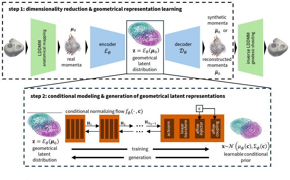
The load-bearing mechanism is the two-step decoupling of representation learning from conditional density estimation. Anatomies are encoded as LDDMM initial momenta $\mu_0 \in \mathbb{R}^{3 \times 720}$; an unregularized autoencoder compresses these momenta into a 44-dimensional latent $z$, and a Glow-style conditional normalizing flow $f_\phi(z,c)$ learns the metadata-dependent density of $z$, with a learnable conditional base $\mathcal{N}(\mu_\phi(c), \Sigma_\phi(c))$, affine injectors, and conditional affine coupling layers. At generation time the flow samples from the conditional base, the inverse flow maps it to $\tilde{z}$, and geodesic shooting converts decoded momenta back into surface meshes. This design is what lets the model vary the latent distribution with metadata without forcing a shared prior during representation learning.
What would settle it
Recompute the KL divergences, Wasserstein distances, point-cloud coverage, and PCA variability ratios using only the 15% held-out test subjects as the real reference population. If CAN-FLOW's advantage over the cVAE baselines shrinks or reverses on out-of-training anatomies, the paper's central distributional-fidelity claim is refuted.
Extended reading notes
Core claim
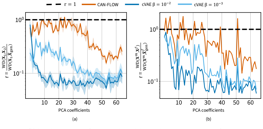
The paper's central claim is that the reason previous conditional anatomy generators underrepresent population variability is architectural: conditional variational autoencoders tie representation learning to a fixed, metadata-agnostic Gaussian prior, so the latent space is regularized toward a single shared distribution. CAN-FLOW breaks that coupling. It first learns an unconstrained, geometry-only latent representation of diffeomorphic shape momenta, then fits a conditional normalizing flow that maps sex, age, and BMI to a learnable Gaussian base distribution and an invertible transformation of that base. On a healthy cohort of 2,208 UK Biobank biventricular meshes, the paper reports that CAN-FLOW outperformed the strongest cVAE baselines on KL divergence for all clinical phenotypes, on most Wasserstein-distance comparisons, on sex- and age-conditioned phenotype distributions, on point-cloud coverage, on within-subgroup spatial variability, and on PCA-based shape-momenta variability, while cVAEs achieved lower minimum matching distance but produced visibly more homogeneous cohorts.
Load-bearing premise
The evaluation assumes that comparing generated cohorts to the full 2,208-subject training cohort measures generalization; if cohort-level metrics are not restricted to the held-out test split, part of the reported distributional agreement could reflect the model matching anatomies it was trained on.
Editorial extensions
If this is right
- Targeted virtual subgroups, such as older male cohorts, can be generated with within-group anatomical variability close to the real subgroup, which is what device and in silico trial studies need.
- A trained CAN-FLOW can produce synthetic biventricular anatomies on demand without sharing individual image-derived meshes, easing privacy and data-access constraints.
- The reported results imply that cVAE-style generators, despite closer nearest-neighbor fidelity, systematically underrepresent tail phenotypes and subgroup variability that matter for cohort-level predictions.
- The conditional healthy-anatomy distribution could be used as a normative reference to score how far an individual heart deviates from expected shape for its sex, age, and BMI, pending validation in diseased cohorts.
Reading between the lines
- Editorial inference: Because the paper's evaluation uses the same post-outlier cohort for training and reference, the distributional metrics may partly reflect memorization; a held-out-only re-analysis is the natural check.
- Editorial inference: Removing 66 Mahalanobis outliers before training likely deletes the most extreme legitimate morphologies, so the claim of preserved tails applies to the retained distribution, not to the rarest real anatomies.
- Editorial inference: A stronger test of generalizability would train on one imaging site or population and evaluate on an external cohort; otherwise the learned healthy-anatomy distribution remains UK Biobank-specific.
- Editorial inference: The static end-diastolic scope means CAN-FLOW does not yet address motion or phase-consistent deformation, so extending it to four-chamber or time-resolved anatomy is an open problem rather than an immediate corollary.
Editorial analysis
A structured set of objections, weighed in public.
Referee Report
Summary. The paper introduces CAN-FLOW, a two-stage conditional generative model for biventricular cardiac anatomy. In the first stage, an autoencoder compresses LDDMM momenta into a geometry-only latent space. In the second stage, a conditional normalizing flow with a metadata-dependent prior, affine injectors, and conditional coupling layers models the distribution of these latents given sex, age, and BMI. The authors compare CAN-FLOW against cVAEs trained over a range of β values on a healthy UK Biobank cohort of 2,208 subjects. Evaluation covers visual plausibility, clinical phenotype distributions (KL divergence, Wasserstein distance), metadata-stratified phenotype trends, subgroup variability, PCA-based shape variability in momenta space, and point-cloud MMD/coverage. The paper reports that CAN-FLOW outperforms cVAEs on most distributional fidelity metrics, with ablations in Appendix B.4 and B.5 showing robustness to architectural choices.
Significance. If the distributional fidelity results survive a proper held-out evaluation, CAN-FLOW is a valuable contribution to virtual cohort generation for cardiac digital twins. The two-step design—decoupling representation learning from conditional density estimation—is a conceptually clean alternative to cVAEs, and the paper evaluates it with a broad set of complementary metrics (KL, Wasserstein, MMD, coverage, PCA ratios). The ablation studies in Appendix B.4 and B.5 are a genuine strength, as they show the advantage is not tied to a single fine-tuned configuration. The paper also clearly defines a train/validation/test split in Section 4.1, indicating awareness of generalization. The central weakness is that the reported quantitative comparisons appear to use the full post-outlier cohort, including the training split, as the real reference distribution, which undermines the generalization claim until re-run on held-out data.
major comments (3)
- [Section 4.1 and Appendix A.5] The distributional evaluation uses the full post-outlier cohort as the real reference, not the held-out test split. Section 4.1 defines a 70/15/15 train/validation/test split, but Sections 2.2 through 2.6 and Appendix A.5 never state that the real cohort subsets are restricted to the test set—for example, the 600 subsampled anatomies in Figure 9 and Appendix A.5.2, the two PCA subsets in Appendix A.5.4, and the subgroup in Section 2.2. Since a flexible normalizing flow with 15 Glow blocks can overfit the training data, the reported KL divergences, Wasserstein distances, MMD/coverage scores, and PCA variability ratios likely reflect, at least in part, memorization of training anatomies rather than generalization to new subjects. The authors should re-run all cohort-level metrics against the held-out test subjects, or at minimum report the train and test metrics separately, and show that CAN-FLOW's advantage over cVAEs persists. This is load-bearing for the paper's headline claim that CAN-FLOW better reproduces the real population distribution.
- [Appendix A.2 and Discussion (tail preservation)] The paper motivates preserving rare but plausible tail anatomies (Introduction, Section 2.3), yet it removes 66 Mahalanobis outliers before training (Appendix A.2) and then uses the same trimmed cohort as the reference in every evaluation. Consequently, the reported metrics only measure fidelity to the post-trimming distribution; they cannot validate preservation of the original extreme tails. The authors should explicitly state whether the removed outliers are included in any reference distribution, and either include them in an additional tail-focused analysis or temper the tail-preservation claim accordingly.
- [Appendix A.5 and Sections 2.3, 2.6] The overall-population comparisons sample age and BMI uniformly from the ranges observed in the real cohort, separately by sex, while the real cohort reference retains its natural joint metadata distribution. This means the synthetic and real cohorts have different metadata marginals, so the reported KL and Wasserstein distances for the whole cohort confound anatomical fidelity with metadata-marginal mismatch. The authors should either sample the synthetic metadata from the real joint metadata distribution (for example, by bootstrapping real metadata vectors) or restrict the overall distributional claims to the metadata-stratified analyses, which are less sensitive to this issue.
minor comments (5)
- [Section 4.3, Eq. (1)] The notation for the composition of flow blocks is typeset awkwardly; consider writing f_phi = f_phi^{(N_b)} ∘ ... ∘ f_phi^{(1)} to make the composition order explicit.
- [Appendix A.1 vs Section 4.1] Appendix A.1 defines N_p = 2274 as the number of anatomies, while Section 4.1 reports 2208 after outlier removal; please clarify whether N_p refers to the pre- or post-outlier count and make the numbering consistent throughout.
- [Section 2.2, Figure 3] The subgroup 'males older than 58 years' is described as containing 674 subjects; please state whether this is the full cohort count or a split-specific count, and report the synthetic cohort size used for comparison.
- [Appendix B.4, Tables B.2–B.4] The phrase 'the largest value in each column is marked in blue' is confusing because lower KL and Wasserstein values are better; presumably the marking indicates the worst value. Please reword to avoid ambiguity.
- [Throughout] Minor typographical and formatting issues include inconsistent number formatting (e.g., '2,208' in the abstract vs '2208' in Section 4.1) and several superscript/subscript renderings in Section 4.3 that should be cleaned up.
Circularity Check
No significant circularity: the model's objectives and the evaluation metrics are not equivalent by construction, and the self-citations are methodological rather than load-bearing.
full rationale
CAN-FLOW's derivation chain is self-contained. The two-stage generator, a geometry-only autoencoder on LDDMM momenta followed by a conditional normalizing flow on the 44-dimensional latent, is trained with standard negative log-likelihood and ELBO losses (Eqs. 1 and 3), while the reported evaluations use independent distributional metrics: histogram-based KL divergence, Wasserstein distance, Chamfer-distance MMD and coverage, and PCA-coefficient Wasserstein ratios (Eqs. A.8-A.16). None of these metrics equals the training objective or a fitted parameter by construction, so the central claim that CAN-FLOW better preserves cohort-level variability is not a renamed fit. The self-citations to Moscoloni et al. [39] supply the LDDMM preprocessing protocol and template settings, but they are methodological and not load-bearing for the novelty claim; no uniqueness theorem or ansatz is imported through self-citation. The Discussion explicitly defers external validation, stating that future work 'will require validation in external cohorts, diseased populations, and underrepresented subgroups,' which is consistent with a limited but non-circular claim. The one validity caveat, that Sections 2.2 to 2.6 and Appendix A.5 compare synthetic cohorts to the full real cohort without stating exclusion of the 70% training split defined in Section 4.1, is a potential in-sample bias in the evaluation, but it does not make any reported quantity equivalent to an input by definition, so it is a correctness risk rather than circularity.
Assumptions & free parameters
free parameters (8)
- Latent dimensionality d_z =
44
- LDDMM control point count N_q =
720
- Normalizing flow blocks N_b =
15
- Metadata embedding dimension g(c) =
12
- LDDMM kernel widths lambda_V and lambda_W =
10
- cVAE regularization beta for main baselines =
10^-2 and 10^-3
- Subgroup stratification thresholds =
age 61, BMI 22
- Outlier removal threshold =
p = 1 - 10^-13 in chi-squared Mahalanobis test
assumptions (6)
- domain assumption The 44-dimensional MSE-trained autoencoder latent space retains all clinically and geometrically relevant variability needed to reproduce phenotypes and metadata trends.
- domain assumption LDDMM momenta with 720 control points and kernel width 10 provide sufficient anatomical correspondence and shape representation.
- domain assumption The healthy UK Biobank subset, after exclusions and outlier removal, is an appropriate normative reference for virtual cohort generation.
- domain assumption The upstream segmentation, mesh fitting, and correspondence pipeline is accurate enough that generated anatomies are physiologically plausible.
- standard math PCA scores are approximately multivariate Gaussian so the chi-squared Mahalanobis threshold is valid for outlier detection.
- ad hoc to paper Comparing synthetic cohorts with uniformly sampled age and BMI to the real cohort with its actual metadata marginal is a valid distributional comparison.
Cite this review
Pith. "Pith review of Flow-based conditional cardiac anatomy generation for virtual cohorts." pith.science (2026). https://pith.science/paper/TM63XFKJ
@misc{pith2026260809460,
  author       = {Pith},
  title        = {Pith review of: Flow-based conditional cardiac anatomy generation for virtual cohorts},
  year         = {2026},
  howpublished = {\url{https://pith.science/paper/TM63XFKJ}},
  note         = {Machine review of arXiv:2608.09460}
}
read the original abstract
Cardiac digital twin research is moving from subject-specific anatomical replicas toward virtual cohorts that represent clinically relevant population subgroups. Yet access to representative imaging-derived anatomy datasets remains limited by cohort size, subgroup sparsity, and data-sharing constraints. Conditional generative models could help address this gap, but virtual cohorts are useful only if they preserve realistic, metadata-dependent anatomical variability. Existing cardiac anatomy generators largely rely on conditional variational autoencoders (cVAEs), which couple representation learning and metadata conditioning through a shared regularized latent prior. We introduce CAN-FLOW, a two-step Conditional ANatomy generation framework based on normalizing FLOWs that first learns geometry-only latent representations of diffeomorphic cardiac shape momenta and then models their sex-, age-, and body-mass-index-dependent distribution with a conditional normalizing flow. We trained CAN-FLOW on 2,208 healthy UK Biobank subjects and compared it with cVAEs across regularization strengths. CAN-FLOW generated plausible stochastic biventricular anatomies that better reproduced clinical phenotype distributions, metadata-dependent trends, subgroup variability, point-cloud coverage, and high-dimensional shape variability. Together, these results establish CAN-FLOW as a shareable framework for generating realistic, stochastically varying, metadata-conditioned biventricular anatomies for virtual cohort construction and in silico clinical trial workflows.
Figures


Figures from the paper (6 more)



Reference graph
Works this paper leans on
-
[35]
H. Dou, N. Ravikumar, A. F. Frangi, A conditional flow variational autoencoder for controllable synthesis of virtual populations of anatomy, in: International Conference on Medical Image Computing and Computer-Assisted Intervention, Springer, 2023, pp. 143–152.doi:10.1007/978-3-031-43990-2_14
-
[1]
Z. Jiang, H. Abbas, K. J. Jang, M. Beccani, J. Liang, S. Dixit, R. Mangharam, In-silico pre-clinical trials for implantable cardioverter defibrillators, in: 2016 38th Annual International Conference of the IEEE Engineering in Medicine and Biology Society (EMBC), IEEE, 2016, pp. 169–172.doi:10.1109/embc.2016.7590667
arXiv 2016
-
[2]
C. M. Augustin, A. Neic, M. Liebmann, A. J. Prassl, S. A. Niederer, G. Haase, G. Plank, Anatomically accurate high resolution modeling of human whole heart electromechanics: A strongly scalable algebraic multigrid solver method for nonlinear deformation, Journal of Computational Physics 305 (2016) 622–646.doi:10.1016/j.jcp.2015.10.045
-
[3]
Corral-Acero, F
J. Corral-Acero, F. Margara, M. Marciniak, C. Rodero, F. Loncaric, Y. Feng, A. Gilbert, J. F. Fernandes, H. A. Bukhari, A. Wajdan, M. V. Martinez, M. S. Santos, M. Shamohammdi, H. Luo, P. Westphal, P. Leeson, P. DiAchille, V. Gurev, M. Mayr, L. Geris, P. Pathmanathan, T. Morrison, R. Cornelussen, F. Prinzen, T. Delhaas, A. Doltra, M. Sitges, E. J. Vigmond...
2020
-
[4]
M. Peirlinck, F. Sahli Costabal, E. Kuhl, Sex differences in drug-induced arrhythmogenesis, Frontiers in Physiology 12 (Aug. 2021).doi:10.3389/fphys.2021.708435
-
[5]
M. Peirlinck, F. S. Costabal, J. Yao, J. M. Guccione, S. Tripathy, Y. Wang, D. Ozturk, P. Segars, T. M. Morrison, S. Levine, E. Kuhl, Precision medicine in human heart modeling: Perspectives, challenges, and opportunities, Biomechanics and Modeling in Mechanobiology 20 (3) (2021) 803–831.doi:10.1007/s10237-021-01421-z
-
[6]
A. Zingaro, M. Bucelli, R. Piersanti, F. Regazzoni, L. Dede’, A. Quarteroni, An electromechanics-driven fluid dynamics model for the simulation of the whole human heart, Journal of Computational Physics 504 (2024) 112885.doi:10.1016/ j.jcp.2024.112885
arXiv 2024
- [7]
Show all 73 references
-
[8]
O. Z. Tikeno˘ gulları, M. Peirlinck, H. Chubb, A. M. Dubin, E. Kuhl, A. L. Marsden, Effects of cardiac growth on electrical dyssynchrony in the single ventricle patient, Computer Methods in Biomechanics and Biomedical Engineering 27 (8) (2023) 1011–1027.doi:10.1080/10255842.20...
2023
-
[9]
Salvador, F
M. Salvador, F. Kong, M. Peirlinck, D. W. Parker, H. Chubb, A. M. Dubin, A. L. Marsden, Digital twinning of cardiac electrophysiology for congenital heart disease, Journal of The Royal Society Interface 21 (215) (Jun. 2024).doi:10.1098/ rsif.2023.0729
2024
-
[10]
S. Qian, D. Ugurlu, E. Fairweather, L. Dal Toso, Y. Deng, M. Strocchi, L. Cicci, R. E. Jones, H. Zaidi, S. Prasad, B. P. Halliday, D. Hammersley, X. Liu, G. Plank, E. Vigmond, R. Razavi, A. A. Young, P. Lamata, M. J. Bishop, S. A. Niederer, Developing cardiac digital twin popu...
2025 doi
-
[11]
S. A. Niederer, Y. Aboelkassem, C. D. Cantwell, C. Corrado, S. Coveney, E. M. Cherry, T. Delhaas, F. H. Fenton, A. V. Panfilov, P. Pathmanathan, G. Plank, M. Riabiz, C. H. Roney, R. W. dos Santos, L. Wang, Creation and application of virtual patient cohorts of heart models, Ph...
2020
-
[12]
K. I. Aycock, T. Battisti, A. Peterson, J. Yao, S. Kreuzer, C. Capelli, S. Pant, P. Pathmanathan, D. M. Hoganson, S. M. Levine, B. A. Craven, Toward trustworthy medical device in silico clinical trials: a hierarchical framework for establishing credibility and strategies for o...
2024 doi
-
[13]
Pathmanathan, K
P. Pathmanathan, K. Aycock, A. Badal, R. Bighamian, J. Bodner, B. A. Craven, S. Niederer, Credibility assessment of in silico clinical trials for medical devices, PLOS Computational Biology 20 (8) (2024) e1012289.doi:10.1371/journal. pcbi.1012289
2024 doi
-
[14]
J. R. Dillon, C. Mauger, D. Zhao, Y. Deng, S. E. Petersen, A. D. McCulloch, A. A. Young, M. P. Nash, An open-source end-to-end pipeline for generating 3D+ t biventricular meshes from cardiac magnetic resonance imag- ing, in: International Conference on Functional Imaging and M...
2025 doi
-
[15]
Ugurlu, S
D. Ugurlu, S. Qian, E. Fairweather, C. Mauger, B. Ruijsink, L. D. Toso, Y. Deng, M. Strocchi, R. Razavi, A. Young, P. Lamata, S. Niederer, M. Bishop, Cardiac digital twins at scale from MRI: Open tools and representative models from 55000 UK Biobank participants, PLOS ONE 20 (...
2025 doi
-
[16]
Doste, J
R. Doste, J. Camps, Z. J. Wang, L. A. Berg, M. Holmes, H. Smith, M. Beetz, L. Li, A. Banerjee, V. Grau, B. Rodriguez, An automated computational pipeline for generating large-scale cohorts of patient-specific ventricu- lar models in electromechanical in silico trials, Computer...
2026
-
[17]
Gonzalez-Martin, M
P. Gonzalez-Martin, M. Webber, F. Chan, S. De, C. C. Topriceanu, D. Falconer, A. Zingaro, J. M. Pozo, C. Butakoff, Y. Rudy, A. D. Hughes, M. Orini, M. Vazquez, J. Aguado-Sierra, G. Captur, A pipeline for developing digital cardiac twins integrating cardiovascular magnetic reso...
2024 doi
-
[18]
M. A. Gsell, B. A. Kl¨ ockl, A. Neic, B. Mautner, C. M. Augustin, E. Zappon, G. Plank, PyMeshTool — a framework for building efficient automated image-based cardiac anatomical twinning workflows in python, Computer Methods and Programs in Biomedicine 285 (2026) 109476.doi:10.1...
2026
-
[19]
Govil, B
S. Govil, B. T. Crabb, Y. Deng, L. Dal Toso, E. Puyol-Ant´ on, K. Pushparajah, S. Hegde, J. C. Perry, J. H. Omens, A. Hsiao, A. A. Young, A. D. McCulloch, A deep learning approach for fully automated cardiac shape modeling in tetralogy of Fallot, Journal of Cardiovascular Magn...
2023 doi
-
[20]
Peloquin, M
D. Peloquin, M. DiMaio, B. Bierer, M. Barnes, Disruptive and avoidable: GDPR challenges to secondary research uses of data, European Journal of Human Genetics 28 (6) (2020) 697–705.doi:10.1038/s41431-020-0596-x
2020 doi
-
[21]
T. J. Littlejohns, J. Holliday, L. M. Gibson, S. Garratt, N. Oesingmann, F. Alfaro-Almagro, J. D. Bell, C. Boultwood, R. Collins, M. C. Conroy, et al., The UK Biobank imaging enhancement of 100,000 participants: rationale, data collection, management and future directions, Nat...
2020 doi
-
[22]
F. Kong, S. Stocker, P. S. Choi, M. Ma, D. B. Ennis, A. L. Marsden, SDF4CHD: Generative modeling of cardiac anatomies with congenital heart defects, Medical Image Analysis 97 (2024) 103293.doi:10.1016/j.media.2024.103293
2024
-
[23]
Sørensen, P
K. Sørensen, P. Diez, J. Margeta, Y. El Youssef, M. Pham, J. J. Pedersen, T. K¨ uhl, O. de Backer, K. Kofoed, O. Camara, R. Paulsen, Spatio-temporal neural distance fields for conditional generative modeling of the heart, in: proceedings of Medical Image Computing and Computer...
2024 doi
-
[24]
Kadry, S
K. Kadry, S. Gupta, F. R. Nezami, E. R. Edelman, Probing the limits and capabilities of diffusion models for the anatomic editing of digital twins, npj Digital Medicine 7 (2024) 354.doi:10.1038/s41746-024-01332-0
2024 doi
-
[25]
M. Qiao, S. Wang, H. Qiu, A. De Marvao, D. P. O’Regan, D. Rueckert, W. Bai, CHeart: A conditional spatio-temporal generative model for cardiac anatomy, IEEE Transactions on Medical Imaging 43 (3) (2024) 1259–1269.doi:10.1109/ TMI.2023.3331982
2024
-
[26]
M. Qiao, K. A. McGurk, S. Wang, P. M. Matthews, D. P. O’Regan, W. Bai, A personalized time-resolved 3D mesh generative model for unveiling normal heart dynamics, Nature Machine Intelligence 7 (5) (2025) 800–811.doi:10.1038/ s42256-025-01035-5
2025
-
[27]
J. L. Bruse, K. McLeod, G. Biglino, H. N. Ntsinjana, C. Capelli, T.-Y. Hsia, M. Sermesant, X. Pennec, A. M. Taylor, S. Schievano, et al., A statistical shape modelling framework to extract 3D shape biomarkers from medical imaging data: assessing arch morphology of repaired coa...
2016
-
[28]
Beetz, J
M. Beetz, J. Corral Acero, A. Banerjee, I. Eitel, E. Zacur, T. Lange, T. Stiermaier, R. Evertz, S. J. Backhaus, H. Thiele, et al., Interpretable cardiac anatomy modeling using variational mesh autoencoders, Frontiers in Cardiovascular Medicine 9 (2022) 983868.doi:10.3389/fcvm....
2022
- [29]
-
[30]
K. Sohn, H. Lee, X. Yan, Learning structured output representation using deep conditional generative models, Advances in neural information processing systems 28 (2015)
2015
-
[31]
Beetz, A
M. Beetz, A. Banerjee, V. Grau, Multi-domain variational autoencoders for combined modeling of MRI-based biventricular anatomy and ECG-based cardiac electrophysiology, Frontiers in Physiology 13 (2022) 886723.doi:10.3389/fphys.2022. 886723
2022 doi
-
[32]
V. V. Kuznetsov, V. A. Moskalenko, D. V. Gribanov, N. Y. Zolotykh, Interpretable feature generation in ECG using a variational autoencoder, Frontiers in Genetics 12 (2021) 638191.doi:10.3389/fgene.2021.638191
2021
-
[33]
J. Peng, M. Beetz, A. Banerjee, M. Chen, V. Grau, Generating virtual populations of 3D cardiac anatomies with Snowflake- Net, in: International Workshop on Statistical Atlases and Computational Models of the Heart, Springer, 2023, pp. 163–173.doi:10.1007/978-3-031-52448-6_16
2023 doi
-
[34]
Beetz, A
M. Beetz, A. Banerjee, V. Grau, Generating subpopulation-specific biventricular anatomy models using conditional point cloud variational autoencoders, in: International Workshop on Statistical Atlases and Computational Models of the Heart, Springer, 2021, pp. 75–83.doi:10.1007...
2021 doi
- [36]
-
[37]
Stein, U
G. Stein, U. Seljak, V. B¨ ohm, G. Aldering, P. Antilogus, C. Aragon, S. Bailey, C. Baltay, S. Bongard, K. Boone, et al., A probabilistic autoencoder for Type Ia supernova spectral time series, The Astrophysical Journal 935 (1) (2022) 5. doi:10.3847/1538-4357/ac7c08
2022 doi
-
[38]
Kaechele, M
F. Kaechele, M. Coblenz, O. Grothe, A comparison of latent space modeling techniques in a plain-vanilla autoencoder setting, Machine Learning 114 (7) (2025) 151.doi:10.1007/s10994-025-06784-3
2025 doi
-
[39]
Moscoloni, C
B. Moscoloni, C. Beeche, J. A. Chirinos, P. Segers, M. Peirlinck, Unveiling sex dimorphism in the healthy cardiac anatomy: fundamental differences between male and female heart shapes, The Journal of Physiology 604 (2025) 5512–5534.doi: 10.1113/JP288667
2025 doi
-
[40]
S. R. St. Pierre, M. Peirlinck, E. Kuhl, Sex matters: A comprehensive comparison of female and male hearts, Frontiers in Physiology 13 (Mar. 2022).doi:10.3389/fphys.2022.831179
2022
-
[41]
Arjovsky, S
M. Arjovsky, S. Chintala, L. Bottou, Wasserstein generative adversarial networks, in: International Conference on Machine Learning, Vol. 70, PMLR, 2017, pp. 214–223
2017
-
[42]
T. M. Cover, J. A. Thomas, Elements of information theory, John Wiley & Sons, 2006
2006
- [43]
-
[44]
Moscoloni, P
B. Moscoloni, P. Segers, M. Peirlinck, Large Deformation Diffeomorphic Cardiac Strain Mapping, Springer Nature Switzer- land, 2025, pp. 207–218.doi:10.1007/978-3-031-94562-5_19
2025 doi
-
[45]
Pentenga, A
P. Pentenga, A. Stroh, W. van Genuchten, W. A. Helbing, M. Peirlinck, Shape Morphing and Slice Shift Correc- tion in Congenital Heart Defect Model Generation, Springer Nature Switzerland, 2023, pp. 347–355.doi:10.1007/ 978-3-031-35302-4_36
2023
-
[46]
S. E. Petersen, P. M. Matthews, J. M. Francis, M. D. Robson, F. Zemrak, R. Boubertakh, A. A. Young, S. Hudson, P. Weale, S. Garratt, et al., UK Biobank’s cardiovascular magnetic resonance protocol, Journal of Cardiovascular Magnetic Resonance 18 (1) (2016) 8.doi:10.1186/s12968...
2016 doi
-
[47]
Mauger, K
C. Mauger, K. Gilbert, A. Suinesiaputra, B. Pontre, J. Omens, A. McCulloch, A. Young, An iterative diffeomorphic algorithm for registration of subdivision surfaces: application to congenital heart disease, in: 2018 40 th Annual In- ternational Conference of the IEEE Engineerin...
2018
-
[48]
Zhang, Iterative point matching for registration of free-form curves and surfaces, International Journal of Computer Vision 13 (2) (1994) 119–152.doi:10.1007/BF01427149
Z. Zhang, Iterative point matching for registration of free-form curves and surfaces, International Journal of Computer Vision 13 (2) (1994) 119–152.doi:10.1007/BF01427149
1994 doi
-
[49]
Schroeder, K
W. Schroeder, K. M. Martin, W. E. Lorensen, The visualization toolkit an object-oriented approach to 3D graphics, Prentice-Hall, Inc., 1998
1998
-
[50]
Durrleman, M
S. Durrleman, M. Prastawa, N. Charon, J. R. Korenberg, S. Joshi, G. Gerig, A. Trouv´ e, Morphometry of anatomical shape complexes with dense deformations and sparse parameters, NeuroImage 101 (2014) 35–49.doi:10.1016/j.neuroimage. 2014.06.043
2014 doi
-
[51]
Bˆ one, M
A. Bˆ one, M. Louis, B. Martin, S. Durrleman, Deformetrica 4: an open-source software for statistical shape analysis, in: Shape in medical imaging: international workshop, ShapeMI 2018, held in conjunction with MICCAI 2018, granada, Spain, september 20, 2018, proceedings, Spri...
2018 doi
- [52]
- [53]
- [54]
-
[55]
Paszke, S
A. Paszke, S. Gross, F. Massa, A. Lerer, J. Bradbury, G. Chanan, T. Killeen, Z. Lin, N. Gimelshein, L. Antiga, A. Des- maison, A. K¨ opf, E. Yang, Z. DeVito, M. Raison, A. Tejani, S. Chilamkurthy, B. Steiner, L. Fang, J. Bai, S. Chin- tala, PyTorch: An imperative style, high-p...
- [56]
-
[57]
Charon, A
N. Charon, A. Trouv´ e, The varifold representation of nonoriented shapes for diffeomorphic registration, SIAM Journal on Imaging Sciences 6 (4) (2013) 2547–2580.doi:10.1137/130918885
2013 doi
-
[58]
P. C. Mahalanobis, Reprint of: On the generalised distance in statistics, Sankhy¯ a: The Indian Journal of Statistics, Series A (2008-) 80 (2018) 1–7.doi:10.1007/s13171-019-00164-5
2018 doi
- [59]
- [60]
- [61]
- [62]
-
[63]
Lugmayr, M
A. Lugmayr, M. Danelljan, L. Van Gool, R. Timofte, SRFlow: Learning the super-resolution space with normalizing flow, in: Computer vision–ECCV 2020: 16 th European conference, glasgow, UK, August 23–28, 2020, proceedings, part v 16, Springer, 2020, pp. 715–732.doi:10.48550/arX...
- [64]
-
[65]
Sullivan, A
B. Sullivan, A. Kaszynski, PyVista: 3D plotting and mesh analysis through a streamlined interface for the Visualization Toolkit (VTK), Journal of Open Source Software 4 (37) (2019) 1450.doi:10.21105/joss.01450
2019 doi
-
[66]
Nakamori, H
S. Nakamori, H. Ismail, L. H. Ngo, W. J. Manning, R. Nezafat, Left ventricular geometry predicts ventricular tach- yarrhythmia in patients with left ventricular systolic dysfunction: a comprehensive cardiovascular magnetic resonance study, Journal of Cardiovascular Magnetic Re...
2017 doi
-
[67]
Medrano-Gracia, B
P. Medrano-Gracia, B. R. Cowan, B. Ambale-Venkatesh, D. A. Bluemke, J. Eng, J. P. Finn, C. G. Fonseca, J. A. Lima, A. Suinesiaputra, A. A. Young, Left ventricular shape variation in asymptomatic populations: the multi-ethnic study of atherosclerosis, Journal of Cardiovascular ...
2014 doi
-
[68]
Virtanen, R
P. Virtanen, R. Gommers, T. E. Oliphant, M. Haberland, T. Reddy, D. Cournapeau, E. Burovski, P. Peterson, W. Weckesser, J. Bright, S. J. van der Walt, M. Brett, J. Wilson, K. J. Millman, N. Mayorov, A. R. J. Nelson, E. Jones, R. Kern, E. Larson, C. J. Carey, ˙I. Polat, Y. Feng...
2020
-
[69]
Van der Maaten, G
L. Van der Maaten, G. Hinton, Visualizing data using t-SNE., Journal of Machine Learning Research 9 (11) (2008)
2008
- [70]
- [71]
- [72]
-
[73]
Elfwing, E
S. Elfwing, E. Uchibe, K. Doya, Sigmoid-weighted linear units for neural network function approximation in reinforcement learning, Neural Networks 107 (2018) 3–11.doi:10.1016/j.neunet.2017.12.012. 32
2018 doi
Reviewed August 11, 2026 · model on record in the stance chip above.
Discussion (0). Continue with ORCID to comment.