REVIEW 4 major objections 6 minor 75 references
Disordered Photosynthetic Aggregates Can Host Functional Vibronic Couplings At Room Temperature
T0 review · 4 major / 6 minor · reviewed 2026-08-06 · deepseek-v4-flash
Pith's one-line read The paper reports direct evidence that large disordered photosynthetic aggregates can host functional vibronic couplings at room temperature, with energetic disorder as the enabling ingredient.
desk verdict Careful P-2D experiments on porphyrin nanotubes give solid evidence for room-temperature Qx-Qy vibronic features, but the disorder-as-vital-ingredient mechanism rests on a sigma borrowed from chlorosomes and is never tested against the 2D observables. read the letter →
The pith
A machine-rendered reading of the paper's core claim, the machinery that carries it, and where it could break.
The reading
What carries the argument
The load-bearing experimental object is the polarization sequence $P = (0^\circ, 0^\circ, +60^\circ, -60^\circ)$, equivalent to $(PA - 3PE)/4$, which cancels isotropic transition-dipole pathways such as $\langle xxxx \rangle$ and leaves only mixed $Q_x$–$Q_y$ pathways such as $\langle xyxy \rangle$ and $\langle xxyy \rangle$. In the 2D and pump-probe experiments, this sequence isolates the signal that reports $Q_x$–$Q_y$ vibronic mixing; the surviving anisotropic beats at 432 and 880 cm$^{-1}$, assigned to out-of-plane pyrrole deformations of the protonated porphyrin ring, are the direct readout. On the theory side, the mechanism is a vibronic exciton Hamiltonian with one explicit quantum vibration, treated without the Born–Oppenheimer approximation, with energetic disorder added directly to the site energies. The argument runs: disorder creates $Q_x$–$Q_y$ electronic mixing, and electronic mixing is the necessary prerequisite for near-resonant vibrations to mediate vibronic coupling and internal conversion.
What would settle it
Measure the site-energy disorder of TPPS nanotubes directly, for example by single-nanotube absorption or low-temperature hole-burning spectroscopy. If the distribution is substantially narrower than $\sigma \approx 200$ cm$^{-1}$, the predicted $Q_x$–$Q_y$ electronic mixing of 22–49% drops toward zero, leaving the proposed disorder mechanism unable to explain the observed cross-peaks and beats. One could also fabricate a low-disorder porphyrin aggregate and check whether the $P$-selected cross-peaks and the 432 and 880 cm$^{-1}$ beats disappear.
Extended reading notes
Core claim
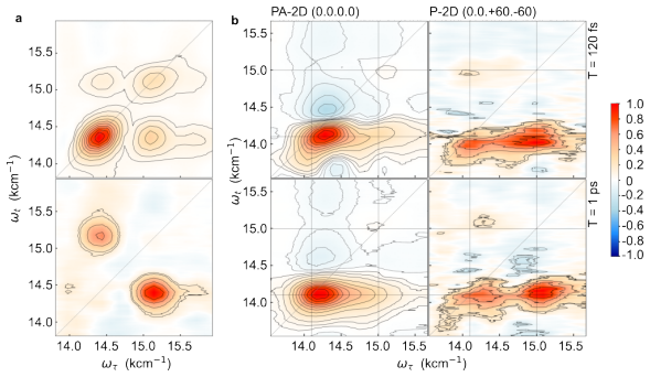
The central claim is that the overlapping vibrational-electronic bands of porphyrin nanotubes, which are chlorophyll-like photosynthetic aggregates, host functional vibronic couplings at room temperature. The evidence is threefold: cross-peaks appear by $T = 30$ fs, they broaden within roughly 150–250 fs indicating rapid exciton delocalization, and anisotropic vibrational quantum beats at 432 and 880 cm$^{-1}$ survive the polarization sequence that selects only mixed $Q_x$–$Q_y$ states, while the 236 and 309 cm$^{-1}$ spectator beats are suppressed. The paper further argues that the mechanism is disorder: with Gaussian energetic disorder of $\sigma = 200$ cm$^{-1}$ per site, the $Q_x$ and $Q_y$ bands acquire 22–49% mixed electronic character across the $Q$ band, and 0–1 vibrational mixing rises from 5% to 19% for the 880 cm$^{-1}$ mode and from 20% to 55% for the 440 cm$^{-1}$ mode. This is presented as the first demonstration that fast internal conversion within the $Q$ band of a large disordered aggregate is driven by robust, resonance-insensitive vibronic couplings at physiological temperature.
Load-bearing premise
The theoretical claim that disorder enables the $Q_x$–$Q_y$ vibronic mixing assumes each porphyrin site carries Gaussian energetic disorder with standard deviation $\sigma = 200$ cm$^{-1}$, a value imported from chlorosome single-particle studies rather than measured on these TPPS nanotubes; if the real disorder is much smaller, the model predicts almost no $Q_x$–$Q_y$ mixing.
Editorial extensions
If this is right
- Room-temperature internal conversion in chlorosome-like aggregates can be driven by vibronic couplings, extending cryogenic evidence for functional vibronic states to physiological conditions.
- Energetic disorder of the order of dense low-frequency Raman-active vibrations with small Huang–Rhys factors may be a usable design principle for artificial light-harvesting antennas.
- Artificial templates built from chlorophyll-like chromophores should reproduce photosynthetic $Q_x$–$Q_y$ physics better than cyanine-based nanotubes.
- The anisotropic quantum-beat signature gives a spectroscopic fingerprint for identifying which vibrations actually promote $Q_x$–$Q_y$ mixing, as opposed to spectator modes.
- Disorder plays a dual role: it enables electronic mixing and widens the resonance range over which vibrations couple, going beyond models that treat disorder only as line broadening.
Reading between the lines
- Editorial inference: the model's quantitative prediction rests on an unmeasured assumption that TPPS nanotubes carry roughly 200 cm$^{-1}$ of Gaussian site disorder; measuring that disorder directly would test the proposed mechanism before it is used as a design rule.
- Editorial inference: the same polarization-selection protocol could be applied to chlorosomes or other chlorophyll aggregates at room temperature, and the paper's mechanism predicts they would show the same surviving anisotropic beats.
- Editorial inference: because the surviving beats are assigned to ground-state wavepackets, the $P$ sequence may serve as a general screening tool for non-adiabatic coupling activity in any multichromophoric assembly, not only nanotubes.
- Editorial inference: the observed 238 fs relaxation between states separated by 315 cm$^{-1}$ within the $Q$ band suggests energetic disorder also creates low-lying dark states, so engineering the disorder distribution could tune excited-state lifetimes in artificial systems.
Signed reviews
Editorial analysis
A structured set of objections, weighed in public.
Referee Report
Summary. This manuscript reports polarization-controlled two-dimensional electronic spectroscopy (P-2DES) and spectrally resolved pump-probe (P-SRPP) measurements on TPPS porphyrin nanotubes at room temperature. The authors observe early-time cross-peaks between the main Q band and its vibronic shoulders, rapid 2D peak broadening, a 238 fs intraband relaxation with a dispersive decay-associated spectrum, and Qx-Qy-specific anisotropic quantum beats at 432 and 880 cm^-1 that survive the P polarization sequence. They interpret these observations as evidence for room-temperature functional vibronic couplings in large chlorophyll-like aggregates. A vibronic exciton Hamiltonian with Gaussian energetic disorder (sigma = 200 cm^-1) is used to show that disorder can induce Qx-Qy electronic mixing and enhance 0-1 vibrational-electronic mixing, leading to the proposed design principle that energetic disorder comparable to dense low-frequency Raman vibrations enables disorder-enhanced vibronic mixing.
Significance. If the central mechanistic claim holds, the paper would be an important contribution: it would extend polarization-based evidence for functional vibronic couplings from cryogenic dimers to room-temperature, large, disordered photosynthetic aggregates, and it would identify energetic disorder as a potentially design-relevant ingredient rather than a purely detrimental factor. The experimental work has notable strengths: the P-sequence suppression is validated on a control molecule (H2Pc), extinction ratios are reported, the key beats are reproducible across multiple trials with error bands, and the vibronic model parameters are constrained by several independent measurements (H-dimer absorption, ring absorption, nanotube diameter from ultracentrifugation). However, the theoretical mechanism is currently supported only by simulations of the linear absorption spectrum and oscillator-strength-weighted electronic/vibrational characters; the nonlinear observables that constitute the paper's central experimental evidence are not computed from the same Hamiltonian.
major comments (4)
- [Disorder enhances Qx − Qy vibronic mixing / Fig. 7] The mechanistic claim that disorder is 'the vital ingredient' is supported only by simulations of the linear absorption spectrum (Fig. 7b,c; Section S5, Eqs. S7–S8). The experiments that carry the central message—early-time cross-peaks, the 238 fs dispersive relaxation, and the 432/880 cm^-1 anisotropic beats—are never computed from the same vibronic exciton Hamiltonian. As a result, the causal link between the disorder-containing model and the observed P-2D cross-peak amplitudes, SRPP kinetics, or beat patterns is asserted rather than demonstrated. The paper should compute at least the relevant third-order response for the model (e.g., P-2D spectra or P-SRPP beat amplitudes), or explicitly delimit the claim to a consistency argument based on absorption alone.
- [Disorder enhances Qx − Qy vibronic mixing / Fig. 7b] The enhancement depends entirely on the value sigma = 200 cm^-1, which is imported from single-chlorosome studies (ref. 65) and not measured for the TPPS nanotubes. Figure 7b shows no Qx-Qy mixing at sigma = 0 and substantial mixing at sigma = 200, so a smaller actual disorder would eliminate the proposed mechanism. A sensitivity scan (e.g., sigma = 0, 50, 100, 200 cm^-1) and, ideally, an independent estimate of energetic disorder in these nanotubes (from single-tube spectroscopy or from the 2D lineshape) are needed before 'disorder is the vital ingredient' can be regarded as established.
- [Selection of mixed Qx − Qy states / Fig. 4c,d] The P-SRPP data were globally fitted with the time constants obtained from the MA-SRPP fit rather than with freely floated parameters. Because the dispersive 238 fs P-DAS line shape is the basis for the 315 cm^-1 intraband gap and the internal-conversion assignment in Figure 5, the analysis should show that the 238 fs component and its dispersive shape are recovered when the P data are fitted independently (or by a global fit with shared rates but P-specific amplitudes); the current constraint may artificially enforce the MA-derived rates.
- [Vibronic Exciton Model / Methods and Section S5] The nanotube simulations use the one-particle approximation (1PA), which the authors acknowledge underestimates resonant vibronic mixing effects in H-aggregates (ref. 63; Fig. S17). Since the central quantitative support for disorder-enhanced 0-1 mixing (19% and 55% values in Fig. 7c and Table S5) is obtained within 1PA, the numbers are likely lower bounds; the paper should state this limitation in the main text and, if possible, test whether the trend survives with a 3PA calculation for a smaller representative aggregate (e.g., a stack of rings) before using the percentages as evidence.
minor comments (6)
- [Abstract and Conclusions] The phrases 'conclusively demonstrate' and 'vital ingredient' overstate the support provided by the current analysis; more cautious language (e.g., 'indicate' or 'suggest') would better match the fact that the nonlinear observables are not simulated from the proposed model.
- [Conclusions] The claim that this is the first instance where Qx-Qy vibronic coupling and fast intraband relaxation in porphyrin nanotubes are revealed by polarization-controlled spectroscopy should be checked against the full literature; as written, the novelty claim is stronger than what is demonstrated.
- [Introduction / Reference 52] The sentence on modes implicated in B-Q Herzberg-Teller coupling cites ref. 52 (Baltuška et al., Opt. Lett. 2002, on visible pulse compression), which appears unrelated; please verify and correct the citation.
- [Figure 2] Given the reported instrument response function FWHM of ~37 fs, the early-time cross-peaks at T = 30 fs should be discussed in relation to pulse-overlap and coherent artifacts; please clarify how these contributions were separated.
- [Polarization scheme / Section S1.3] The relation P = (PA - 3PE)/4 is stated without derivation; a brief derivation or a more explicit reference would help readers verify the orientational averages for the nanotube geometry with distributed dipole orientations.
- [Data and Code Availability] The data are said to be in the paper and SI but are not deposited in a repository, and the code is available only 'upon reasonable request'; archiving the analysis code and processed data would strengthen reproducibility.
Circularity Check
No significant circularity: the P-2D observations are experimental, and the disorder parameter is imported from external chlorosome studies rather than fitted to the target 2D data.
full rationale
The paper's central experimental claims—early-time cross-peaks, rapid broadening, and surviving anisotropic beats—are direct measurements from P-2D/P-SRPP spectroscopies, independent of the theoretical model. The theoretical claim that disorder enhances Qx–Qy vibronic mixing is a forward simulation: the disorder magnitude (σ = 200 cm−1) is taken from single-particle chlorosome studies (ref. 65), not fitted to the present 2D spectra. The model parameters (HR factors, structural angles, ring size) are constrained by independent absorption data for the H-dimer and ring aggregate, and the nanotube simulations then predict vibronic character. Nowhere does the paper define the predicted mixing in terms of the observed beats or cross-peaks, nor does it fit the 2D data and then call that a prediction. Self-citations (refs. 36, 37, 63, 66) supply the vibronic Hamiltonian and polarization formalism, but these are published, independently derived results; they are not used as an unverified premise to force the present conclusion. The main weakness—that the model is not directly tested against the nonlinear 2D spectra—is a validation gap, not a circular reduction. No equation is equivalent to its input by construction, and no fitted parameter is renamed as a prediction. The paper is therefore not circular in the sense defined here; at most it contains minor, non-load-bearing self-citations, warranting a low score.
Assumptions & free parameters
free parameters (4)
- Gaussian energetic disorder sigma =
200 cm^-1
- Huang-Rhys factors and transition dipole strengths =
not stated explicitly
- Tangential and axial angles alpha and beta =
not stated explicitly
- Explicit vibration frequencies =
1250, 880, 440 cm^-1
assumptions (5)
- domain assumption One-particle approximation (1PA) for nanotube calculations
- domain assumption Nearest-neighbor electronic interactions only
- domain assumption Qx-Qy transition dipole angle fixed at 75 degrees
- ad hoc to paper Gaussian energetic disorder with sigma = 200 cm^-1 applies to porphyrin nanotube sites
- domain assumption All modeled vibrational modes can tune the donor-acceptor energy gap equally
Cite this review
Pith. "Pith review of Disordered Photosynthetic Aggregates Can Host Functional Vibronic Couplings At Room Temperature." pith.science (2026). https://pith.science/paper/35OOEGVY
@misc{pith2026250711007,
  author       = {Pith},
  title        = {Pith review of: Disordered Photosynthetic Aggregates Can Host Functional Vibronic Couplings At Room Temperature},
  year         = {2026},
  howpublished = {\url{https://pith.science/paper/35OOEGVY}},
  note         = {Machine review of arXiv:2507.11007}
}
read the original abstract
Photosynthesis relies on a network of chlorophyll-like molecules which together lead to efficient long-range energy funneling. Evidence at cryogenic temperatures suggests that mechanistic details of energy/charge transfer must invoke delocalized vibronic states. Whether these survive at physiological temperature in large photosynthetic aggregates is an open question. Parallel research on artificial templates has relied on cyanines which are unlike chlorophylls. We report two-dimensional electronic spectra of porphyrin nanotubes where we selectively probe mixed Qx-Qy states through polarization control. Early time cross-peaks, their rapid broadening and survival of anisotropic Qx-Qy quantum beats conclusively demonstrate that overlapping vibrational-electronic bands of photosynthetic aggregates indeed host functional vibronic couplings at room temperature. Calculations reveal that disorder is the vital ingredient that dramatically enhances Qx-Qy vibronic mixing across the entire Q band. The parameter regime where energetic disorder is of the order of dense Raman-active vibrations with weak reorganization energies may be the key design principle.
Figures


Figures from the paper (4 more)


Reference graph
Works this paper leans on
-
[1]
Robinson, G. W.; Frosch, R. P. Theory of Electronic Energy Relaxation in the Solid Phase.The Journal of Chemical Physics1962, 37, 1962–1973
work page 1962
-
[2]
E.Molecular mechanisms of photosynthesis; John Wiley & Sons, 2021
Blankenship, R. E.Molecular mechanisms of photosynthesis; John Wiley & Sons, 2021
work page 2021
-
[3]
Kundu, S.; Dani, R.; Makri, N. Tight inner ring architecture and quantum motion of nuclei enable efficient energy transfer in bacterial light harvesting.Science Advances 2022, 8, eadd0023
work page 2022
-
[4]
O’Reilly, E. J.; Olaya-Castro, A. Non-classicality of the molecular vibrations assisting exciton energy transfer at room temperature.Nature Communications 2014, 5, 3012
work page 2014
-
[5]
Cao, J. et al. Quantum biology revisited.Science Advances 2020, 6
work page 2020
-
[6]
Scholes, G. D.; Fleming, G. R.; Chen, L. X.; Aspuru-Guzik, A.; Buchleitner, A.; Coker, D. F.; Engel, G. S.; Van Grondelle, R.; Ishizaki, A.; Jonas, D. M., et al. Using coherence to enhance function in chemical and biophysical systems.Nature 2017, 543, 647–656
work page 2017
-
[7]
Brixner, T.; Hildner, R.; Köhler, J.; Lambert, C.; Würthner, F. Exciton Transport in Molecular Ag- gregates – From Natural Antennas to Synthetic Chromophore Systems.Advanced Energy Materials 2017, 7, 1–33
work page 2017
-
[8]
Jing, H.; Magdaong, N. C. M.; Diers, J. R.; Kirmaier, C.; Bocian, D. F.; Holten, D.; Lindsey, J. S. Dyads with tunable near-infrared donor-acceptor excited-state energy gaps: molecular design and Förster analysis for ultrafast energy transfer.Physical Chemistry Chemical Physics 2023, 1827– 1847
work page 2023
Show all 75 references
-
[9]
R.; Doria, S.; Eisele, D
Caram, J. R.; Doria, S.; Eisele, D. M.; Freyria, F. S.; Sinclair, T. S.; Rebentrost, P.; Lloyd, S.; Bawendi, M. G. Room-Temperature Micron-Scale Exciton Migration in a Stabilized Emissive Molec- ular Aggregate.Nano Letters 2016, 16, 6808–6815
2016
-
[10]
M.; Chen, W
Hart, S. M.; Chen, W. J.; Banal, J. L.; Bricker, W. P.; Dodin, A.; Markova, L.; Vyborna, Y.; Willard, A. P.; Häner, R.; Bathe, M.; Schlau-Cohen, G. S. Engineering couplings for exciton trans- port using synthetic DNA scaffolds.Chem 2021, 7, 752–773
2021
-
[11]
Jansen, T. L. C.; Günther, L. M.; Knoester, J.; Köhler, J. Electronically excited states in cylindrical molecular aggregates: Exciton delocalization, dynamics, and optical response. Chemical Physics Reviews 2024, 5, 041305
2024
-
[12]
F.; Milota, F
Sperling, J.; Nemeth, A.; Hauer, J.; Abramavicius, D.; Mukamel, S.; Kauffmann, H. F.; Milota, F. Excitons and Disorder in Molecular Nanotubes: A 2D Electronic Spectroscopy Study and First 22 Comparison to a Microscopic Model.The Journal of Physical Chemistry A2010, 114, 8179–8...
-
[13]
S.; Jumde, V
Kriete, B.; Bondarenko, A. S.; Jumde, V. R.; Franken, L. E.; Minnaard, A. J.; Jansen, T. L.; Knoester, J.; Pshenichnikov, M. S. Steering Self-Assembly of Amphiphilic Molecular Nanostructures via Halogen Exchange.Journal of Physical Chemistry Letters2017, 8, 2895–2901
-
[14]
T.; Van Amerongen, H.; Boekema, E
Oostergetel, G. T.; Van Amerongen, H.; Boekema, E. J. The chlorosome: a prototype for efficient light harvesting in photosynthesis.Photosynthesis research 2010, 104, 245–255
2010
-
[15]
Spectra of porphyrins.Journal of Molecular Spectroscopy1961, 6, 138–163
Gouterman, M. Spectra of porphyrins.Journal of Molecular Spectroscopy1961, 6, 138–163
-
[16]
K.; Dunietz, B
Song, Y.; Schubert, A.; Maret, E.; Burdick, R. K.; Dunietz, B. D.; Geva, E.; Ogilvie, J. P. Vibronic structure of photosynthetic pigments probed by polarized two-dimensional electronic spectroscopy and ab initio calculations.Chem. Sci. 2019, 10, 8143–8153
2019
-
[17]
Y.; Joo, T
Kim, S. Y.; Joo, T. Coherent Nuclear Wave Packets in Q States by Ultrafast Internal Conversions in Free Base Tetraphenylporphyrin.The Journal of Physical Chemistry Letters2015, 6, 2993–2998
-
[18]
R.; Bocian, D
Diers, J. R.; Bocian, D. F. Qy-excitation resonance Raman spectra of bacteriochlorophyll observed under fluorescence-free conditions. Implications for cofactor structure in photosynthetic proteins. Journal of the American Chemical Society1995, 117, 6629–6630
-
[19]
R.; Freiberg, A
Rätsep, M.; Cai, Z.-L.; Reimers, J. R.; Freiberg, A. Demonstration and interpretation of significant asymmetry in the low-resolution and high-resolution Qy fluorescence and absorption spectra of bacteriochlorophyll a.The Journal of Chemical Physics2011, 134
-
[20]
K.; Jonas, D
Tiwari, V.; Peters, W. K.; Jonas, D. M. Electronic resonance with anticorrelated pigment vibrations drives photosynthetic energy transfer outside the adiabatic framework.Proceedings of the National Academy of Sciences2013, 110, 1203–1208
-
[21]
Jonas, D. M. Vibrational and Nonadiabatic Coherence in 2D Electronic Spectroscopy, the Jahn–Teller Effect, and Energy Transfer.Annual Review of Physical Chemistry2018, 69, 327–352
-
[22]
K.; Jonas, D
Tiwari, V.; Peters, W. K.; Jonas, D. M. Vibronic coherence unveiled.Nature Chemistry 2014, 6, 173–175
2014
-
[23]
Löhner, A.; Kunsel, T.; Röhr, M. I. S.; Jansen, T. L. C.; Sengupta, S.; Würthner, F.; Knoester, J.; Köhler, J. Spectral and Structural Variations of Biomimetic Light-Harvesting Nanotubes.The Jour- nal of Physical Chemistry Letters2019, 10, 2715–2724. 23
-
[24]
Keil, E.; Kumar, A.; Bäuml, L.; Reiter, S.; Thyrhaug, E.; Moser, S.; Duffy, C. D. P.; de Vivie- Riedle, R.; Hauer, J. Reassessing the role and lifetime of Qx in the energy transfer dynamics of chlorophyll a.Chem. Sci. 2025, 16, 1684–1695
2025
-
[25]
S.; Quintela, F.; Russo, M.; Moretti, L.; Moore, A.; Moore, T.; Gust, D.; Prezzi, D.; Scholes, G
Petropoulos, V.; Rukin, P. S.; Quintela, F.; Russo, M.; Moretti, L.; Moore, A.; Moore, T.; Gust, D.; Prezzi, D.; Scholes, G. D.; Molinari, E.; Cerullo, G.; Troiani, F.; Rozzi, C. A.; Maiuri, M. Vibronic Coupling Drives the Ultrafast Internal Conversion in a Functionalized Free...
-
[26]
Shape of the Q band in the absorption spectra of porphyrin nanotubes: Vibronic coupling or exciton effects?Journal of Chemical Physics2010, 133
Stradomska, A.; Knoester, J. Shape of the Q band in the absorption spectra of porphyrin nanotubes: Vibronic coupling or exciton effects?Journal of Chemical Physics2010, 133
-
[27]
The Journal of Physical Chemistry1994, 98, 3612–3618
Akins,D.L.; Zhu,H.-R.; Guo,C.AbsorptionandRamanScatteringbyAggregatedmeso-Tetrakis(p- sulfonatophenyl)porphine. The Journal of Physical Chemistry1994, 98, 3612–3618
-
[28]
S.; Bhat, V
Thomas, A. S.; Bhat, V. N.; Tiwari, V. Rapid scan white light two-dimensional electronic spec- troscopy with 100 kHz shot-to-shot detection.The Journal of Chemical Physics2023, 159
-
[29]
Jonas, D. M. Two-dimensional femtosecond spectroscopy.Annual review of physical chemistry2003, 54, 425–463
-
[30]
W.; Mohan T
Sil, S.; Tilluck, R. W.; Mohan T. M., N.; Leslie, C. H.; Rose, J. B.; Domínguez-Martín, M. A.; Lou, W.; Kerfeld, C. A.; Beck, W. F. Excitation energy transfer and vibronic coherence in intact phycobilisomes. Nature Chemistry 2022, 14, 1286–1294
2022
-
[31]
D.; Ogilvie, J
Fuller, F. D.; Ogilvie, J. P. Experimental Implementations of Two-Dimensional Fourier Transform Electronic Spectroscopy.Annual Review of Physical Chemistry2015, 66, 667–690
-
[32]
L.; Schlau-Cohen, G
Read, E. L.; Schlau-Cohen, G. S.; Engel, G. S.; Wen, J.; Blankenship, R. E.; Fleming, G. R. Visual- ization of excitonic structure in the Fenna-Matthews-Olson photosynthetic complex by polarization- dependent two-dimensional electronic spectroscopy.Biophysical journal 2008, 95...
2008
-
[33]
M.; Yang, N.; Zanni, M
Farrell, K. M.; Yang, N.; Zanni, M. T. A polarization scheme that resolves cross-peaks with transient absorption and eliminates diagonal peaks in 2D spectroscopy.Proceedings of the National Academy of Sciences 2022, 119, e2117398119
2022
-
[34]
T.; Hitchcock, A.; MacGregor-Chatwin, C.; Iwanicki, A.; Ghosh, I.; Shen, Q.; Hunter, C
Sohoni, S.; Lloyd, L. T.; Hitchcock, A.; MacGregor-Chatwin, C.; Iwanicki, A.; Ghosh, I.; Shen, Q.; Hunter, C. N.; Engel, G. S. Phycobilisome’s exciton transfer efficiency relies on an energetic funnel driven by chromophore–linker protein interactions.Journal of the American Ch...
-
[35]
Quantum coherence as a witness of vibroni- cally hot energy transfer in bacterial reaction center.Science Advances 2017, 3, e1603141
Paleček, D.; Edlund, P.; Westenhoff, S.; Zigmantas, D. Quantum coherence as a witness of vibroni- cally hot energy transfer in bacterial reaction center.Science Advances 2017, 3, e1603141
2017
-
[36]
Bhattacharyya, A.; Sahu, A.; Patra, S.; Tiwari, V. Low-and high-frequency vibrations synergisti- cally enhance singlet exciton fission through robust vibronic resonances.Proceedings of the National Academy of Sciences2023, 120, e2310124120
-
[37]
Isolating non-adiabatically enhanced ground state quantum beats through two-dimensional electronic spectroscopy.The Journal of Chemical Physics2025, 162
Sahu, A.; Tiwari, V. Isolating non-adiabatically enhanced ground state quantum beats through two-dimensional electronic spectroscopy.The Journal of Chemical Physics2025, 162
-
[38]
Dynamic intensity borrowing in porphyrin J-aggregates revealed by sub-5-fs spectroscopy.Springer Series in Chemical Physics2001, 66, 604–606
Kano, H.; Saito, T.; Kobayashi, T. Dynamic intensity borrowing in porphyrin J-aggregates revealed by sub-5-fs spectroscopy.Springer Series in Chemical Physics2001, 66, 604–606
-
[39]
D.; Wiederrecht, G
Wan, Y.; Stradomska, A.; Fong, S.; Guo, Z.; Schaller, R. D.; Wiederrecht, G. P.; Knoester, J.; Huang, L. Exciton level structure and dynamics in tubular porphyrin aggregates.Journal of Physical Chemistry C 2014, 118, 24854–24865
2014
-
[40]
Principles of Nonlinear Optical Spectroscopy; Oxford University Press: New York, 1995; Vol
Mukamel, S. Principles of Nonlinear Optical Spectroscopy; Oxford University Press: New York, 1995; Vol. 6; p 543
1995
-
[41]
Two-Dimensional ElectronicSpectroscopyRevealsUltrafastEnergyDiffusioninChlorosomes
Dostál, J.; Mančal, T.; Augulis, R.-n.; Vácha, F.; Pšenčík, J.; Zigmantas, D. Two-Dimensional ElectronicSpectroscopyRevealsUltrafastEnergyDiffusioninChlorosomes. Journal of the American Chemical Society 2012, 134, 11611–11617
2012
-
[42]
Kriete, B.; Lüttig, J.; Kunsel, T.; Malý, P.; Jansen, T. L. C.; Knoester, J.; Brixner, T.; Pshenich- nikov, M. S. Interplay between structural hierarchy and exciton diffusion in artificial light harvesting. Nature Communications 2019, 10, 4615
2019
-
[43]
Coherent Oscillations in Chlorosome Elucidated by Two-Dimensional Electronic Spectroscopy.The Journal of Physical Chemistry Letters 2014, 5, 1386–1392
Jun, S.; Yang, C.; Isaji, M.; Tamiaki, H.; Kim, J.; Ihee, H. Coherent Oscillations in Chlorosome Elucidated by Two-Dimensional Electronic Spectroscopy.The Journal of Physical Chemistry Letters 2014, 5, 1386–1392
2014
-
[44]
W.; McHale, J
Leishman, C. W.; McHale, J. L. Light-Harvesting Properties and Morphology of Porphyrin Nanos- tructures Depend on Ionic Species Inducing Aggregation.Journal of Physical Chemistry C2015, 119, 28167–28181
-
[45]
W.; McHale, J
Leishman, C. W.; McHale, J. L. Morphologically Determined Excitonic Properties of Porphyrin Aggregates in Alcohols with Variable Acidity.Journal of Physical Chemistry C2016, 120, 15496– 15508
-
[46]
M.; Augulis, R.; Stuart, M
Vlaming, S. M.; Augulis, R.; Stuart, M. C.; Knoester, J.; Van Loosdrecht, P. H. Exciton spectra and the microscopic structure of self-assembled porphyrin nanotubes.Journal of Physical Chemistry B 2009, 113, 2273–2283. 25
2009
-
[47]
I.; Zhu, Y.; Lin, S.; Blankenship, R
Savikhin, S.; van Noort, P. I.; Zhu, Y.; Lin, S.; Blankenship, R. E.; Struve, W. S. Ultrafast en- ergy transfer in light-harvesting chlorosomes from the green sulfur bacterium Chlorobium tepidum. Chemical physics 1995, 194, 245–258
1995
-
[48]
Time-resolved fluorescence and absorption spectroscopies of porphyrin J-aggregates
Kano, H.; Kobayashi, T. Time-resolved fluorescence and absorption spectroscopies of porphyrin J-aggregates. Journal of Chemical Physics2002, 116, 184–195
-
[49]
Akins,D.L.; Özçelik,S.; Zhu,H.-R.; Guo,C.Fluorescencedecaykineticsandstructureofaggregated tetrakis (p-sulfonatophenyl) porphyrin.The Journal of Physical Chemistry1996, 100, 14390–14396
-
[50]
R.; Buda, F.; Sevink, G
Eric, V.; Li, X.; Dsouza, L.; Huijser, A.; Holzwarth, A. R.; Buda, F.; Sevink, G. A.; de Groot, H. J.; Jansen, T. L. Observation of Dark States in Two-Dimensional Electronic Spectra of Chlorosomes. The Journal of Physical Chemistry B2024, 128, 3575–3584
-
[51]
Tang, J.; Albrecht, A. C. InRaman Spectroscopy: Theory and Practice; Szymanski, H. A., Ed.; Springer US: Boston, MA, 1970; pp 33–68
1970
-
[52]
Visible pulse compression to 4 fs by optical parametric ampli- fication and programmable dispersion control.Opt
Baltuška, A.; Fuji, T.; Kobayashi, T. Visible pulse compression to 4 fs by optical parametric ampli- fication and programmable dispersion control.Opt. Lett. 2002, 27, 306–308
2002
-
[53]
Resonance Raman spectroscopy study of protonated porphyrin
Gorski, A.; Starukhin, A.; Stavrov, S.; Gawinkowski, S.; Waluk, J. Resonance Raman spectroscopy study of protonated porphyrin. Spectrochimica Acta Part A: Molecular and Biomolecular Spec- troscopy 2017, 173, 350–355
2017
-
[54]
S.; Kruk, M
Starukhin, A. S.; Kruk, M. M.; Gladkova, O. L.; Maes, W. Transformations in the fluorescence line narrowing spectra of porphine upon the formation of diprotonated species.Macroheterocycles 2011, 4, 85–88
2011
-
[55]
Stone, A.; Fleischer, E. B. The molecular and crystal structure of porphyrin diacids.Journal of the American Chemical Society1968, 90, 2735–2748
-
[56]
L.; Starukhin, A
Gladkova, O. L.; Starukhin, A. S.; Kruk, M. M. Influence of spin-orbit coupling on vibronic spectra of metalocomplexes porphin: Manifestation of out-of-plane vibrations.Optics and Spectroscopy2011, 110, 234–241
-
[57]
M.; Kincaid, J
Burke, J. M.; Kincaid, J. R.; Spiro, T. G. Resonance Raman spectra and vibrational modes of iron(III) tetraphenylporphine .mu.-oxo dimer. Evidence for phenyl interaction and lack of dimer splitting. Journal of the American Chemical Society1978, 100, 6077–6083
-
[58]
N.; Patra, S.; Tiwari, V
Sahu, A.; Bhat, V. N.; Patra, S.; Tiwari, V. High-sensitivity fluorescence-detected multidimen- sional electronic spectroscopy through continuous pump–probe delay scan.The Journal of Chemical Physics 2023, 158, 024201. 26
2023
-
[59]
M.; Fleming, G
Jean, J. M.; Fleming, G. R. Competition between energy and phase relaxation in electronic curve crossing processes.The Journal of Chemical Physics1995, 103, 2092–2101
-
[60]
Tiwari,V.; Peters,W.K.; Jonas,D.M.Electronicenergytransferthroughnon-adiabaticvibrational- electronic resonance. I. Theory for a dimer.The Journal of Chemical Physics2017, 147, 154308
-
[61]
Tiwari, V.; Jonas, D. M. Electronic energy transfer through non-adiabatic vibrational-electronic resonance. II. 1D spectra for a dimer.The Journal of Chemical Physics2018, 148, 84308
-
[62]
Sahu, A.; Tiwari, V. Vibrations that do not promote vibronic coupling can dominate observed lineshapes in two-dimensional electronic spectroscopy.The Journal of Physical Chemistry Letters 2023, 14, 4617–4624
2023
-
[63]
S.; Tiwari, V
Sahu, A.; Kurian, J. S.; Tiwari, V. Vibronic resonance is inadequately described by one-particle basis sets.The Journal of Chemical Physics2020, 153, 224114
-
[64]
V.; Richard, A
Hollingsworth, J. V.; Richard, A. J.; Vicente, M. G. H.; Russo, P. S. Characterization of the self- assembly of meso-tetra(4-sulfonatophenyl) porphyrin (H 2TPPS 4-) in aqueous solutions.Biomacro- molecules 2012, 13, 60–72
2012
-
[65]
M.; Jendrny, M.; Bloemsma, E
Gunther, L. M.; Jendrny, M.; Bloemsma, E. A.; Tank, M.; Oostergetel, G. T.; Bryant, D. A.; Knoester, J.; Köhler, J. Structure of Light-Harvesting Aggregates in Individual Chlorosomes.The Journal of Physical Chemistry B2016, 120, 5367–5376
-
[66]
Vibronic resonance along effective modes mediates selective energy transfer in excitonically coupled aggregates.The Journal of Chemical Physics2022, 156, 184115
Patra, S.; Tiwari, V. Vibronic resonance along effective modes mediates selective energy transfer in excitonically coupled aggregates.The Journal of Chemical Physics2022, 156, 184115
-
[67]
P.; Zheng, W.; Chuang, C.; Bailey, A
Deshmukh, A. P.; Zheng, W.; Chuang, C.; Bailey, A. D.; Williams, J. A.; Sletten, E. M.; Egel- man, E. H.; Caram, J. R. Near-atomic-resolution structure of J-aggregated helical light-harvesting nanotubes. Nature Chemistry 2024, 16, 800–808
2024
-
[68]
L.; Li, X.; Dsouza, L.; Frehan, S
Eric, V.; Castro, J. L.; Li, X.; Dsouza, L.; Frehan, S. K.; Huijser, A.; Holzwarth, A. R.; Buda, F.; Sevink, G. J. A.; de Groot, H. J. M.; Jansen, T. L. C. Ultrafast Anisotropy Decay Reveals Structure and Energy Transfer in Supramolecular Aggregates.The Journal of Physical Che...
-
[69]
Unraveling the nature of coherent beatings in chlorosomes.The Journal of Chemical Physics2014, 140, 115103
Dostál, J.; Mančal, T.; Vácha, F.; Pšenčík, J.; Zigmantas, D. Unraveling the nature of coherent beatings in chlorosomes.The Journal of Chemical Physics2014, 140, 115103
-
[70]
M.; Moran, A
Womick, J. M.; Moran, A. M. Vibronic Enhancement of Exciton Sizes and Energy Transport in Photosynthetic Complexes.J. Phys. Chem. B2011, 115, 1347–1356. 27
-
[71]
M.; Chowdhury, R.; Murto, P.; Gillett, A
Ghosh, P.; Alvertis, A. M.; Chowdhury, R.; Murto, P.; Gillett, A. J.; Dong, S.; Sneyd, A. J.; Cho, H.-H.; Evans, E. W.; Monserrat, B.; Li, F.; Schnedermann, C.; Bronstein, H.; Friend, R. H.; Rao, A. Decoupling excitons from high-frequency vibrations in organic molecules.Nature...
2024
-
[72]
K.; Moix, J
Chuang, C.; Lee, C. K.; Moix, J. M.; Knoester, J.; Cao, J. Quantum Diffusion on Molecular Tubes: Universal Scaling of the 1D to 2D Transition.Phys. Rev. Lett.2016, 116, 196803
2016
-
[73]
Yang, X.; Bittner, E. R. Intramolecular Charge- and Energy-Transfer Rates with Reduced Modes: Comparison to Marcus Theory for Donor–Bridge–Acceptor Systems.The Journal of Physical Chem- istry A 2014, 118, 5196–5203, PMID: 24983415
2014
-
[74]
N.; Thomas, A
Bhat, V. N.; Thomas, A. S.; Bhattacharyya, A.; Tiwari, V. Rapid scan white light pump–probe spectroscopy with 100 kHz shot-to-shot detection.Optics Continuum 2023, 2, 1981–1995
2023
-
[75]
Two-dimensional electronic spectroscopy with birefringent wedges.Review of Scientific Instruments2014, 85, 123107
Réhault, J.; Maiuri, M.; Oriana, A.; Cerullo, G. Two-dimensional electronic spectroscopy with birefringent wedges.Review of Scientific Instruments2014, 85, 123107. Acknowledgements A.S.T. acknowledges the Prime Ministers’ Research Fellowship, MoE, India. V.N.B. acknowledges th...
2023
Reviewed August 6, 2026 · model on record in the stance chip above.
Discussion (0). Continue with ORCID to comment.