Prototype Memory-Guided Training-Free Anomaly Classification and Localization in Prenatal Ultrasound
Pith reviewed 2026-07-02 14:32 UTC · model grok-4.3
The pith
A training-free framework uses a memory bank of multi-granular prototypes to classify and localize prenatal ultrasound anomalies with only a few reference images per class.
A machine-rendered reading of the paper's core claim, the machinery that carries it, and where it could break.
Core claim
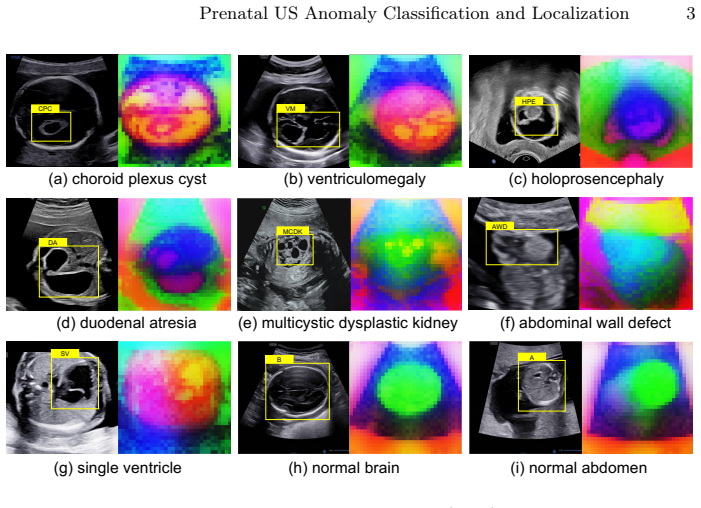
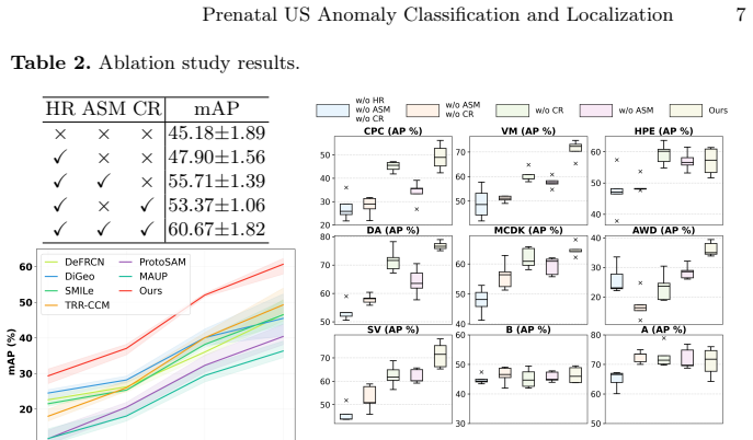
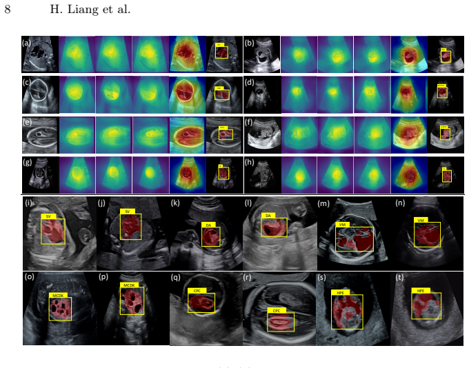
The paper claims that a training-free framework built around a memory bank of multi-granular prototypes, a prototype-driven soft merging mechanism for region detection, and a class-aware refinement step based on prototype consistency can achieve multi-class anomaly classification and localization in prenatal ultrasound using only a few reference images per class, and that this framework outperforms existing methods on a multi-center dataset of 1,149 cases comprising 2,357 images across 9 categories.
What carries the argument
Memory bank of multi-granular prototypes that stores class-level semantics and anomaly characteristics, paired with soft merging to aggregate features for localization and consistency-based refinement for classification.
If this is right
- The framework can be applied directly in clinical settings where collecting large annotated datasets is impractical.
- New anomaly categories can be added by supplying a few reference images without retraining the system.
- Both classification and localization outputs are produced from the same prototype-based process.
- Performance depends on the representativeness of the chosen reference prototypes rather than overall dataset scale.
Where Pith is reading between the lines
- The same prototype memory structure could be tested on other scarce-data medical imaging tasks such as rare tumor detection in MRI.
- If the prototypes prove stable across ultrasound machines, the method could reduce the need for site-specific retraining in multi-hospital deployments.
- Extending the refinement step to handle temporal sequences of scans might improve detection of evolving fetal anomalies.
- The approach suggests that explicit prototype storage can substitute for learned embeddings in other few-shot anomaly tasks.
Load-bearing premise
A small set of reference images per class is enough to capture the necessary class semantics and anomaly variation without any model training or fine-tuning.
What would settle it
Running the method on the described multi-center dataset with the stated few reference images per class and finding that it does not outperform the competitors in classification accuracy or localization metrics.
Figures

read the original abstract
Prenatal anomaly classification and localization is of critical importance for fetal health and pregnancy management. Although ultrasound (US) is the primary modality for prenatal screening, accurate diagnosis remains challenging due to the low prevalence and high heterogeneity of anomalies. Existing deep learning methods for prenatal tasks rely on large-scale annotated datasets, which are difficult to obtain in practice. Although few-shot learning alleviates data scarcity, it typically requires fine-tuning for new categories, limiting its practicality in resource-limited clinical settings. To address these challenges, we propose a training-free framework for multi-class prenatal US anomaly classification and localization that operates with only a few reference images per class, representing the first exploration of this setting. Our framework comprises three key components: (1) a memory bank with multi-granular prototypes that explicitly models both class-level semantics and anomaly characteristics; (2) a prototype-driven soft merging mechanism that aggregates discriminative features to detect the anomaly region; and (3) a class-aware refinement strategy that leverages prototype consistency to improve category prediction. Extensively validated on a multi-center prenatal US dataset containing 1,149 cases, with a total of 2,357 images and 9 categories, our proposed method outperforms the competitors.
Editorial analysis
A structured set of objections, weighed in public.
Referee Report
Summary. The manuscript proposes a training-free framework for multi-class anomaly classification and localization in prenatal ultrasound images. It constructs a memory bank of multi-granular prototypes from a few reference images per class to model both class semantics and anomaly characteristics, applies prototype-driven soft merging to aggregate features for anomaly region detection, and uses class-aware refinement based on prototype consistency for final category prediction. The central claim is that this approach outperforms existing methods on a multi-center dataset of 1,149 cases (2,357 images across 9 categories) without any training or fine-tuning.
Significance. If the empirical results hold, the training-free design with explicit multi-granular prototype modeling represents a practical advance for data-scarce medical imaging domains, particularly prenatal screening where large annotated datasets are hard to obtain. The avoidance of fine-tuning for new categories directly addresses a key limitation of few-shot learning methods in clinical settings.
minor comments (2)
- [Abstract] Abstract: The statement that the method 'outperforms the competitors' is unsupported by any metrics, baselines, or statistical details. Adding at least the primary quantitative results (e.g., accuracy, AUC, or localization IoU) would make the abstract self-contained and allow immediate evaluation of the central claim.
- [Introduction] The manuscript states it is 'the first exploration' of the training-free few-reference setting for this task. A brief comparison to the closest prior training-free or prototype-based methods in medical imaging would clarify the novelty boundary.
Simulated Author's Rebuttal
We thank the referee for the positive summary of our manuscript, the recognition of its significance for data-scarce medical imaging domains, and the recommendation for minor revision. The referee's description accurately reflects the proposed training-free framework, multi-granular prototypes, soft merging, and class-aware refinement, as well as the validation on the 1,149-case multi-center dataset.
Circularity Check
No significant circularity
full rationale
The paper describes a training-free prototype memory framework for prenatal US anomaly classification and localization, with claims of outperformance on a 1,149-case multi-center dataset. The abstract and visible text contain no equations, derivations, parameter-fitting steps, or mathematical predictions. No load-bearing steps reduce by construction to inputs, self-definitions, or self-citations. The central claims are empirical and descriptive rather than derived, making the derivation chain self-contained with no circularity.
Axiom & Free-Parameter Ledger
Reference graph
Works this paper leans on
-
[1]
arXiv preprint arXiv:2407.07042 (2024)
Ayzenberg, L., Giryes, R., Greenspan, H.: Protosam: One-shot medical image seg- mentation with foundational models. arXiv preprint arXiv:2407.07042 (2024)
-
[2]
Ultrasound in Obstetrics and Gynecology61(1) (2023)
Bilardo, C., Chaoui, R., Hyett, J., Kagan, K., Karim, J., Papageorghiou, A., Poon, L., Salomon, L., et al.: Isuog practice guidelines (updated): performance of 11–14- week ultrasound scan. Ultrasound in Obstetrics and Gynecology61(1) (2023)
2023
-
[3]
In: Proceedings of the IEEE/CVF international conference on computer vision
Caron, M., Touvron, H., Misra, I., Jégou, H., Mairal, J., Bojanowski, P., Joulin, A.: Emerging properties in self-supervised vision transformers. In: Proceedings of the IEEE/CVF international conference on computer vision. pp. 9650–9660 (2021)
2021
-
[4]
arXiv preprint arXiv:2506.11136 (2025)
Couairon, P., Chambon, L., Serrano, L., Haugeard, J.E., Cord, M., Thome, N.: Ja- far: Jack up any feature at any resolution. arXiv preprint arXiv:2506.11136 (2025)
-
[5]
Computer Methods and Programs in Biomedicine p
Dou, H., Huang, Y., Huang, Y., Yang, X., Zhen, C., Zhang, Y., Xiong, Y., Huang, W., Ni, D.: Standard plane localization using denoising diffusion model with multi- scale guidance. Computer Methods and Programs in Biomedicine p. 108619 (2025)
2025
-
[6]
In: International Conference on Medical Image Com- puting and Computer-Assisted Intervention
Huang, Y., Xu, Y., Dou, H., Deng, J., Yang, X., Zheng, H., Ni, D.: Uncertainty- aware diffusion and reinforcement learning for joint plane localization and anomaly diagnosis in 3d ultrasound. In: International Conference on Medical Image Com- puting and Computer-Assisted Intervention. pp. 650–660. Springer (2025)
2025
-
[7]
data from two birth-defect reg- istries
Laurichesse Delmas, H., Kohler, M., Doray, B., Lémery, D., Francannet, C., Quistrebert, J., Marie, C., Perthus, I.: Congenital unilateral renal agenesis: preva- lence, prenatal diagnosis, associated anomalies. data from two birth-defect reg- istries. Birth defects research109(15), 1204–1211 (2017)
2017
-
[8]
In: International Conference on Medical Image Computing and Computer-Assisted Intervention
Liang, H., Xu, J., Zhang, Y., Huang, Y., Zhang, Y., Yang, X., Li, R., Deng, X., Liu, Y., Tao, G., et al.: Medical-knowledge driven multiple instance learning for classifying severe abdominal anomalies on prenatal ultrasound. In: International Conference on Medical Image Computing and Computer-Assisted Intervention. pp. 344–354. Springer (2025)
2025
-
[9]
Liang et al
Lin, M., He, X., Guo, H., He, M., Zhang, L., Xian, J., Lei, T., Xu, Q., Zheng, J., Feng,J.,etal.:Useofreal-timeartificialintelligenceindetectionofabnormalimage 10 H. Liang et al. patterns in standard sonographic reference planes in screening for fetal intracranial malformations. Ultrasound in Obstetrics & Gynecology59(3), 304–316 (2022)
2022
-
[10]
arXiv preprint arXiv:2509.06467 (2025)
Liu, C., Chen, Y., Shi, H., Lu, J., Jian, B., Pan, J., Cai, L., Wang, J., et al.: Does dinov3 set a new medical vision standard? benchmarking 2d and 3d classification, segmentation, and registration. arXiv preprint arXiv:2509.06467 (2025)
-
[11]
In: Interna- tional Conference on Medical Image Computing and Computer-Assisted Interven- tion
Liu, Y., Xiao, H., Chai, J., Zhang, Y., Wang, R., et al.: Synpo: Boosting training- free few-shot medical segmentation via high-quality negative prompts. In: Interna- tional Conference on Medical Image Computing and Computer-Assisted Interven- tion. pp. 594–603. Springer (2025)
2025
-
[12]
In: Proceedings of the AAAI conference on artificial intelligence
Lu, X., Diao, W., Mao, Y., Li, J., et al.: Breaking immutable: Information-coupled prototype elaboration for few-shot object detection. In: Proceedings of the AAAI conference on artificial intelligence. vol. 37, pp. 1844–1852 (2023)
2023
-
[13]
Ma, J., Niu, Y., Xu, J., et al.: Digeo: Discriminative geometry-aware learning for generalizedfew-shotobjectdetection.In:ProceedingsoftheIEEE/CVFconference on computer vision and pattern recognition. pp. 3208–3218 (2023)
2023
-
[14]
In: European Conference on Computer Vision
Majee, A., Sharp, R., Iyer, R.: Smile: Leveraging submodular mutual information for robust few-shot object detection. In: European Conference on Computer Vision. pp. 350–366. Springer (2024)
2024
-
[15]
DINOv2: Learning Robust Visual Features without Supervision
Oquab, M., Darcet, T., Moutakanni, T., Vo, H., Szafraniec, M., Khalidov, V., Fernandez, P., Haziza, D., Massa, F., El-Nouby, A., et al.: Dinov2: Learning robust visual features without supervision. arXiv preprint arXiv:2304.07193 (2023)
work page internal anchor Pith review Pith/arXiv arXiv 2023
-
[16]
Frontiers in Surgery9, 891896 (2022)
Pechriggl, E., Blumer, M., Tubbs, R.S., Olewnik, Ł., Konschake, M., Fortélny, R., Stofferin, H., et al.: Embryology of the abdominal wall and associated malforma- tions—a review. Frontiers in Surgery9, 891896 (2022)
2022
-
[17]
In: Proceedings of the IEEE/CVF international conference on computer vision
Qiao, L., Zhao, Y., Li, Z., Qiu, X., Wu, J., Zhang, C.: Defrcn: Decoupled faster r- cnn for few-shot object detection. In: Proceedings of the IEEE/CVF international conference on computer vision. pp. 8681–8690 (2021)
2021
-
[18]
In: International conference on machine learning
Radford, A., Kim, J.W., Hallacy, C., Ramesh, A., Goh, G., Agarwal, S., Sastry, G., Askell, A., Mishkin, P., Clark, J., et al.: Learning transferable visual models from natural language supervision. In: International conference on machine learning. pp. 8748–8763. PmLR (2021)
2021
-
[19]
Ultrasound in Obstetrics and Gynecology59(6), 840–856 (2022)
Salomon, L., Alfirevic, Z., Berghella, V., Bilardo, C., Chalouhi, G., Costa, F.D.S., Hernandez-Andrade, E., Malinger, G., Munoz, H., Paladini, D., et al.: Isuog prac- tice guidelines (updated): performance of the routine mid-trimester fetal ultra- sound scan. Ultrasound in Obstetrics and Gynecology59(6), 840–856 (2022)
2022
-
[20]
Siméoni, O., Vo, H.V., Seitzer, M., Baldassarre, F., Oquab, M., Jose, C., Khali- dov, V., Szafraniec, M., Yi, S., Ramamonjisoa, M., et al.: Dinov3. arXiv preprint arXiv:2508.10104 (2025)
work page internal anchor Pith review Pith/arXiv arXiv 2025
-
[21]
BMC Public Health25(1), 449 (2025)
Xie, X., Pei, J., et al.: Global birth prevalence of major congenital anomalies: a systematic review and meta-analysis. BMC Public Health25(1), 449 (2025)
2025
-
[22]
Medical Image Analysis72, 102119 (2021)
Yang, X., Huang, Y., Huang, R., Dou, H., Li, R., Qian, J., Huang, X., Shi, W., Chen, C., Zhang, Y., et al.: Searching collaborative agents for multi-plane local- ization in 3d ultrasound. Medical Image Analysis72, 102119 (2021)
2021
-
[23]
arXiv preprint arXiv:2508.11032 (2025)
Yang, Y., Su, G., Hu, J., Sammarco, F., Geiping, J., Wolfers, T.: Medsamix: A training-free model merging approach for medical image segmentation. arXiv preprint arXiv:2508.11032 (2025)
-
[24]
3d ai-enhanced ultrasound for fetal crown-rump length evaluation in the first trimester
Zhang, Y., Huang, Y., Chen, C., Hu, X., Pan, W., Luo, H., Huang, Y., Wang, H., Cao, Y., Yi, Y., et al.: Comparative study of 2d vs. 3d ai-enhanced ultrasound for fetal crown-rump length evaluation in the first trimester. BMC Pregnancy and Childbirth25(1), 766 (2025) Prenatal US Anomaly Classification and Localization 11
2025
-
[25]
Nature Communications (2026)
Zhang, Y., Huang, Y., Dou, H., Zhu, X., Ling, C., Yang, Z., Liang, L., Li, J., Liang, S., Li, R., et al.: Artificial intelligence for detecting fetal orofacial clefts and advancing medical education. Nature Communications (2026)
2026
-
[26]
Radiology: Artificial Intelligence7(4), e240498 (2025)
Zhang, Y., Yang, X., Ji, C., Hu, X., Cao, Y., Chen, C., Sui, H., Li, B., Zhen, C., Huang, W., et al.: Deep learning model for real-time nuchal translucency assess- ment at prenatal us. Radiology: Artificial Intelligence7(4), e240498 (2025)
2025
-
[27]
Zhu, Y., Zhang, H.: Maup: Training-free multi-center adaptive uncertainty-aware promptingforcross-domainfew-shotmedicalimagesegmentation.In:International Conference on Medical Image Computing and Computer-Assisted Intervention. pp. 326–336. Springer (2025)
2025
-
[28]
In: International Conference on Medical Image Computing and Computer-Assisted Intervention
Zhu, Y., Liang, B., Li, N., Zhao, L., Li, X., Li, H., Yang, F., Pu, B.: Anatomical structure few-shot detection utilizing enhanced human anatomy knowledge in ul- trasound images. In: International Conference on Medical Image Computing and Computer-Assisted Intervention. pp. 35–45. Springer (2025)
2025
discussion (0)
Sign in with ORCID, Apple, or X to comment. Anyone can read and Pith papers without signing in.