REVIEW 3 major objections 6 minor 54 references
This paper claims that the nitrogen-injection protocol during CVD growth is the dominant controllable factor behind interfacial disorder in preferentially aligned NV-diamond layers, and that smooth mass-flow delivery suppresses that disorde
Reviewed by Pith at T0; open to challenge. T0 means a machine referee read the full paper against a public rubric. the ladder, T0–T4 →
Smooth nitrogen delivery during CVD diamond growth suppresses interfacial strain and extra spin noise in thin preferentially aligned NV layers, enabling nanoscale proton NMR detection.
T0 review reviewed 2026-08-04 challenge →
load-bearing objection Solid layer-resolved growth study: the MFC-vs-pulsed nitrogen comparison is real and useful, but the strain attribution is overreach and the single-growth-pair design limits how strongly you can push the central claim. the 3 major comments →
Revealing and reducing growth-induced interfacial disorder in preferentially aligned nitrogen-vacancy centers in diamond
The pith
A machine-rendered reading of the paper's core claim, the machinery that carries it, and where it could break.
The reading
Core claim
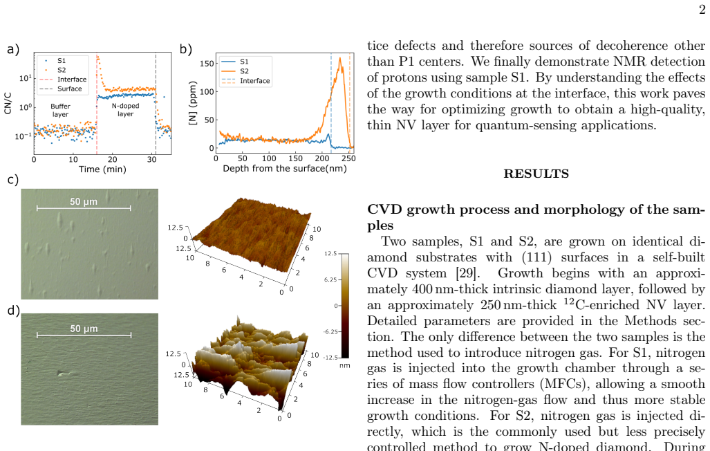
On the paper's own terms, the discovery is that interfacial disorder in thin (111)-oriented 12C-enriched NV diamond films is largely caused by the nitrogen-injection protocol, not by the NV layer itself. Pulsed injection creates a nitrogen overshoot in a 60–80 nm interfacial region; this region shows elevated decay rates, a strain component that grows toward the interface and persists even after 250 nm of overgrowth, and spin noise in excess of what P1 centers can explain. Smooth injection through mass flow controllers suppresses the overshoot and the strain, leaving measured dephasing rates equal, within uncertainty, to the DQR spin-noise contribution. The implied causal chain is: injection
What carries the argument
The central analytic device is the dephasing-rate budget Γ2* ≈ ΓDQR/2 + Γstrain. Double-quantum Ramsey (DQR) suppresses common-mode strain and electric-field dephasing, so it isolates the spin-noise contribution; subtracting it from the single-quantum Ramsey rate yields the strain contribution. DEER (double electron-electron resonance), with an additional π pulse at 218 MHz, selectively recouples on-axis P1 centers, allowing the P1 concentration to be measured (1/12 of the full bath at 115 G) and compared with total nitrogen from ToF-SIMS. Step-etching combined with weighted-average analysis converts region measurements into layer-resolved concentrations and decay rates.
Load-bearing premise
The argument rests on inferring lattice strain as whatever dephasing remains after subtracting the P1 spin-bath contribution, an inference that assumes double-quantum Ramsey isolates all magnetic noise and that no other magnetic noise sources are present, with only one sample per injection method compared.
What would settle it
Measure lattice strain directly, for example by X-ray diffraction, Raman mapping, or the strain splitting of NV energy levels, across the 60–80 nm interface in a pulsed-injection sample. If the apparent strain residual is absent while the dephasing remains, or is fully explained by magnetic noise from nitrogen-related defects, the mechanistic claim would be refuted. Growing additional S1/S2 pairs would also test whether the MFC advantage is reproducible rather than sample-specific.
If this is right
- Smooth MFC nitrogen injection should be used when growing thin NV layers, because pulsed injection leaves a 60–80 nm defective zone that dominates dephasing.
- Thin (50 nm) preferentially aligned NV layers grown this way are good enough for nanoscale NMR, with a measured sensitivity of 17.66 nT/√Hz.
- Nitrogen atoms near the interface that do not become P1 centers still contribute spin noise, so total nitrogen content, not P1 content, is the relevant predictor of dephasing in interfacial layers.
- Strain from the pulsed-injection interface persists after 250 nm of overgrowth, so interfacial disorder can degrade layers much thicker than the overshoot region itself.
- The DQR-minus-strain decomposition gives a practical route to separate magnetic and non-magnetic decoherence sources in NV ensembles.
Where Pith is reading between the lines
- If the interfacial overshoot is causal, a systematic ramp-rate series should show a monotonic relation between nitrogen-injection slope and interfacial strain; this is directly testable in any CVD reactor.
- The persistence of strain after 250 nm of overgrowth hints that the overshoot alters growth morphology, such as grain structure and roughness, meaning the damage may be structural memory rather than only a dopant spike; a growth interrupt or modified temperature profile might heal it.
- The same overshoot mechanism may apply to other dopants in diamond CVD, so smooth delivery could be a general requirement for sharp doped interfaces, not just nitrogen-doped NV layers.
- The measurement-time dependence of effective NV depth noted in the supplement implies that reported ensemble depths and volume-normalized sensitivities carry a protocol dependence that future comparisons should state explicitly.
Editorial analysis
A structured set of objections, weighed in public.
Referee Report
Summary. The paper compares two nitrogen-injection procedures for CVD growth of preferentially oriented NV layers on (111) diamond: a smooth mass-flow-controller (MFC) delivery (S1) and a pulsed injection (S2). Using ToF-SIMS, step-etching, Ramsey/DQR, spin-echo, and DEER measurements, the authors report that pulsed injection creates a pronounced interfacial nitrogen overshoot (60–80 nm), leading to enhanced spin noise and a large residual dephasing contribution attributed to lattice strain, whereas the MFC-grown sample shows nearly no residual strain and a T2 close to the P1-limited value. A 50-nm-thick NV layer from S1 is used to demonstrate NMR detection of protons. The central claim is that the nitrogen-injection protocol dominates interfacial disorder in thin (111) NV-diamond layers.
Significance. If the conclusions hold, the work identifies a practical, controllable growth parameter that governs the quality of thin, preferentially oriented NV ensembles—an important issue for nanoscale quantum sensing. The combination of independent SIMS, DEER, and coherence measurements is methodologically strong, and the demonstration of NMR with a 50-nm layer adds practical value. The strain attribution, however, rests on a residual measurement whose interpretation depends on assumptions about the DQR spin-noise contribution, and the comparison between growth protocols is based on one sample each. The paper is relevant for the diamond quantum-sensing community and the CVD diamond growth community.
major comments (3)
- [§'Analysis of decoherence sources', Eq. (2), Fig. 2(c)] The central mechanistic claim that S2 contains large interfacial strain is based on the residual Γ2* − ΓDQR/2. This subtraction is valid only if ΓDQR/2 exactly equals the magnetic-noise contribution to Γ2*, including magnetic noise from non-P1 defects. The paper's own data (Fig. 4(e,f)) show that in interfacial layers the measured Γ2 tracks [N] rather than [P1], implying the existence of non-P1 magnetic defects. While DQR should in principle sense all magnetic noise with a factor-of-two enhancement, this relation is an assumption that deserves explicit validation, especially at the high defect densities of S2's overshoot region. I recommend adding a direct strain probe (e.g., magnetic-field-dependent ODMR line positions) or a control measurement with a known spin bath to confirm that the residual is not contaminated by a spin-noise component that is not captured by DQR at the factor-of-2
- [Methods, 'DEER fitting and [P1] calculation'; Fig. 4(b),(f)] The layer-resolved values for S2 R0 and R1 are obtained from biexponential fits in which the slow component Td2 is fixed to the value obtained for R2. This assumes that the slow-decaying layer in R0/R1 is identical to R2; if the spin environment varies smoothly with depth, fixing Td2 can bias the fast component and, consequently, the derived layer-resolved [P1] and Γ2. The weighted-average analysis (Eq. (3)) additionally assumes that decay rates are additive across layers. The authors should quantify the sensitivity of the extracted layer-resolved strain and [P1] to the fixed-Td2 assumption (e.g., by freeing Td2 or varying it within its uncertainty).
- [Overall comparison, Figs. 1–4] The manuscript compares one S1 and one S2 sample. The conclusion that MFC-based nitrogen delivery 'substantially suppresses interfacial disorder' relative to pulsed injection is presented as a general property of the growth protocol, but with n=1 per condition the difference could include uncontrolled run-to-run variability. A replicate growth for at least S2 (or a statement of repeated growths in the Methods) would materially strengthen the claim. As written, this is a load-bearing limitation for the practical recommendation in the abstract and discussion.
minor comments (6)
- [Fig. 2(c)] No error bars are visible in the bar chart; please specify whether they are omitted or smaller than the marker size, and provide the numerical values and uncertainties in the text or SI.
- [Methods, 'DEER fitting and [P1] calculation'] The fitting function for the biexponential is written with two components but the stretching exponent 'p' is not defined in that equation; clarify whether p is the same for both components or fitted separately.
- [Supplementary Note 5, Eq. (7)] The expression for K(Nτ) is stated without specifying its domain or the assumption of infinitely narrow resonance; a brief justification would help readers reproduce the sensitivity estimate.
- [Supplementary Table 1] The normalized weights wi for S2 (0.071, 0.291, 0.354, 0.284) do not sum exactly to 1; please check rounding and describe how the normalization was performed.
- [Results, 'Demonstration of nanoscale quantum sensing'] The effective depth d_NV is determined to be 75.30 nm for R0 and 37.71 nm for R2, which are not the geometric centers of the respective layer thicknesses. The explanation in the SI is reasonable, but the statement should appear in the main text to avoid confusion when the reader compares with the layer thicknesses.
- [General notation] The symbol Γ2 is used both for the spin-echo decay rate and, via Γ2*, for the free-induction decay rate in Eq. (2). To avoid confusion, define Γ2* and Γ2 explicitly in the figure captions and text.
Circularity Check
No significant circularity: DEER, SIMS, Ramsey, and DQR are independent; strain attribution is a residual model assumption, not a built-in loop.
full rationale
The claimed derivation chain is not circular. The central comparison is between two growth protocols; the evidence chain is: (i) SIMS gives total [N] profiles independently of any spin measurement; (ii) DEER difference decays give [P1] through a literature coupling coefficient (132 kHz/ppm, Ref. [31]); (iii) Ramsey and DQR give Γ2* and ΓDQR/2; (iv) Eq. (2) attributes the difference Γ2* − ΓDQR/2 to strain. Steps (i)-(iii) are independent measurements with stated literature calibrations. Step (iv) is a residual attribution: 'strain' is not directly measured but is what remains after subtracting the DQR spin-bath contribution, under assumptions (electric-field noise small, DQR cancels strain/electric noise). Those assumptions may be questioned — in particular, non-P1 nitrogen-related spin defects could in principle contribute magnetic noise that is not captured by ΓDQR/2 as the P1-only bath — but an incorrect or debatable attribution is a correctness/validity risk, not circularity: the paper does not define strain as the residual by construction and then use that definition as evidence; it cites external control-sequence and dephasing results for the cancellation and small electric-field terms. The only mild internal loop is that the 'P1-projected' Γ2 curve in Fig. 4 reuses the DEER-derived [P1] and the same spin-bath model, so it is a consistency check rather than an independent prediction; the paper's interfacial conclusion is instead anchored by the independent SIMS-projected [N] curve and by the raw excess of Γ2* over ΓDQR/2 in S2. No load-bearing self-citation chain is apparent from the manuscript text; references to the self-built reactor [29] and the dephasing model [40] are background/methods citations. Therefore no circular step is established.
Axiom & Free-Parameter Ledger
free parameters (3)
- Stretching exponent p in decay fits =
not reported (free for spin echo; fixed to 1 for DEER)
- Biexponential Td1 and amplitudes for S2 R0/R1 =
not reported explicitly beyond Td1
- Effective NV depth d_NV for NMR sensitivity =
75.30 nm (R0), 37.71 nm (R2)
axioms (5)
- domain assumption Additive decomposition of dephasing rates and DQR strain cancellation (Eq. 2)
- domain assumption DEER conversion factors: 1/12 of P1 bath and 132 kHz/ppm coupling
- domain assumption Weighted-average deconvolution of layer properties from step-etch regions
- standard math Semi-infinite spin-bath and Poisson photon statistics for sensitivity
- ad hoc to paper One growth per injection mode is representative
Cite this review
Pith. "Pith review of Revealing and reducing growth-induced interfacial disorder in preferentially aligned nitrogen-vacancy centers in diamond." pith.science (2026). https://pith.science/paper/42KNVR4Z
@misc{pith2026260802350,
  author       = {Pith},
  title        = {Pith review of: Revealing and reducing growth-induced interfacial disorder in preferentially aligned nitrogen-vacancy centers in diamond},
  year         = {2026},
  howpublished = {\url{https://pith.science/paper/42KNVR4Z}},
  note         = {Machine review of arXiv:2608.02350}
}
abstract
Nitrogen-vacancy (NV) centers in chemical-vapor-deposition (CVD) diamond can form preferentially oriented ensembles with high sensing performance and low densities of lattice defects. Thin films of this material are a cornerstone of various imaging modalities. However, nitrogen injection needed to produce such films can transiently drive growth out of equilibrium, generating interfacial strain and spin defects that degrade NV coherence. Here, we investigate this disorder in $^{12}\text{C}$-enriched, preferentially oriented NV layers grown on (111) diamond using two nitrogen-injection procedures, combined with nanometer-scale selective plasma etching and NV spin-coherence measurements. Pulsed nitrogen injection produces a pronounced nitrogen overshoot within a 60--80 nm interfacial region, generating excessive amounts of defects. By contrast, smooth nitrogen delivery through mass flow controllers substantially suppresses interfacial disorder, yielding coherence properties close to the theoretical limit imposed by spin-bath noise. A 50-nm NV layer is used to demonstrate proton nuclear magnetic resonance detection. This work reveals the role of interfacial disorder associated with the nitrogen-doping procedure and provides a route to growing high-quality, thin NV-doped layers for quantum-sensing applications.
Figures


Reference graph
Works this paper leans on
-
[1]
D., Hanson, R., Wrachtrup, J
Awschalom, D. D., Hanson, R., Wrachtrup, J. & Zhou, B. B. Quantum technologies with optically interfaced solid-state spins.Nature Photonics12, 516–527 (2018)
2018
-
[2]
W.et al.The nitrogen-vacancy colour centre in diamond.Physics Reports528, 1–45 (2013)
Doherty, M. W.et al.The nitrogen-vacancy colour centre in diamond.Physics Reports528, 1–45 (2013)
2013
-
[3]
M., Jarmola, A., Budker, D
Bar-Gill, N., Pham, L. M., Jarmola, A., Budker, D. & Walsworth, R. L. Solid-state electronic spin coherence time approaching one second.Nature Communications 4, 1743 (2013)
2013
-
[4]
Taylor, J.M.et al.High-sensitivitydiamondmagnetome- ter with nanoscale resolution.Nature Physics4, 810–816 (2008)
2008
-
[5]
Bian, K.et al.Nanoscale electric-field imaging based on a quantum sensor and its charge-state control under ambi- ent condition.Nature Communications12, 2457 (2021)
2021
-
[6]
Jayaram, S.et al.Probing Vortex Dynamics in 2D Super- conductors with Scanning Quantum Microscope.Physi- cal Review Letters135, 126001 (2025)
2025
-
[7]
C.et al.Super-moiré spin textures in twisted two-dimensional antiferromagnets.Nature Nanotechnol- ogy21, 359–365 (2026)
Wong, K. C.et al.Super-moiré spin textures in twisted two-dimensional antiferromagnets.Nature Nanotechnol- ogy21, 359–365 (2026)
2026
-
[8]
Chigusa, S.et al.Nuclear spin metrology with nitrogen vacancy center in diamond for axion dark matter detec- tion.Physical Review D111, 075028 (2025)
2025
-
[9]
& Degen, C
Loretz, M., Pezzagna, S., Meijer, J. & Degen, C. L. Nanoscale nuclear magnetic resonance with a 1.9-nm- deep nitrogen-vacancy sensor.Applied Physics Letters 104, 033102 (2014)
2014
-
[10]
Aslam, N.et al.Nanoscale nuclear magnetic resonance with chemical resolution.Science357, 67–71 (2017)
2017
-
[11]
Arunkumar, N.et al.Micron-Scale NV-NMR Spec- troscopy with Signal Amplification by Reversible Ex- change.PRX Quantum2, 010305 (2021)
2021
-
[12]
Abendroth, J.M.et al.Single-Nitrogen–VacancyNMRof Amine-Functionalized Diamond Surfaces.Nano Letters 22, 7294–7303 (2022)
2022
-
[13]
O.et al.Magnetic Resonance Detection of Individual Proton Spins Using Quantum Reporters
Sushkov, A. O.et al.Magnetic Resonance Detection of Individual Proton Spins Using Quantum Reporters. Physical Review Letters113, 197601 (2014)
2014
-
[14]
Shi, F.et al.Single-protein spin resonance spectroscopy under ambient conditions.Science347, 1135–1138 (2015)
2015
-
[15]
Lovchinsky, I.et al.Nuclear magnetic resonance detec- tion and spectroscopy of single proteins using quantum logic.Science351, 836–841 (2016)
2016
-
[16]
Balasubramanian, G.et al.Ultralongspincoherencetime in isotopically engineered diamond.Nature Materials8, 383–387 (2009)
2009
-
[17]
D.et al.Ultra-long coherence times amongst room-temperature solid-state spins.Nature Communications10, 3766 (2019)
Herbschleb, E. D.et al.Ultra-long coherence times amongst room-temperature solid-state spins.Nature Communications10, 3766 (2019)
2019
-
[18]
Kim, S.et al.Scalable nanoscale positioning of highly coherent color centers in prefabricated diamond nanos- tructures.Nature Communications16, 9803 (2025)
2025
-
[19]
Yamamoto, T.et al.Ten-Second Electron-Spin Co- herence in Isotopically Engineered Diamond (2026). 2604.07439
Pith/arXiv arXiv 2026
-
[20]
Michl, J.et al.Perfect alignment and preferential orien- tation of nitrogen-vacancy centers during chemical vapor deposition diamond growth on (111) surfaces.Applied Physics Letters104, 102407 (2014)
2014
-
[21]
Götze, A.et al.Preferential Placement of Aligned Ni- trogen Vacancy Centers in Chemical Vapor Deposition Overgrown Diamond Microstructures.physica status so- lidi (RRL) – Rapid Research Letters16, 2100373 (2022)
2022
-
[22]
Osterkamp, C.et al.Engineering preferentially-aligned nitrogen-vacancy centre ensembles in CVD grown dia- mond.Scientific Reports9, 5786 (2019)
2019
-
[23]
M.et al.Enhanced metrology using preferen- tial orientation of nitrogen-vacancy centers in diamond
Pham, L. M.et al.Enhanced metrology using preferen- tial orientation of nitrogen-vacancy centers in diamond. Physical Review B86, 121202 (2012)
2012
-
[24]
& Moustakas, T
Jin, S. & Moustakas, T. D. Effect of nitrogen on the growth of diamond films.Applied Physics Letters65, 403–405 (1994)
1994
-
[25]
& Lux, B
Bohr, S., Haubner, R. & Lux, B. Influence of nitrogen additions on hot-filament chemical vapor deposition of diamond.Applied Physics Letters68, 1075–1077 (1996)
1996
-
[26]
Very high growth rate chemical vapor deposition of single- crystal diamond.Proceedings of the National Academy of Sciences99, 12523–12525 (2002)
Yan, C.-s., Vohra, Y.K., Mao, H.-k.&Hemley, R.J. Very high growth rate chemical vapor deposition of single- crystal diamond.Proceedings of the National Academy of Sciences99, 12523–12525 (2002)
2002
-
[27]
Chayahara, A.et al.The effect of nitrogen addition dur- ing high-rate homoepitaxial growth of diamond by mi- crowave plasma CVD.Diamond and Related Materials 13, 1954–1958 (2004)
1954
-
[28]
Chemical Vapor Deposition of Homoepitaxial Diamond Films
Teraji, T. Chemical Vapor Deposition of Homoepitaxial Diamond Films. InPhysics and Applications of CVD Diamond, chap.3, 29–75(JohnWiley&Sons, Ltd, 2008). 9
2008
-
[29]
Schätzle, P.et al.A Chemical Vapor Deposition Di- amond Reactor for Controlled Thin-Film Growth with Sharp Layer Interfaces.physica status solidi (a)220, 2200351 (2023)
2023
-
[30]
F.et al.Sensitivity optimization for NV- diamond magnetometry.Reviews of Modern Physics92, 015004 (2020)
Barry, J. F.et al.Sensitivity optimization for NV- diamond magnetometry.Reviews of Modern Physics92, 015004 (2020)
2020
-
[32]
Bauch, E.et al.Ultralong Dephasing Times in Solid- State Spin Ensembles via Quantum Control.Physical Review X8, 031025 (2018)
2018
-
[33]
& Awschalom, D
Hanson, R., Gywat, O. & Awschalom, D. D. Room- temperature manipulation and decoherence of a single spin in diamond.Physical Review B74, 161203 (2006)
2006
-
[34]
E., Martineau, P
Glover, C., Newton, M. E., Martineau, P. M., Quinn, S. & Twitchen, D. J. Hydrogen Incorporation in Diamond: The Vacancy-Hydrogen Complex.Physical Review Let- ters92, 135502 (2004)
2004
-
[35]
J.et al.Importance of Quantum Tunneling in Vacancy-Hydrogen Complexes in Diamond.Physical Review Letters95, 105502 (2005)
Shaw, M. J.et al.Importance of Quantum Tunneling in Vacancy-Hydrogen Complexes in Diamond.Physical Review Letters95, 105502 (2005)
2005
-
[36]
Yamamoto, T.et al.Extending spin coherence times of diamond qubits by high-temperature annealing.Physical Review B88, 075206 (2013)
2013
-
[37]
P., Briddon, P
Goss, J. P., Briddon, P. R., Papagiannidis, S. & Jones, R. Interstitial nitrogen and its complexes in diamond. Physical Review B70, 235208 (2004)
2004
-
[38]
Ashfold, M. N. R.et al.Nitrogen in Diamond.Chemical Reviews120, 5745–5794 (2020)
2020
-
[39]
& Seo, H
Park, H., Lee, J., Han, S., Oh, S. & Seo, H. Decoherence ofnitrogen-vacancyspinensemblesinanitrogenelectron- nuclear spin bath in diamond.npj Quantum Information 8, 95 (2022)
2022
-
[40]
Bauch, E.et al.Decoherence of ensembles of nitrogen- vacancy centers in diamond.Physical Review B102, 134210 (2020)
2020
-
[41]
M., Álvarez, G
Souza, A. M., Álvarez, G. A. & Suter, D. Robust Dynam- ical Decoupling for Quantum Computing and Quantum Memory.Physical Review Letters106, 240501 (2011)
2011
-
[42]
Tetienne, J.-P.et al.Spinpropertiesofdensenear-surface ensembles of nitrogen-vacancy centers in diamond.Phys- ical Review B97, 085402 (2018)
2018
-
[43]
F.et al.Optical magnetic detection of single- neuron action potentials using quantum defects in dia- mond.Proceedings of the National Academy of Sciences 113, 14133–14138 (2016)
Barry, J. F.et al.Optical magnetic detection of single- neuron action potentials using quantum defects in dia- mond.Proceedings of the National Academy of Sciences 113, 14133–14138 (2016)
2016
-
[44]
Maier, R.et al.Readout of a solid state spin ensemble at the projection noise limit.Nature Communications17, 4028 (2026)
2026
-
[45]
& Jelezko, F
Fuhrmann, J., Findler, C., Olney-Fraser, M., Kazak, L. & Jelezko, F. Photoactivation of NV Centers in Diamond via Continuous Wave Laser Illumination of Shallow As- Implanted Nitrogen.Advanced Functional Materials36, 2501661 (2026)
2026
-
[46]
Maier, R.et al.Efficient Detection of Statistical RF Fields at High Magnetic Field with a Quantum Sensor (2025)
2025
-
[47]
is used to find the global minimum, and results with fitting errors greater than 30% are discarded. In the bi- exponential fits to R0 and R1 of S2,Td2 is fixed to the values obtained from single-exponential fits to R2 of S2: 2.019±0.036µsfor spin echo and1.194±0.012µsfor DEER (see the Supplementary Information). The fitted value ofTd1 is obtained for R0* ...
-
[48]
& Stariolo, D
Tsallis, C. & Stariolo, D. A. Generalized simulated an- nealing.Physica A: Statistical Mechanics and its Appli- cations233, 395–406 (1996)
1996
-
[49]
M.et al.NMR technique for determining the depth of shallow nitrogen-vacancy centers in diamond
Pham, L. M.et al.NMR technique for determining the depth of shallow nitrogen-vacancy centers in diamond. Physical Review B93, 045425 (2016). Supplementary Information for Revealing and reducing growth-induced interfacial disorder in preferentially aligned nitrogen-vacancy centers in diamond Cheng-I Ho*1,2, Marina Davydova3, Patrik Straˇn´ak3, Felix Hoffma...
2016
-
[50]
& Stals, L
Vandevelde, T., Nesladek, M., Quaeyhaegens, C. & Stals, L. Optical emission spectroscopy of the plasma during CVD diamond growth with nitrogen addition.Thin Solid Films290–291, 143–147 (1996)
1996
-
[51]
& Choo, J
Baek, S.-J., Park, A., Ahn, Y .-J. & Choo, J. Baseline correction using asymmetrically reweighted penalized least squares smoothing.The Analyst140, 250–257 (2015)
2015
-
[52]
Zhang, J.et al.Unraveling quantum dephasing of nitrogen-vacancy center ensembles in diamond.npj Quantum Materials11, 27 (2026)
2026
-
[53]
M.et al.NMR technique for determining the depth of shallow nitrogen-vacancy centers in diamond
Pham, L. M.et al.NMR technique for determining the depth of shallow nitrogen-vacancy centers in diamond. Physical Review B93, 045425 (2016)
2016
-
[54]
M.et al.Single-Nitrogen–Vacancy NMR of Amine-Functionalized Diamond Surfaces.Nano Letters22, 7294–7303 (2022)
Abendroth, J. M.et al.Single-Nitrogen–Vacancy NMR of Amine-Functionalized Diamond Surfaces.Nano Letters22, 7294–7303 (2022)
2022
-
[55]
& Degen, C
Loretz, M., Pezzagna, S., Meijer, J. & Degen, C. L. Nanoscale nuclear magnetic resonance with a 1.9-nm-deep nitrogen-vacancy sensor.Applied Physics Letters104, 033102 (2014). 7
2014
This paper was first reviewed by deepseek-v4-flash on August 4, 2026.
discussion (0)
Sign in with ORCID, Apple, or X to comment. Anyone can read and Pith papers without signing in.