REVIEW 3 major objections 5 minor 51 references
From biting to engulfment: Target mechanics determines modes of phagocytosis through curvature--actin coupling
T0 review · 3 major / 5 minor · reviewed 2026-08-03 · deepseek-v4-flash
Pith's one-line read A two-vesicle simulation predicts that target stiffness dictates whether a phagocyte bites, pushes, or engulfs, via feedback between target deformation and the orientation of actin-driven forces.
desk verdict The paper's real contribution is a two-vesicle simulation showing target deformability selects engulfment vs pushing; the 'biting/trogocytosis' label overstates what is simulated, since fission is explicitly disallowed. read the letter →
The pith
A machine-rendered reading of the paper's core claim, the machinery that carries it, and where it could break.
The reading
What carries the argument
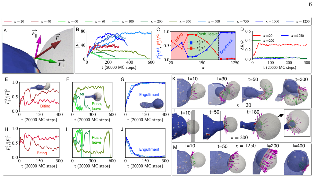
The central object is a pair of dynamically triangulated, self-avoiding vesicles interacting through adhesion and through local forces applied by curved membrane protein complexes (CMC). The CMC have an intrinsic curvature that recruits actin-like active forces, and the delivered force on a target node is decomposed into tangential and normal components relative to the target surface. The load-bearing mechanism is the shape-force feedback: target deformation changes the local normal, which reorients the active force, which in turn either promotes spreading (tangential dominance) or arrests it (normal dominance). The same phase behaviour is obtained by tuning bending rigidity or internal pres
What would settle it
Measure the same three interaction modes while independently changing cortical tension without altering membrane bending rigidity—for example, using micropipette aspiration or drugs that disrupt the actin cortex on target cells. If pushing still occurs for low-bending targets, or if engulfment fails for cortex-free but stiff GUVs, then the proposed mechanical phase boundaries are wrong. A more direct test is to track the tangential versus normal force fraction at the leading edge: the model predicts a high normal fraction in the pushing phase and a high tangential fraction in engulfment, which
Extended reading notes
Core claim
The central claim is that the outcome of phagocytosis of a deformable target is set by a feedback loop between the target's shape change and the orientation of active protrusive forces exerted by the cell's leading edge. In the simulations, curved membrane complexes (CMC) recruit actin-like normal forces, and the target vesicle deforms in response. For a stiff target, the CMC cluster aligns tangentially at the leading edge, driving efficient engulfment. For intermediate stiffness, the target deforms enough that the leading-edge force stays partially normal, arresting cup spreading and ending in a push-then-detach event. For the softest targets, large deformation keeps both normal and tangent
Load-bearing premise
The target is modeled as a pure lipid-bilayer vesicle, so its resistance comes only from membrane bending; real cells resist deformation mainly through their cortical actin network and internal structures, and if cortical tension rather than membrane bending sets target deformability, the predicted biting/pushing/engulfment thresholds may not transfer to living cells.
Editorial extensions
If this is right
- If the central claim is correct, target stiffness alone can switch an immune cell between engulfment, pushing, and trogocytosis, making mechanics a functional input to immune clearance.
- The push-and-detach regime predicts that intermediate-stiffness targets can survive contact by being displaced rather than internalized, which could explain how some apoptotic or tumor cells evade clearance.
- The model implies that raising target tension or rigidity—for example through crosslinking or osmotic stress—could convert a biting interaction into full engulfment, suggesting a mechanical route to enhance phagocytosis.
- Confinement or tethering of a target changes the outcome from pushing to engulfment or biting, so the same cell-target pair may behave differently in tissue versus suspension.
- Because weaker active forces suffice to engulf soft targets, the model predicts that excess protrusive force can be counterproductive for soft-target clearance.
Reading between the lines
- A testable extension is to measure the rigidity of individual lymphoma cells before contact and correlate it with the observed macrophage behavior; the model predicts a sharp stiffness threshold between pushing and engulfment.
- The phase diagram suggests a potential immune-evasion strategy: a target that softens itself (e.g., by loosening its cortex) could shift from being engulfed to being bitten or pushed, which may be relevant to tumor cell heterogeneity.
- The equivalence of bending rigidity and internal pressure in the model hints that cellular osmotic regulation could be a fast, reversible way for targets to modulate their own fate during immune attack.
- If the force-orientation feedback is the real driver, then therapies that alter actin organization at the leading edge rather than target stiffness should also shift the phase boundaries, a prediction that could be probed with cytoskeletal drugs.
Signed reviews
Editorial analysis
A structured set of objections, weighed in public.
Referee Report
Summary. The paper extends a previously published Monte Carlo triangulated-vesicle model of curvature–actin coupling to two interacting vesicles, treating both the phagocyte-like vesicle and the target as deformable membranes. After benchmarking against analytic adhesion results and the rigid-sphere limit, the authors vary the target's bending rigidity (or internal pressure) and report three dynamical regimes: 'biting' (stalled partial engulfment with adhered-area fraction below 0.5), 'pushing' (transient contact followed by detachment), and full engulfment. They argue that the regime boundary is set by feedback between target deformation and the tangential/normal orientation of active forces. Qualitative comparison is made with macrophage engulfment of GUVs, lymphoma cells, and deformable microparticles. The abstract frames the work as predicting that target stiffness governs distinct phagocytic modes, including trogocytosis.
Significance. If the central claim stands, the paper provides a mechanistic, parameter-light explanation for why immune cells engulf stiff targets but push away or partially engulf soft ones, and it connects the simulation observables to force-orientation feedback. The manuscript has real strengths: the code/algorithm is benchmarked against analytic adhered-vesicle results and the rigid-sphere limit; the parameters are mostly taken from prior work rather than fitted to the new experiments; the κ- and pressure-variation phase sequences are internally consistent; and the experimental imaging provides qualitative support for the pushing and engulfment regimes. However, the headline 'biting/trogocytosis' regime is not actually simulated: fission is explicitly disallowed, so the low-stiffness outcome is a stalled partial engulfment. This gap directly affects the abstract's main claim and must be addressed before the paper can be accepted.
major comments (3)
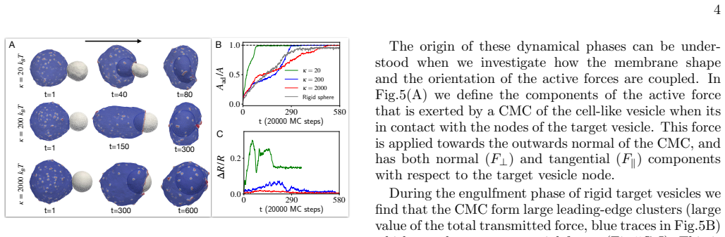
- [Abstract; Fig. 4C and caption; Discussion] The abstract states the model 'predicts three mechanical regimes ... (i) biting (trogocytosis), where part of the target is extracted,' but the simulations never extract a part: Fig. 4's caption says 'we do not allow the vesicles to undergo fission,' and the Discussion says 'so the biting behaviour in the simulations is arrested.' What is actually computed in the low-κ regime is a stalled partial engulfment with Aad/A<0.5. No scission mechanism is supplied, and no evidence rules out eventual retraction (as in the pushing regime) or slower full engulfment. The claim that the model predicts trogocytosis therefore outruns the simulation; either a topological fission step must be added or the 'biting' label must be changed to 'stalled partial engulfment,' with the trogocytosis connection presented only as a speculative extrapolation.
- [Theoretical Model, Eq. (1); p.2] The interaction energy Wint applies an active force from the cell-like vesicle to target vertices without an explicit reaction force on the cell-like vesicle; the text acknowledges 'we do not explicitly maintain force balance' and argues that free vesicles are effectively balanced in the center-of-mass frame. Because the central mechanism is the feedback between target deformation and force orientation, the absence of local momentum conservation could in principle alter the cup-shape dynamics and the stall that is labeled 'biting.' The authors should either implement reaction forces and show that the three regimes are unchanged, or provide a specific argument why the missing reaction force cannot affect the phase boundaries.
- [Comparison to experiments; Fig. 7] The simulations vary target bending rigidity κ or osmotic pressure p, but the cellular targets (lymphoma cells, apoptotic cells) resist deformation primarily through cortical actomyosin tension and internal structure, not membrane bending. The manuscript asserts an equivalence ('effectively stiffening the vesicle') without a quantitative mapping between the simulated κ/p values and cortical stiffness. This is load-bearing for the claim that target mechanics—rather than a particular model mechanism—determines the regimes. At minimum, the authors should estimate the physical ranges of κ and p used here, or include an explicit surface-tension/cortical-tension term and test whether the regime order is preserved.
minor comments (5)
- [Title] Typo: 'phagocytosis o f soft, deformable targets' should be 'phagocytosis of soft, deformable targets.'
- [Supplementary Eq. (S12)] The citation placeholder '[ ? ]' appears after the osmotic-pressure energy term; the missing reference should be supplied.
- [Methods and SI] Units of the active force are inconsistent: the main text gives F = 2 kBT/lmin and the SI Fig. S6 caption gives F = 2 kBT lmin^-2. Please unify and define the dimensionality of F in Eq. (S3).
- [Fig. 5 labels] Panel labels in Fig. 5 use inconsistent capitalization ('H)-j)'); also 'Ffraction' appears in Fig. 4B. Please clean up the axis labels and panel callouts.
- [Results, Fig. 2 and Methods] Volume is not conserved by default, and the symmetry-breaking transition in Fig. 2 involves large volume changes. The text should state explicitly which production runs conserve volume and which do not, and how this choice affects the interpretation of the engulfment/pushing regimes for real cells.
Circularity Check
No significant circularity: the regime structure is emergent from the simulations, but the 'biting' label is an extrapolation rather than a simulated topological outcome.
full rationale
The central phase structure is not fitted to the new experiments: the simulation parameters (F = 2 kBT/lmin, w = 1 kBT, Ead = 2 kBT, cell bending rigidity 20 kBT) are fixed, and the target bending rigidity and internal pressure are scanned; the phase boundaries are emergent model outputs. The model is benchmarked against independent analytic results for adhering vesicles (SI S5-S6) and against external experiments, e.g. soft-bead engulfment rates (Refs. 17, 19), low-tension GUV trogocytosis (Ref. 20), and embryonic epithelial engulfment/pushing (Ref. 38). The self-citations to the authors' earlier CMC framework (Refs. 8, 14-16) are continuity rather than load-bearing circularity: the same curvature-actin ansatz is also anchored to external experimental observations (Refs. 9-13), and no uniqueness theorem is imported. The paper itself flags the key limitation of the 'biting' regime: 'Note that we do not allow the vesicles to undergo fission' (Fig. 4 caption) and 'so the biting behaviour in the simulations is arrested' (Discussion). Because the simulated low-kappa state is only a stalled partial engulfment, the abstract's wording 'predicts ... biting (trogocytosis), where part of the target is extracted' overstates what is computed. However, this is an unsupported extrapolation and a terminology decision, not a case where the predicted quantity is identical by construction to an input or fitted parameter. The post-hoc mapping of experimental osmolarity/tension to simulation pressure and of lymphoma mode to kappa is qualitative, but the model parameters were not tuned to reproduce those observations. Overall no significant circularity; the score of 2 reflects only the mild anchoring to the authors' own previously published CMC framework.
Assumptions & free parameters
free parameters (7)
- Cell-like vesicle bending rigidity κ_cell =
20 kBT
- Adhesion energy E_ad =
2 kBT per adhered vertex pair
- Active force F =
2 kBT/l_min
- CMC spontaneous curvature c0 =
1 l_min^-1
- Protein–protein interaction w =
1 kBT
- CMC density ρ =
4.8% (150/3127 vertices) for engulfment; 6.93% in Fig.2
- Internal pressure p (target) =
0, 0.1, 0.5, 10 kBT l_min^-3
assumptions (6)
- domain assumption The cell and target are represented as dynamically triangulated, self-avoiding vesicles with area conserved via bond-length constraints (l_min < l < 1.7 l_min).
- standard math Membrane bending energy follows a Helfrich-type expression with spontaneous curvature c0 at CMC sites (Eq. S1).
- domain assumption Curvature-sensing CMC complexes recruit actin, modeled as an outward normal active force at each CMC node (Eqs. S3, S8), plus binding between CMC nodes (Eq. S2).
- domain assumption Vesicle–vesicle adhesion is a constant energy −E_ad for every vertex pair within l_min (Eq. 2).
- domain assumption Vesicle volume is not conserved unless an osmotic pressure term p·dV is imposed (SI S7, S12).
- ad hoc to paper A stalled partial engulfment (Aad/A<0.5) in the soft-target regime is interpreted as biting/trogocytosis, even though fission is not allowed in the simulation.
Cite this review
Pith. "Pith review of From biting to engulfment: Target mechanics determines modes of phagocytosis through curvature--actin coupling." pith.science (2026). https://pith.science/paper/4MAX4BFC
@misc{pith2026260120719,
  author       = {Pith},
  title        = {Pith review of: From biting to engulfment: Target mechanics determines modes of phagocytosis through curvature--actin coupling},
  year         = {2026},
  howpublished = {\url{https://pith.science/paper/4MAX4BFC}},
  note         = {Machine review of arXiv:2601.20719}
}
read the original abstract
Phagocytosis is a core innate immune process that clears targets spanning a wide range of mechanical properties, yet the role of target mechanics in recognition and engulfment remains unclear. Here, we combine theoretical modeling and experiments to reveal how target stiffness governs distinct modes of phagocyte--target interaction. We develop a membrane-based simulation framework in which both the engulfing cell and its target are deformable and undergo large shape changes, while actin-driven protrusions are regulated by curvature-sensitive membrane complexes. The model predicts three mechanical regimes with increasing target stiffness: (i) biting (trogocytosis), where part of the target is extracted; (ii) pushing, where the target is displaced rather than engulfed; and (iii) complete engulfment. We validate these predictions in epithelial clearance of apoptotic targets in vivo and macrophage engulfment of Giant Unilamellar Vesicles (GUVs) and lymphoma cells. Together, our results identify target mechanics as a key regulator of clearance and cell--cell interactions.
Figures


Figures from the paper (4 more)



Reference graph
Works this paper leans on
-
[1]
R. S. Flannagan, V. Jaumouill´ e, and S. Grinstein, The cell biology of phagocytosis, Annual Review of Pathology: Mechanisms of Disease 7, 61 (2012)
2012
-
[2]
Uribe-Querol and C
E. Uribe-Querol and C. Rosales, Phagocytosis: our cur- rent understanding of a universal biological process, Frontiers in immunology 11, 1066 (2020)
2020
-
[3]
J. A. Swanson, Shaping cups into phagosomes and macropinosomes, Nature reviews Molecular cell biology 9, 639 (2008)
2008
-
[4]
Mylvaganam, S
S. Mylvaganam, S. A. Freeman, and S. Grinstein, The cytoskeleton in phagocytosis and macropinocytosis, Cur- rent Biology 31, R619 (2021)
2021
-
[5]
A. K. Panah and V. Jaumouill´ e, Signaling of phagocyto- sis, in Encyclopedia of Immunobiology (Second Edition) , edited by P. M. Kaye (Academic Press, Oxford, 2026) second edition ed., pp. 651–669
2026
-
[6]
Vorselen, R
D. Vorselen, R. L. D. Labitigan, and J. A. Theriot, A me- chanical perspective on phagocytic cup formation, Cur- rent opinion in cell biology 66, 112 (2020)
2020
-
[7]
Jaumouill´ e and C
V. Jaumouill´ e and C. M. Waterman, Physical constraints and forces involved in phagocytosis, Frontiers in im- munology 11, 1097 (2020)
2020
-
[8]
R. K. Sadhu, S. R. Barger, S. Peniˇ c, A. Igliˇ c, M. Kren- del, N. C. Gauthier, and N. S. Gov, A theoretical model of efficient phagocytosis driven by curved membrane pro- teins and active cytoskeleton forces, Soft Matter 19, 31 (2023)
2023
Show all 51 references
-
[9]
Scita, S
G. Scita, S. Confalonieri, P. Lappalainen, and S. Suet- sugu, Irsp53: crossing the road of membrane and actin dynamics in the formation of membrane protrusions, Trends in cell biology 18, 52 (2008)
2008
-
[10]
Linkner, G
J. Linkner, G. Witte, H. Zhao, A. Junemann, B. Nord- holz, P. Runge-Wollmann, P. Lappalainen, and J. Faix, The inverse bar domain protein ibara drives membrane remodeling to control osmoregulation, phagocytosis and cytokinesis, Journal of Cell Science 127, 1279 (2014)
2014
-
[11]
Begemann, T
I. Begemann, T. Saha, L. Lamparter, I. Rathmann, D. Grill, L. Golbach, C. Rasch, U. Keller, B. Trappmann, M. Matis, et al. , Mechanochemical self-organization determines search pattern in migratory cells, Nature Physics 15, 848 (2019)
2019
-
[12]
Pipathsouk, R
A. Pipathsouk, R. M. Brunetti, J. P. Town, B. R. Graziano, A. Breuer, P. A. Pellett, K. Marchuk, N.-H. T. Tran, M. F. Krummel, D. Stamou, et al. , The wave com- plex associates with sites of saddle membrane curvature, Journal of Cell Biology 220, e202003086 (2021)
2021
-
[13]
M. Wu, R. K. Sadhu, K. Meyer, Z. Tang, P. Marchando, D. N. Woolfson, N. S. Gov, and O. D. Weiner, Wave complex forms linear arrays at negative membrane cur- vature to instruct lamellipodia formation, Journal of Cell Biology 224, e202410098 (2025)
2025
-
[14]
R. K. Sadhu, A. Igliˇ c, and N. S. Gov, A minimal cell model for lamellipodia-based cellular dynamics and mi- gration, Journal of Cell Science 136, jcs260744 (2023)
2023
-
[15]
R. K. Sadhu, M. Luciano, W. Xi, C. Martinez-Torres, M. Schr¨ oder, C. Blum, M. Tarantola, S. Villa, S. Peniˇ c, A. Igliˇ c,et al. , A minimal physical model for curvotaxis driven by curved protein complexes at the cell’s leading edge, Proceedings of the National Academy of Sci...
2024
-
[16]
Sadhukhan, C
S. Sadhukhan, C. Martinez-Torres, S. Peniˇ c, C. Beta, A. c. v. Igliˇ c, and N. Gov, Modeling how lamellipodia- driven cells maintain persistent migration and interact with external barriers, Phys. Rev. Res. 7, 013319 (2025)
2025
-
[17]
K. A. Beningo and Y.-l. Wang, Fc-receptor-mediated phagocytosis is regulated by mechanical properties of the target, Journal of cell science 115, 849 (2002)
2002
-
[18]
Vorselen, Y
D. Vorselen, Y. Wang, M. M. de Jesus, P. K. Shah, M. J. Footer, M. Huse, W. Cai, and J. A. Theriot, Mi- croparticle traction force microscopy reveals subcellular force exertion patterns in immune cell–target interac- tions, Nature Communications 11, 10.1038/s41467-019- 13804-z (2020)
2020 doi
-
[19]
A. H. Settle, B. Y. Winer, M. M. de Jesus, L. Seeman, Z. Wang, E. Chan, Y. Romin, Z. Li, M. M. Miele, R. C. Hendrickson, et al. , β2 integrins impose a mechanical checkpoint on macrophage phagocytosis, Nature Com- munications 15, 8182 (2024)
2024
-
[20]
C. E. Cornell, A. Chorlay, D. Krishnamurthy, N. R. Mar- tin, L. Baldauf, and D. A. Fletcher, Target cell cortical tension regulates macrophage trogocytosis, Nature Cell Biology , 1 (2025)
2025
-
[21]
K. R. Rollins, S. Fiaz, I. Datta, and M. A. Morris- sey, Target cell adhesion limits macrophage phagocytosis and promotes trogocytosis, Journal of Cell Biology 224, e202502034 (2025)
2025
-
[22]
J. Shen, X. Li, G. Hide, Z.-R. Lun, and Z. Wu, Trogocy- tosis: revealing new insights into parasite–host interac- tions, Trends in Parasitology (2025)
2025
-
[23]
Foˇ snariˇ c, S
M. Foˇ snariˇ c, S. Peniˇ c, A. Igliˇ c, V. Kralj-Igliˇ c, M. Drab, and N. S. Gov, Theoretical study of vesicle shapes driven by coupling curved proteins and active cytoskele- tal forces, Soft Matter 15, 5319 (2019)
2019
-
[24]
R. K. Sadhu, S. Peniˇ c, A. Igliˇ c, and N. S. Gov, Modelling cellular spreading and emergence of motility in the pres- ence of curved membrane proteins and active cytoskele- ton forces, The European Physical Journal Plus 136, 495 (2021)
2021
-
[25]
Pavliˇ c, T
J. Pavliˇ c, T. Mareˇ s, J. Beˇ ster, V. Janˇ sa, M. Daniel, and A. Igliˇ c, Encapsulation of small spherical liposome into larger flaccid liposome induced by human plasma pro- teins, Computer Methods in Biomechanics and Biomed- ical Engineering 12, 147 (2009)
2009
-
[26]
Tordeux, J.-B
C. Tordeux, J.-B. Fournier, and P. Galatola, Analytical characterization of adhering vesicles, Physical Review E 65, 041912 (2002)
2002
-
[27]
Frank, M
M. Frank, M. Manˇ cek-Keber, M. Krˇ zan, S. Sodin-ˇSemrl, R. Jerala, A. Igliˇ c, B. Rozman, and V. Kralj-Igliˇ c, Pre- vention of microvesiculation by adhesion of buds to the mother cell membrane—a possible anticoagulant effect of healthy donor plasma, Autoimmunity Reviews 7, 2...
2008
-
[28]
Mareˇ s, M
T. Mareˇ s, M. Daniel, A. Igliˇ c, V. Kralj-Igliˇ c, and M. Foˇ snariˇ c, Determination of the strength of adhe- sion between lipid vesicles, The Scientific World Journal 2012, 146804 (2012)
2012
-
[29]
Urbanija, B
J. Urbanija, B. Babnik, M. Frank, N. Tomˇ siˇ c, B. Roz- man, V. Kralj-Igliˇ c, and A. Igliˇ c, Attachment of β2- glycoprotein i to negatively charged liposomes may pre- vent the release of daughter vesicles from the par- ent membrane, European Biophysics Journal 37, 1085 (2008). 11
2008
-
[30]
Torres-S´ anchez, M
A. Torres-S´ anchez, M. Kerr Winter, and G. Sal- breux, Interacting active surfaces: A model for three- dimensional cell aggregates, PLOS Computational Biol- ogy 18, e1010762 (2022)
2022
-
[31]
Vorselen, Y
D. Vorselen, Y. Wang, M. M. de Jesus, P. K. Shah, M. J. Footer, M. Huse, W. Cai, and J. A. Theriot, Micropar- ticle traction force microscopy reveals subcellular force exertion patterns in immune cell–target interactions, Na- ture communications 11, 20 (2020)
2020
-
[32]
Motahari and A
F. Motahari and A. Carlsson, Actin based pulling forces in endocytosis, Biophysical Journal 112, 561a (2017)
2017
-
[33]
J. C. Herron, S. Hu, T. Watanabe, A. T. Nogueira, B. Liu, M. E. Kern, J. Aaron, A. Taylor, M. Pablo, T.- L. Chew, et al. , Actin nano-architecture of phagocytic podosomes, Nature communications 13, 4363 (2022)
2022
-
[34]
Sopelniak, R
K. Sopelniak, R. Batlouni, Q.-f. Sun, P. Cervero, and S. Linder, Phagocytic podosomes enable efficient up- take of candida auris by primary human macrophages, bioRxiv , 2025 (2025)
2025
-
[35]
Rianna, M
C. Rianna, M. Radmacher, and S. Kumar, Direct evi- dence that tumor cells soften when navigating confined spaces, Molecular biology of the cell 31, 1726 (2020)
2020
-
[36]
Lekka, Discrimination between normal and cancerous cells using afm, Bionanoscience 6, 65 (2016)
M. Lekka, Discrimination between normal and cancerous cells using afm, Bionanoscience 6, 65 (2016)
2016
-
[37]
M. Yang, Y. Yang, L. Liu, and M. Li, Single-cell par- allel plate mechanics by side-view optical microscopy- assisted atomic force microscopy, Nanoscale Advances 7, 2158 (2025)
2025
-
[38]
Hoijman, H.-M
E. Hoijman, H.-M. H¨ akkinen, Q. Tolosa-Ramon, S. Jimenez-Delgado, C. Wyatt, M. Miret-Cuesta, M. Ir- imia, A. Callan-Jones, S. Wieser, and V. Ruprecht, Co- operative epithelial phagocytosis enables error correction in the early embryo, Nature 590, 618 (2021)
2021
-
[39]
Miyake and H
K. Miyake and H. Karasuyama, The role of trogocytosis in the modulation of immune cell functions, Cells 10, 1255 (2021)
2021
-
[40]
Vorselen, Dynamics of phagocytosis mediated by phosphatidylserine, Biochemical Society Transactions 50, 1281 (2022)
D. Vorselen, Dynamics of phagocytosis mediated by phosphatidylserine, Biochemical Society Transactions 50, 1281 (2022)
2022
-
[41]
Barbera, M
S. Barbera, M. J. Schuiling, N. A. Sanjaya, I. Pietil¨ a, T. Sar´ en, M. Essand, and A. Dimberg, Trogocytosis of chimeric antigen receptors between t cells is regulated by their transmembrane domains, Science immunology 10, eado2054 (2025)
2025
-
[42]
T. W. Gadella Jr, L. Van Weeren, J. Stouthamer, M. A. Hink, A. H. Wolters, B. N. Giepmans, S. Aumonier, J. Dupuy, and A. Royant, mscarlet3: a brilliant and fast- maturing red fluorescent protein, Nature methods 20, 541 (2023)
2023
-
[43]
Vorselen, S
D. Vorselen, S. Barger, J. Theriot, N. Gauthier, and M. Krendel, Phagocytic microscopy and mp-tfm assay with raw macrophages upon treatment with cytoskeletal inhibitors (2021)
2021
-
[44]
Vorselen, S
D. Vorselen, S. R. Barger, Y. Wang, W. Cai, J. A. Theriot, N. C. Gauthier, and M. Krendel, Phagocytic ‘teeth’ and myosin-ii ‘jaw’ power target constriction dur- ing phagocytosis, eLife 10, 10.7554/elife.68627 (2021)
2021 doi
-
[45]
A. Mali, Y. Peeters, R. Rodrigues de Mercado, A. H. Settle, M. J. Footer, M. Srinivas, J. A. Theriot, and D. Vorselen, Using tunable hydrogel micropar- ticles to measure cellular forces, Nature Protocols 10.1038/s41596-025-01281-2 (2025). Supplementary material: From biting to...
2025 doi
-
[46]
We placed 847 points on a sphere of radius 10 lmin
is the golden angle and the integer index i runs from 1 to N T . We placed 847 points on a sphere of radius 10 lmin. Then, we do the same calculation as before. S5. VERIFICA TION OF RADIUS OF CUR V A TURE NEAR THE CONT ACT WIT H A SINGLE VESICLE A single vesicle is allowed to ...
-
[47]
C. E. Cornell, A. Chorlay, D. Krishnamurthy, N. R. Martin, L. Bal dauf, and D. A. Fletcher. Target cell cortical tension regulates macrophage trogocytosis. Nature Cell Biology , pages 1–11, 2025
2025
-
[48]
Mareˇ s, M
T. Mareˇ s, M. Daniel, A. Igliˇ c, V. Kralj-Igliˇ c, and M. Foˇ snariˇ c. Determination of the strength of adhesion between lipid vesicles. The Scientific World Journal , 2012(1):146804, 2012
2012
-
[49]
R. K. Sadhu, S. R. Barger, S. Peniˇ c, A. Igliˇ c, M. Krendel, N. C. Gauthier, and N. S. Gov. A theoretical model of efficient phagocytosis driven by curved membrane proteins and active cyto skeleton forces. Soft Matter , 19(1):31–43, 2023
2023
-
[50]
R. K. Sadhu, A. Igliˇ c, and N. S. Gov. A minimal cell model fo r lamellipodia-based cellular dynamics and migration. Journal of Cell Science , 136(14):jcs260744, 2023
2023
-
[51]
R. K. Sadhu, S. Peniˇ c, A. Igliˇ c, and N. S. Gov. Modelling cellular spreading and emergence of motility in the presence of curved membrane proteins and active cytoskeleton forces. The European Physical Journal Plus , 136(5):495, 2021
2021
Reviewed August 3, 2026 · model on record in the stance chip above.
Discussion (0). Continue with ORCID to comment.