REVIEW 3 major objections 5 minor 26 references
CAMEL: A Weakly Supervised Learning Framework for Histopathology Image Segmentation
T0 review · 3 major / 5 minor · reviewed 2026-08-14 · deepseek-v4-flash
Pith's one-line read Image-level labels alone can train tumor segmentation models that match pixel-supervised baselines
desk verdict Useful MIL-based label enrichment for histopathology, but the 'image-level' supervision in the experiments is actually derived from pixel-level masks, so the central claim is untested. read the letter →
The pith
A machine-rendered reading of the paper's core claim, the machinery that carries it, and where it could break.
The reading
What carries the argument
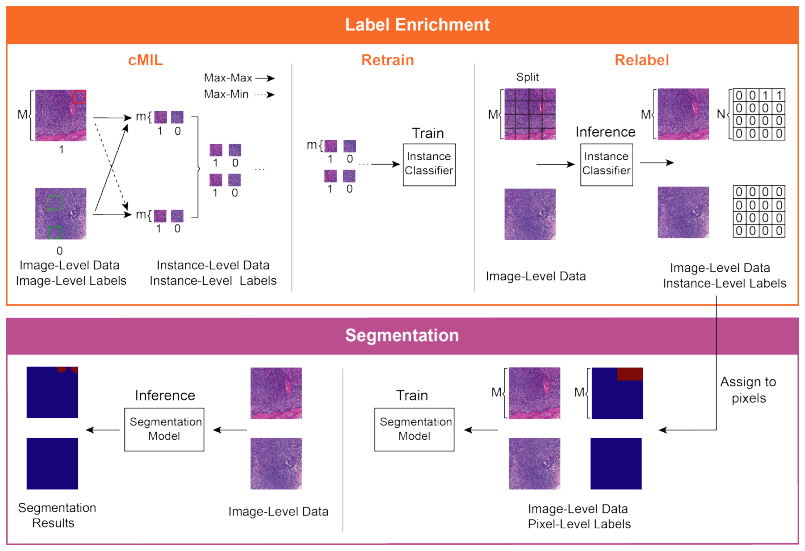
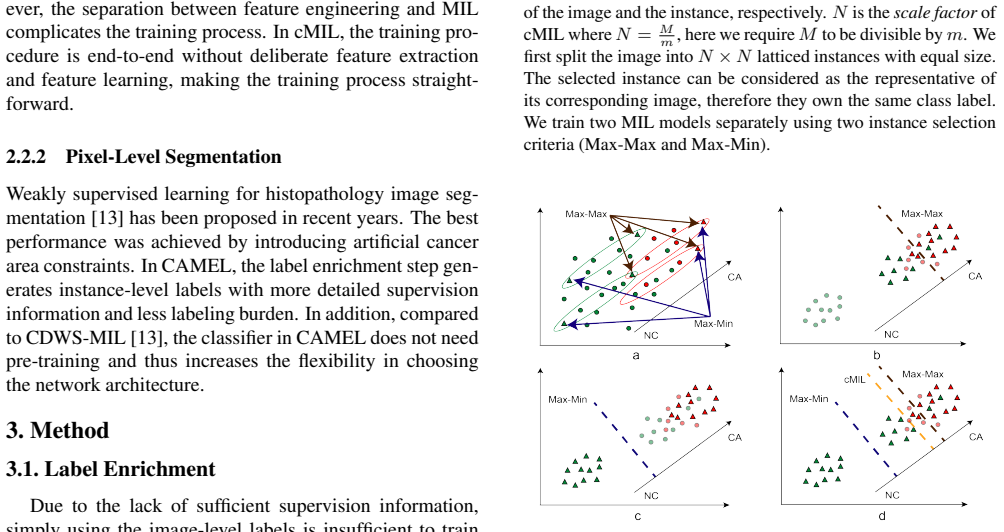
The load-bearing mechanism is cMIL (combined multiple instance learning), defined by two instance-selection criteria applied to latticed image patches. In multiple instance learning, an image is a 'bag' of patches and only the bag label is known; Max-Max picks the patch with the highest cancer response from every image, while Max-Min picks the highest-response patch from cancer images and the lowest-response patch from non-cancer images. Training two classifiers with these complementary criteria and pooling their selected instances rebalances the pseudo-training data; the method then retrains a fully supervised classifier on those instances, relabels every patch in the original images, and assigns each patch label to its pixels. Cascade data enhancement (two-stage cMIL at different scales) and an image-level constraint loss during retraining are supporting mechanisms that recover information lost by selecting only one instance per image.
What would settle it
Reproduce CAMEL's enrichment on a held-out cohort whose whole-slide images have independent pathologist pixel annotations, train DeepLabv2 only on the automatically generated masks, and compare against the same network trained on manual masks; the central claim fails if the weakly supervised IoU falls more than roughly two points below the fully supervised IoU, because that would show the pseudo-labels are not close enough to pixel truth to explain the reported parity.
Extended reading notes
Core claim
On its own terms, the paper's discovery is that a simple self-training loop can bootstrap pixel-level masks from image-level labels. First, a combined multiple instance learning step splits each image into an $N \times N$ grid of instances, trains two classifiers that each select one representative instance per image under different criteria (Max-Max and Max-Min), and uses the selected instances, together with the image label, as a pseudo-labeled instance dataset. The classifier is retrained on these pseudo-labels, then applied to every instance of every training image; the resulting instance labels are copied onto all pixels in each instance to form approximate segmentation masks. With these masks, training DeepLabv2 or U-Net produces pixel-level accuracy, F1, and IoU close to models trained on the true pixel annotations. On CAMELYON16, CAMEL (160) reaches an IoU of 85.4 against 86.3 for the fully supervised DeepLabv2 baseline, and the same pattern holds on the colorectal adenoma dataset.
Load-bearing premise
The load-bearing premise is that the instance labels automatically produced by a classifier trained on its own pseudo-labels are accurate enough to stand in for pixel-level ground truth, even though any instance containing a speck of cancer is labeled wholly positive (the paper's own over-labeling caveat).
Editorial extensions
If this is right
- A single image-level label can be expanded into $N^2$ instance-level labels, so the cost of annotation drops from pixel boundaries to a yes/no decision per image.
- The complementary Max-Max and Max-Min selection is the component that balances sensitivity against specificity; using either criterion alone gives skewed predictions, while the combination approaches the fully supervised trade-off.
- Because the enriched labels already support training of standard segmentation architectures, future improvements in segmentation networks can be plugged into CAMEL without changing the labeling step.
- Finer instance granularity (160 versus 320) improves segmentation and reduces over-labeling, so further refinement of the lattice is a direct path to closing the remaining gap.
- The same label-enrichment pipeline transfers to a second, independently collected histopathology dataset, which supports the paper's claim that the automatic labeling method is general rather than dataset-specific.
Reading between the lines
- The selection-criteria trick is a transferable recipe: use a high-precision positive selector and a high-precision negative selector to build pseudo-labels for any class-imbalanced weakly supervised task, not just histopathology.
- Since the paper acknowledges over-labeling (an instance is positive if it contains any cancer pixel), its own logic suggests that adding boundary refinement, such as CRF post-processing or affinity learning, would push CAMEL's segmentation IoU toward or past the fully supervised baseline.
- A testable extension is to push the cascade to finer lattices with multiple stages; the paper shows gains from 320 to 160, and the trend suggests that finer granularity may keep improving until the instance size approaches the smallest tumor islands.
- The framework's dependence on the retrained classifier's own predictions implies that a poorly calibrated first-stage classifier would propagate errors; measuring enriched-label accuracy on an independent pixel-annotated cohort would tell whether the one-to-two-point gap is stable.
Signed reviews
Editorial analysis
A structured set of objections, weighed in public.
Referee Report
Summary. This paper proposes CAMEL, a weakly supervised learning framework for histopathology image segmentation that claims to use only image-level labels. The method first performs label enrichment: it splits images into latticed instances, trains two multiple-instance-learning classifiers using Max-Max and Max-Min selection criteria, combines the selected instances into a fully supervised instance-level dataset, retrains an instance classifier, and relabels all instances. These instance-level labels are then assigned to pixels to produce approximate segmentation masks, which are used to train DeepLabv2 and U-Net in a fully supervised manner. Experiments on CAMELYON16 and a colorectal adenoma dataset report instance-level classification and pixel-level segmentation performance comparable to fully supervised baselines, and the authors state that the adenoma dataset will be released publicly.
Significance. If the central claim were fully validated, CAMEL would be a valuable contribution because it could substantially reduce annotation cost for histopathology segmentation. The paper has several strengths: the method is clearly described; the label-enrichment idea is intuitive; comparisons include multiple weakly supervised baselines and fully supervised upper bounds; and the authors explicitly discuss the over-labeling limitation in Section 5. However, the experimental protocol does not actually test the stated image-level-label setting, and the reported comparisons lack statistical validation. These issues bear directly on the paper's main claim, so the manuscript needs substantive revision before the contribution can be accepted.
major comments (3)
- [§4.1 and Abstract] The experiments do not actually test the stated 'image-level labels only' setting. In Section 4.1, the 1,280×1,280 training patches are labeled CA/NC by checking whether each patch contains any region marked in the pixel-level tumor masks; the resulting patch-level labels are a coarse aggregation of the very pixel-level ground truth used to train the pixel-level FSB. A WSI-level diagnostic label alone cannot determine which patches in a positive WSI are CA, so the claimed scenario of avoiding pixel-level annotation is not exercised. The abstract and contributions should be revised to describe patch-level weak supervision, or an experiment using true WSI-level labels should be added.
- [Tables 3 and 4] All reported numbers are single-run point estimates with no error bars, confidence intervals, or significance tests. The 'comparable' claim rests on gaps as small as 0.9 percentage points of IoU (85.4 vs 86.3 on CAMELYON16 with DeepLabv2) and 0.2 percentage points of F1 (87.3 vs 87.5 on the adenoma dataset), which could easily be within run-to-run variability. Please report means and variances over multiple runs, or otherwise justify that the differences are stable.
- [Table 3 footnote] The exclusion of Test 114 with the footnote 'because of the duplicate labeling [15]' is a data-selection choice that affects the test set composition. The authors should show that the conclusions are unchanged when Test 114 is included, or provide a principled, pre-specified criterion for its exclusion.
minor comments (5)
- [§3.1.2 and Table 2] The quality of the enriched instance labels is evaluated on the same training set from which the pseudo-labels were generated; an independent validation set would strengthen the claim that the pseudo-labels are generally accurate.
- [§3.3.2, Eq. (4)] The weights w1 and w2 are set equal without sensitivity analysis; since w1=w2 is a hyperparameter, a brief ablation would clarify its role.
- [Figure 2 caption] The caption defines N = M/m and requires M to be divisible by m, but the text does not state how the scale factor is chosen for the adenoma dataset.
- [§4.1] The phrase 'image-level data' for 1,280×1,280 patches conflicts with the histopathology convention where image-level usually means WSI-level; please define the term explicitly at first use.
- [§1] There is a typographical error in Section 1: 'theses fine-grained labels' should read 'these fine-grained labels'.
Circularity Check
No significant circularity: CAMEL's label-enrichment pipeline is evaluated against held-out ground truth, and no derivation step reduces the predicted segmentation to the input labels by construction.
full rationale
I walked the claimed derivation chain: (1) cMIL trains two MIL classifiers with Max-Max and Max-Min selection and uses their predictions to build an instance-level dataset; (2) retrain and relabel trains a classifier on those instances and re-labels all instances; (3) instance labels are assigned to pixels; (4) a standard segmentation model is trained and evaluated on held-out test data. No equation in the paper defines the output in terms of a fitted parameter, and no load-bearing result is imported solely from a self-citation. The retrain/relabel loop is a standard pseudo-labeling procedure; the final segmentation metrics in Tables 3 and 4 are computed against held-out ground truth and are not forced by construction. The one notable concern is experimental validity rather than circularity: Section 4.1 states that 'we regard the 1,280×1,280 patches at 20x magnification in the WSIs as image-level data' and that 'the patches and the instances are labeled as CA if it contains any cancerous region,' meaning the weak supervision inputs are patch-level labels derived from pixel-level masks. This weakens the abstract's wording that CAMEL uses 'only image-level labels,' but it does not make the derivation circular: the patch labels are a coarse aggregation of the ground truth, not a quantity that by construction determines the final pixel-level segmentation, and the test evaluation remains independent. Accordingly, the circularity score is 0.
Assumptions & free parameters
free parameters (3)
- Scale factor N =
16 and 64 (for 320x320 and 160x160 instances)
- Loss weights w1, w2 =
equal (w1 = w2)
- Random crop size =
640x640
assumptions (3)
- domain assumption Every positive image contains at least one positive instance, and every instance in a negative image is negative.
- domain assumption A label assigned to an instance correctly represents all its pixels.
- domain assumption The retrained classifier, trained on a biased subset of instances, generalizes to all instances.
Cite this review
Pith. "Pith review of CAMEL: A Weakly Supervised Learning Framework for Histopathology Image Segmentation." pith.science (2026). https://pith.science/paper/5B7YUODB
@misc{pith2026190810555,
  author       = {Pith},
  title        = {Pith review of: CAMEL: A Weakly Supervised Learning Framework for Histopathology Image Segmentation},
  year         = {2026},
  howpublished = {\url{https://pith.science/paper/5B7YUODB}},
  note         = {Machine review of arXiv:1908.10555}
}
read the original abstract
Histopathology image analysis plays a critical role in cancer diagnosis and treatment. To automatically segment the cancerous regions, fully supervised segmentation algorithms require labor-intensive and time-consuming labeling at the pixel level. In this research, we propose CAMEL, a weakly supervised learning framework for histopathology image segmentation using only image-level labels. Using multiple instance learning (MIL)-based label enrichment, CAMEL splits the image into latticed instances and automatically generates instance-level labels. After label enrichment, the instance-level labels are further assigned to the corresponding pixels, producing the approximate pixel-level labels and making fully supervised training of segmentation models possible. CAMEL achieves comparable performance with the fully supervised approaches in both instance-level classification and pixel-level segmentation on CAMELYON16 and a colorectal adenoma dataset. Moreover, the generality of the automatic labeling methodology may benefit future weakly supervised learning studies for histopathology image analysis.
Figures

Figures from the paper (6 more)



Reference graph
Works this paper leans on
-
[15]
Cancer metastasis detection with neural conditional random field
Yi Li and Wei Ping. Cancer metastasis detection with neural conditional random field. arXiv preprint arXiv:1806.07064, 2018
arXiv 2018
- [1]
-
[2]
Tensorflow: A system for large-scale machine learning
Mart ´ın Abadi, Paul Barham, Jianmin Chen, Zhifeng Chen, Andy Davis, Jeffrey Dean, Matthieu Devin, Sanjay Ghe- mawat, Geoffrey Irving, Michael Isard, et al. Tensorflow: A system for large-scale machine learning. In OSDI, vol- ume 16, pages 265–283, 2016
work page 2016
-
[3]
Weakly super- vised learning of instance segmentation with inter-pixel rela- tions
Jiwoon Ahn, Sunghyun Cho, and Suha Kwak. Weakly super- vised learning of instance segmentation with inter-pixel rela- tions. In Proceedings of the IEEE Conference on Computer Vision and Pattern Recognition, pages 2209–2218, 2019
work page 2019
-
[4]
Jiwoon Ahn and Suha Kwak. Learning pixel-level semantic affinity with image-level supervision for weakly supervised semantic segmentation. In Proceedings of the IEEE Con- ference on Computer Vision and Pattern Recognition, pages 4981–4990, 2018
work page 2018
-
[5]
Babak Ehteshami Bejnordi, Mitko Veta, Paul Johannes Van Diest, Bram Van Ginneken, Nico Karssemeijer, Geert Litjens, Jeroen AWM Van Der Laak, and the CAME- LYON16 Consortium. Diagnostic assessment of deep learn- ing algorithms for detection of lymph node metastases in women with breast cancer. JAMA, 318(22):2199, 2017
work page 2017
-
[6]
Semantic image seg- mentation with deep convolutional nets and fully connected CRFs
Liang-Chieh Chen, George Papandreou, Iasonas Kokkinos, Kevin Murphy, and Alan L Yuille. Semantic image seg- mentation with deep convolutional nets and fully connected CRFs. Computer Science, (4):357–361, 2014
work page 2014
-
[7]
Liang-Chieh Chen, George Papandreou, Iasonas Kokkinos, Kevin Murphy, and Alan L Yuille. DeepLab: Semantic im- age segmentation with deep convolutional nets, atrous con- volution, and fully connected CRFs. IEEE Transactions on Pattern Analysis and Machine Intelligence , 40(4):834–848, 2018
work page 2018
Show all 26 references
-
[8]
BoxSup: Exploit- ing bounding boxes to supervise convolutional networks for semantic segmentation
Jifeng Dai, Kaiming He, and Jian Sun. BoxSup: Exploit- ing bounding boxes to supervise convolutional networks for semantic segmentation. In Proceedings of the IEEE Inter- national Conference on Computer Vision, pages 1635–1643, 2015
2015
-
[9]
WILDCAT: Weakly supervised learning of deep convnets for image classification, pointwise localiza- tion and segmentation
Thibaut Durand, Taylor Mordan, Nicolas Thome, and Matthieu Cord. WILDCAT: Weakly supervised learning of deep convnets for image classification, pointwise localiza- tion and segmentation. In Proceedings of the IEEE Con- ference on Computer Vision and Pattern Recognition, pages 6...
2017
-
[10]
Multi-evidence filtering and fusion for multi-label classification, object de- tection and semantic segmentation based on weakly super- vised learning
Weifeng Ge, Sibei Yang, and Yizhou Yu. Multi-evidence filtering and fusion for multi-label classification, object de- tection and semantic segmentation based on weakly super- vised learning. In Proceedings of the IEEE Conference on Computer Vision and Pattern Recognition , pages...
2018
-
[11]
Deep residual learning for image recognition
Kaiming He, Xiangyu Zhang, Shaoqing Ren, and Jian Sun. Deep residual learning for image recognition. In Proceed- ings of the IEEE Conference on Computer Vision and Pattern Recognition, pages 770–778, 2016
2016
-
[12]
Weakly-supervised semantic segmentation network with deep seeded region growing
Zilong Huang, Xinggang Wang, Jiasi Wang, Wenyu Liu, and Jingdong Wang. Weakly-supervised semantic segmentation network with deep seeded region growing. In Proceedings of the IEEE Conference on Computer Vision and Pattern Recognition, pages 7014–7023, 2018
2018
-
[13]
Constrained deep weak supervision for histopathology im- age segmentation
Zhipeng Jia, Xingyi Huang, Eric I-Chang Chao, and Yan Xu. Constrained deep weak supervision for histopathology im- age segmentation. IEEE Transactions on Medical Imaging, 36(11):2376–2388, 2017
2017
-
[14]
Simple does it: Weakly supervised instance and semantic segmentation
Anna Khoreva, Rodrigo Benenson, Jan Hendrik Hosang, Matthias Hein, and Bernt Schiele. Simple does it: Weakly supervised instance and semantic segmentation. In Proceed- ings of the IEEE Conference on Computer Vision and Pattern Recognition, pages 876–885, 2017
2017
-
[16]
ScribbleSup: Scribble-supervised convolutional networks for semantic segmentation
Di Lin, Jifeng Dai, Jiaya Jia, Kaiming He, and Jian Sun. ScribbleSup: Scribble-supervised convolutional networks for semantic segmentation. In Proceedings of the IEEE Con- ference on Computer Vision and Pattern Recognition, pages 3159–3167, 2016
2016
-
[17]
Fast Scannet: Fast and dense analysis of multi-gigapixel whole-slide images for cancer metastasis detection
Huangjing Lin, Hao Chen, Simon Graham, Qi Dou, Nasir Rajpoot, and Pheng-Ann Heng. Fast Scannet: Fast and dense analysis of multi-gigapixel whole-slide images for cancer metastasis detection. IEEE Transactions on Medical Imag- ing, 38(8):1948–1958, 2019
1948
-
[18]
Detecting cancer metastases on gigapixel pathol- ogy images
Yun Liu, Krishna Gadepalli, Mohammad Norouzi, George E Dahl, Timo Kohlberger, Aleksey Boyko, Subhashini Venu- gopalan, Aleksei Timofeev, Philip Q Nelson, Greg S Cor- rado, et al. Detecting cancer metastases on gigapixel pathol- ogy images. arXiv preprint arXiv:1703.02442, 2017
2017 arXiv
-
[19]
Image analysis and ma- chine learning in digital pathology: Challenges and opportu- nities
Anant Madabhushi and George Lee. Image analysis and ma- chine learning in digital pathology: Challenges and opportu- nities. Medical Image Analysis, 33:170–175, 2016
2016
-
[20]
U- Net: Convolutional networks for biomedical image segmen- tation
Olaf Ronneberger, Philipp Fischer, and Thomas Brox. U- Net: Convolutional networks for biomedical image segmen- tation. In International Conference on Medical Image Com- puting and Computer-Assisted Intervention, pages 234–241. Springer, 2015
2015
-
[21]
Platt, and Cha Zhang
Paul Viola, John C. Platt, and Cha Zhang. Multiple instance boosting for object detection. InInternational Conference on Neural Information Processing Systems , pages 1417–1424, 2005
2005
-
[22]
Object region mining with adversarial erasing: A simple classification to semantic segmentation approach
Yunchao Wei, Jiashi Feng, Xiaodan Liang, Ming-Ming Cheng, Yao Zhao, and Shuicheng Yan. Object region mining with adversarial erasing: A simple classification to semantic segmentation approach. In Proceedings of the IEEE Con- ference on Computer Vision and Pattern Recognition, p...
2017
-
[23]
STC: A simple to complex framework for weakly- supervised semantic segmentation
Yunchao Wei, Xiaodan Liang, Yunpeng Chen, Xiaohui Shen, Ming-Ming Cheng, Jiashi Feng, Yao Zhao, and Shuicheng Yan. STC: A simple to complex framework for weakly- supervised semantic segmentation. IEEE Transactions on Pattern Analysis and Machine Intelligence , 39(11):2314– 2320, 2017
2017
-
[24]
The application of two- level attention models in deep convolutional neural network for fine-grained image classification
Tianjun Xiao, Yichong Xu, Kuiyuan Yang, Jiaxing Zhang, Yuxin Peng, and Zheng Zhang. The application of two- level attention models in deep convolutional neural network for fine-grained image classification. In Proceedings of the IEEE Conference on Computer Vision and Pattern Rec...
2015
-
[25]
Deep learning of feature representation with multiple instance learning for medical image analysis
Yan Xu, Tao Mo, Qiwei Feng, Peilin Zhong, Maode Lai, and Eric I-Chang Chao. Deep learning of feature representation with multiple instance learning for medical image analysis. In IEEE International Conference on Acoustics, Speech and Signal Processing, pages 1626–1630, 2014
2014
-
[26]
Weakly supervised histopathology cancer im- age segmentation and classification
Yan Xu, Jun-Yan Zhu, Eric I-Chang Chao, Maode Lai, and Zhuowen Tu. Weakly supervised histopathology cancer im- age segmentation and classification. Medical Image Analy- sis, 18(3):591–604, 2014
2014
Reviewed August 14, 2026 · model on record in the stance chip above.
Discussion (0). Continue with ORCID to comment.