REVIEW 2 major objections 4 minor 6 references
Streaming Potential in Bio-mimetic Microvessels Mediated by Capillary Glycocalyx
T0 review · 2 major / 4 minor · reviewed 2026-08-14 · deepseek-v4-flash
Pith's one-line read A charged, deformable lining on capillary walls converts pressure-driven blood flow into a streaming potential of about 0.2 millivolts per micrometre, large enough to be a candidate power source for implantable biosensors.
desk verdict Solid but narrowly scoped modeling extension of the EGL streaming-potential framework; the µW-mW device-powering claim is an order-of-magnitude overreach that should be removed or backed by a real load calculation. read the letter →
The pith
A machine-rendered reading of the paper's core claim, the machinery that carries it, and where it could break.
The reading
What carries the argument
The load-bearing object is the volume-charged poroelastic EGL coupled to a two-fluid blood model. The EGL is described by triphasic mixture theory, a charged elastic solid skeleton saturated by an ionic pore fluid, with a constant volumetric fixed charge concentration and zero surface charge; blood is split into a Newtonian plasma layer (region II) and a viscoelastic whole-blood core modeled with the simplified Phan-Thien-Tanner (sPTT) constitutive equation (region I). The streaming potential emerges from the zero-net-current constraint: pressure drags excess counter-ions in the electric double layer downstream (streaming current), and the induced field drives a conduction current back, with the balance enforced by integrating the axial current density to zero across the vessel. The sPTT nonlinearity in the core enters through interfacial stress continuity and thus modulates the potential, which is why the two-fluid treatment is essential to the quantitative claim.
What would settle it
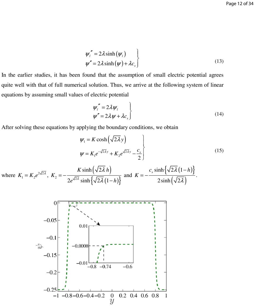
Measure the open-circuit streaming potential per unit length in an ex-vivo perfused microvessel, or in a microchannel lined with a charged porous hydrogel mimicking the EGL, using blood or a hematocrit-matched analog: if the voltage is not near 0.2 mV per micrometre for the stated parameter set, or if it does not vanish when the porous layer is removed, the volume-charge-only, zero-surface-charge model or its claim that the double layer is confined to the plasma layer is wrong. A second check is to probe the potential profile across the vessel: the model predicts the electric potential falls to zero inside the cell-free layer (beyond about 74% of the radius), with no field in the whole-blood core.
Extended reading notes
Core claim
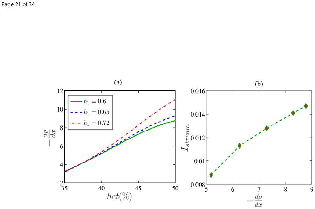
The central claim is that a charged poroelastic glycocalyx converts the mechanical energy of capillary blood flow into a streaming potential of order 0.2 mV/µm (about 0.1 to 0.2 V/mm), and that this value is substantially larger than a single-fluid Newtonian treatment predicts once the two-fluid rheology is included: at the physiological viscosity ratio of 0.078 and Deborah number near 0.7, the induced potential is about 40% higher. The magnitude is fixed by the electroneutrality constraint that the pressure-driven advection of counter-ions inside the thin double layer must be exactly cancelled by the conduction current the induced field drives back. The paper further claims the potential rises linearly with hematocrit up to 45%, reaches 2.5 to 3.5 times its 35%-hematocrit value at 50%, grows as the cell-free plasma layer thins (up to about 3 times the single-fluid value), and increases monotonically with EGL thickness, vanishing when the EGL is absent, which the authors read as a direct signature of the volume-charge model with zero surface charge.
Load-bearing premise
The calculation assumes the glycocalyx is electrically characterized only by a constant volume charge with zero surface charge, and that the roughly 30 nm electric double layer sits entirely inside the cell-free plasma layer, so the viscoelastic whole-blood core never feels the electric field; if either assumption fails, the predicted streaming potential and the two-fluid decoupling change.
Editorial extensions
If this is right
- At roughly 0.2 mV per micrometre, a millimetre-scale implantable conduit would develop on the order of a volt, a range compatible with micro- to milli-watt biosensor power budgets.
- Rheology acts as a multiplier, not a correction: at the physiological viscosity ratio of about 0.078 and Deborah number ~0.7, the two-fluid model predicts streaming potentials roughly 40% higher than a single-fluid Newtonian model.
- Streaming potential rises with hematocrit (2.5 to 3.5 times from 35% to 50%), so the same mechanism that could power a sensor also encodes a physiological state variable.
- Thinner cell-free layers and thicker EGL both raise the potential (up to about 3 times), meaning vascular conditions that remodel the glycocalyx will directly modulate any harvestable voltage.
- A vanishing streaming potential at zero EGL thickness provides a clean experimental signature: with a volume-charge-only EGL, no electrochemical interaction exists without the layer itself.
- The hematocrit sensitivity suggests a possible point-of-care diagnostic that measures streaming potential to flag dehydration or dengue-like hematocrit spikes, an application the paper motivates but does not design.
- Because disease states such as sepsis and diabetes degrade the glycocalyx, the same mechanism that offers power in healthy vessels would predict a falling voltage and falling harvestable power as the EGL thins, a coupling the paper leaves implicit.
- Measuring the streaming potential in an ex-vivo perfused microvessel or a glycocalyx-mimetic charged hydrogel channel would convert the model's ~0.2 mV/µm figure into a tested engineering specification.
Reading between the lines
- An ex-vivo experiment perfusing a microvessel, or a microchannel lined with a charged porous hydrogel, could directly test whether the open-circuit potential per unit length matches the predicted ~0.2 mV/µm scaling.
- The model's zero-potential-at-zero-EGL-thickness prediction is the sharpest signature of the volume-charge-only assumption; a nonzero potential with no porous layer would indicate a surface-charge contribution the model omits.
- The predicted rise of streaming potential with hematocrit suggests a passive point-of-care diagnostic for conditions with abnormal hematocrit, such as dengue, though the paper does not design such a device.
- Because many disease states degrade the glycocalyx, the same mechanism that powers devices in healthy vessels would predict both a falling voltage and a falling harvestable power as the EGL thins, a coupling the paper leaves implicit.
Signed reviews
Editorial analysis
A structured set of objections, weighed in public.
Referee Report
Summary. This paper develops an analytical model of pressure-driven blood flow in a microvessel coated by a charged poroelastic endothelial glycocalyx layer (EGL). The flow is modelled as a two-fluid system: a viscoelastic whole-blood core described by the linear simplified Phan-Thien-Tanner (sPTT) constitutive equation, a Newtonian cell-free plasma layer, and a poroelastic EGL with a constant volumetric fixed charge. The Poisson-Nernst-Planck equations are solved in the thin-Debye-layer limit, and the streaming potential is obtained from the zero-net-axial-current condition. The model reduces to the earlier Newtonian result of Sumets et al. (2015) when the Deborah number is zero, the cell-free layer is absent, and the viscosity ratio is unity. The authors report streaming potentials of order 0.2 mV/um and study their variation with hematocrit, cell-free layer thickness, and EGL thickness, concluding that the induced potential may be useful for powering implantable medical devices and biosensors.
Significance. The analytical derivation is detailed and self-contained, and the reduction to a known Newtonian limit provides a useful consistency check. The two-fluid treatment of blood with a viscoelastic core and a Newtonian cell-free layer, coupled to a charged poroelastic EGL, extends prior electrokinetic models of microvascular flow and yields falsifiable predictions for how the streaming potential depends on physiological parameters. These results could be valuable for label-free sensing or for understanding electrokinetic effects in the microcirculation. However, the central application claim that the induced streaming potential can power implantable devices in the micro-to-milli-Watt range is not derived from the model; the calculation gives only the open-circuit voltage, not the extractable power, and a simple Thévenin estimate places the available power orders of magnitude below the claimed range.
major comments (2)
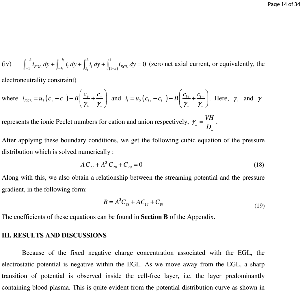
- [Abstract; Section III, 'Induced streaming potential in micro-fluidic systems...'] The claim that the induced streaming potential of order 0.2 mV/um 'may turn out to be substantial towards energizing biosensors and implantable medical devices whose power requirements are typically in the range of micro to milli Watt' is not a consequence of the calculation. The zero-net-axial-current condition (item iv in Section II) determines only the open-circuit streaming potential B. No load resistance, load current, or internal ionic resistance is computed anywhere in the paper. For a streaming-potential source, the maximum extractable power is P_max = V_oc^2 / (4 R_int). Using the paper's own parameters (vessel radius 5 um, blood plasma conductivity ~1.5 S/m, length ~1 mm, hematocrit 45%), the ionic resistance of the conduit is of order 10^7 ohm, so with V_oc ~0.2 V the matched-load power is approximately 1 nW, six orders of magnitude below the stated micro-to-milli-Watt range. Even an optimistic estimate based on the EGL fixed charge (c_s ~0.154 M, velocity ~10^-3 m/s, EGL cross-sectional area ~10^-11 m^2) gives a short-circuit streaming current below 1 uA and P_max below 0.1 uW. The power claim must therefore be supported by an explicit electrical-load model, or it must be removed or substantially qualified.
- [Section II, paragraph beginning 'The electrostatic charge of the EGL is characterized by a constant volumetric charge…] The model assumes that the EGL charge is purely volumetric with zero surface charge on the lumen-EGL interface. This assumption is load-bearing for the reported dependence of streaming potential on EGL thickness: Figure 9(a) shows that the streaming potential tends to zero as EGL thickness goes to zero, which is a direct consequence of setting the surface charge to zero. If a surface charge were present on the endothelial surface, the zero-thickness limit would not vanish, and the thickness dependence would change. The paper should either justify this assumption more strongly with physiological evidence or discuss the sensitivity of the main results to the inclusion of a surface-charge contribution. At present, the statement 'no electrochemical interaction takes place between the whole blood and blood plasma' rests on this assumption, so the reader cannot assess how robust the reported streaming-potential magnitudes are.
minor comments (4)
- [Throughout] The spelling of the reference 'Sumets' is inconsistent ('Sumets', 'Summets', 'Sumetc'), and the reference list contains incomplete entries (e.g., 'Sumetc, P. (2017)' appears only as a thesis URL). Please standardize the citations.
- [Section II and Section III] The notation for the fluid-fluid interface is inconsistent: the governing equations and boundary conditions use y = h, while the discussion and figures refer to h1 as the distance from the centreline. Please define h1 explicitly in the problem formulation and use one symbol consistently throughout.
- [Section III, paragraph on angiogenesis] The statement that the dependence of streaming potential on EGL thickness 'might be one of the key aspects in unlocking the mystery behind the angiogenesis pattern' is highly speculative and not supported by the model, which does not include any biochemical signalling or vascular-growth mechanism. This sentence should be softened or removed.
- [Figure 2 and Section II, text below Eq. (15)] The value of the Debye parameter lambda is reported as '42 10×' which is unclear; the text states that this gives a Debye layer thickness of 30 nm, but the relationship between lambda and the Debye length is not spelled out. Please state the dimensional value of lambda and the corresponding Debye length explicitly, and indicate the locations of the EGL/plasma and plasma/core interfaces on Figure 2.
Circularity Check
No significant circularity: the streaming potential is solved from the zero-net-current constraint, not fitted; self-citations are background and the device-power claim is an extrapolation, not a circular derivation.
full rationale
The core quantity B is an output of the coupled system: after prescribing the flow model and charge distribution, the paper imposes zero net axial current (Section II, item iv) and simultaneously solves Eq. (18) for the pressure gradient and Eq. (19) for B; no target value of B is used as an input, and B is not a fitted parameter renamed as a prediction. The validation in Fig. 4 is a reduction to the Newtonian single-fluid limit (De=0, h1=0, mu_r=1), which is a consistency check rather than circular reasoning. The many self-citations (e.g., Chakraborty 2019 for the implantable-device motivation, Mukherjee et al. for viscoelastic electroosmotic formulations) support background or constitutive choices and do not carry the derivation of the streaming potential. The by-construction statement that zero EGL thickness gives zero streaming potential is explicitly attributed by the authors to their volume-charge model, not presented as an independent result. One application-level claim is not supported by the calculation: Section III, in the paragraph beginning 'Induced streaming potential in micro-fluidic systems...', converts the open-circuit streaming potential (0.2 mV/um, hence ~0.2-1 V over mm) directly into a micro-to-milli-Watt powering capability without computing load resistance, load current, or extractable power. That is an engineering extrapolation and a correctness risk, but it is not a circular reduction of the model's output to its input.
Assumptions & free parameters
free parameters (7)
- Dimensionless fixed charge concentration c_s =
1 (dimensionless, corresponding to order 0.1 M from Silberberg 1991)
- Debye parameter lambda =
Text states lambda = 2e4 with Debye thickness 30 nm, but the relation to vessel radius is not made transparent
- EGL thickness fraction =
0.2 of vessel radius (h=0.8)
- Cell-free layer thickness parameter h1 =
0.6 and 0.72 in Fig 9
- Deborah number De =
0.7
- sPTT extensibility parameter epsilon =
Not stated numerically in the text
- Ionic Peclet numbers gamma+ and gamma- =
Not stated numerically in the text
assumptions (6)
- domain assumption EGL is a poroelastic volume-charge layer with constant volumetric fixed charge and zero surface charge (volume charge model).
- domain assumption The EDL is entirely contained in the cell-free plasma layer, so the viscoelastic whole-blood core is electrically inert and no Maxwell stress is needed at the fluid-fluid interface.
- domain assumption Flow is steady, incompressible, and fully developed; Womersley, Reynolds, and capillary numbers are small enough to neglect pulsatility, inertia, and interface deformation.
- domain assumption The Debye-Huckel linearization (small electric potential) is valid.
- domain assumption EGL solid deformation does not affect the streaming potential; the solid displacement field is decoupled from the streaming potential calculation.
- domain assumption Ion concentrations follow Boltzmann equilibrium with no axial concentration gradient.
Cite this review
Pith. "Pith review of Streaming Potential in Bio-mimetic Microvessels Mediated by Capillary Glycocalyx." pith.science (2026). https://pith.science/paper/7KF2ALAH
@misc{pith2026190801492,
  author       = {Pith},
  title        = {Pith review of: Streaming Potential in Bio-mimetic Microvessels Mediated by Capillary Glycocalyx},
  year         = {2026},
  howpublished = {\url{https://pith.science/paper/7KF2ALAH}},
  note         = {Machine review of arXiv:1908.01492}
}
read the original abstract
Implantable medical devices and biosensors are pivotal in revolutionizing the field of medical technology by opening new dimensions in the field of disease detection and cure. These devices need to harness a biocompatible and physiologically sustainable safe power source instead of relying on external stimuli, overcoming the constraints on their applicability in-vivo. Here, by appealing to the interplay of electromechanics and hydrodynamics in physiologically relevant microvessels, we bring out the role of charged Endothelial Glycocalyx layer (EGL) towards establishing a streaming potential across physiological fluidic conduits. We account for the complex rheology of blood-mimicking fluid by appealing to Newtonian fluid model representing the blood plasma and a viscoelastic fluid model representing the whole blood. We model the EGL as a poroelastic layer with volumetric charge distribution. Our results reveal that for physiologically relevant microflows, the streaming potential induced is typically of the order of 0.1 V/mm, which may turn out to be substantial towards energizing biosensors and implantable medical devices whose power requirements are typically in the range of micro to milli Watt. We also bring out the specific implications of the relevant physiological parameters towards establishment of the streaming potential, with a vision of augmenting the same within plausible functional limits. We further unveil that the dependence of streaming potential on EGL thickness might be one of the key aspects in unlocking the mystery behind the angiogenesis pattern. Our results may open up novel bio-sensing and actuating possibilities in medical diagnostics as well as may provide a possible alternative regarding the development of physiologically safe and biocompatible power sources within the human body.
Figures

Figures from the paper (7 more)




Reference graph
Works this paper leans on
-
[1]
Afonso, A. M., Alves, M. A., & Pinho, F. T. (2009). Analytical solution of mixed electro- osmotic/pressure driven flows of viscoelastic fluids in microchannels. Journal of Non- Newtonian Fluid Mechanics, 159(1–3), 50–63. Akbarzadeh, P. (2016). Pulsatile magneto-hydrodynamic blood flows through porous blood vessels using a third grade non-Newtonian fluids ...
work page 2009
-
[179]
Chakraborty, S. (2005). Dynamics of capillary flow of blood into a microfluidic channel. Lab on a Chip, 5(4),
work page 2005
-
[283]
Poddar, A., Maity, D., Bandopadhyay, A., & Chakraborty, S. (2016). Electrokinetics in polyelectrolyte grafted nanofluidic channels modulated by the ion partitioning effect. Soft Matter, 12(27), 5968–5978. Pries, A. R., Höpfner, M., le Noble, F., Dewhirst, M. W., & Secomb, T. W. (2010). The shunt problem: control of functional shunting in normal and tumour...
work page 2016
-
[321]
paper-and-pencil
Kang, H., Fan, Y., & Deng, X. (2011). Vascular smooth muscle cell glycocalyx modulates shear- induced proliferation, migration, and NO production responses. American Journal of Physiology-Heart and Circulatory Physiology, 300(1), H76–H83. Katanov, D., Gompper, G., & Fedosov, D. A. (2015). Microvascular blood flow resistance: Role of red blood cell migrati...
2011
-
[421]
Chakraborty, S. (2019). Electrokinetics with blood. Electrophoresis, 40(1), 180–189. Damiano, E. R., & Stace, T. M. (2002). A Mechano-Electrochemical Model of Radial Deformation of the Capillary Glycocalyx. Biophysical Journal, 82(3), 1153–1175. Damiano, E. R., & Stace, T. M. (2005). Flow and deformation of the capillary glycocalyx in the Page 30 of 34 31...
work page 2019
-
[3210]
Niu, P., Chapman, P., Riemer, R., & Zhang, X. (n.d.). Evaluation of motions and actuation methods for biomechanical energy harvesting. In 2004 IEEE 35th Annual Power Electronics Specialists Conference (IEEE Cat. No.04CH37551), IEEE, pp. 2100–2106. Page 32 of 34 33 P. Thien, N. (1978). A Nonlinear Network Viscoelastic Model. Journal of Rheology, 22(3), 259–
work page 1978
Reviewed August 14, 2026 · model on record in the stance chip above.
Discussion (0). Continue with ORCID to comment.