REVIEW 3 major objections 7 minor 43 references
DISCo: Deep learning, Instance Segmentation, and Correlations for cell segmentation in calcium imaging
T0 review · 3 major / 7 minor · reviewed 2026-08-14 · deepseek-v4-flash
Pith's one-line read The paper claims that DISCo, by combining segment-wise pixel correlations with a summary image and partitioning a signed graph of learned affinities, outperforms all other methods trained on the Neurofinder calcium-imaging datasets.
desk verdict Solid methods paper with a real pipeline advance, but the 'outperforms all' claim is slightly ahead of the evidence. read the letter →
The pith
A machine-rendered reading of the paper's core claim, the machinery that carries it, and where it could break.
The reading
What carries the argument
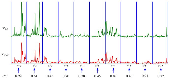
The load-bearing object is the predicted pixel-affinity field combined with the GASP signed-graph partitioner. Affinities $a_{ij}\in[0,1]$ are converted to edge weights $w_{ij}=a_{ij}-0.5$, so positive weights attract pixels into the same cluster and negative weights separate them; GASP then merges clusters while average linkage is positive. This turns instance segmentation into correlation clustering, solved approximately by a greedy agglomerative algorithm. The other essential component is the input representation: segment-wise Pearson correlations ($N=10$ segments, $C=15$ neighbor offsets) aggregated by a 3D CNN, concatenated with a mean-intensity projection.
What would settle it
Rerun the released DISCo code on the Neurofinder training set, train per-series models with several random seeds, and submit to the Neurofinder test set; the claim fails if the average F1 does not reliably stay above 0.66, the score reported for 3dCNN.
Extended reading notes
Core claim
DISCo treats cell segmentation in calcium-imaging videos as a graph-partitioning problem on learned pixel affinities. The video is split into ten temporal segments; for each pixel, Pearson correlations are computed against fifteen neighboring pixels in each segment, producing a stack that a small 3D convolutional network aggregates. This aggregated correlation map is concatenated with the mean-intensity projection over the whole video and passed to a U-Net that predicts, for each pixel, a foreground/background score and affinities to nearby pixels. Affinities are converted to signed edge weights ($w_{ij}=a_{ij}-0.5$) and background edges are removed before the GASP algorithm partitions the graph into cell instances. On the Neurofinder test set, the paper reports that the per-series-trained variant (DISCos) achieves an average F1 of 0.67, outperforming all other methods trained only on the Neurofinder datasets, with 3dCNN at 0.66; the single-model variant (DISCo) reaches 0.63.
Load-bearing premise
The central outperformance claim depends on the Neurofinder leaderboard scores for other methods being current, accurate, and directly comparable; with a winning margin of only 0.01 average F1 over the runner-up 3dCNN, stale leaderboard entries or slight differences in training data would erase the claimed advantage.
Editorial extensions
If this is right
- A single DISCo model, trained once on all five Neurofinder dataset series, attains a 0.63 average F1 on the test set, showing that one network can handle heterogeneous recordings without per-series tuning.
- Per-series training (DISCos) raises the average F1 to 0.67 and edges out the previous best Neurofinder-trained method (3dCNN at 0.66), supporting the claim that the correlation-plus-shape design is competitive with spatiotemporal deep learning.
- The single-video training experiment, with a 0.58 average F1 on test videos from the same series, implies that a neuroscience lab could label one video for a new recording setup and get usable segmentation without retraining on large data.
- The lesion study's ordering of inputs implies that future systems should treat temporal correlations and shape summaries as complements rather than alternatives; dropping either cue costs roughly 0.13-0.14 in average F1.
Reading between the lines
- None of the comparisons reports variance across training runs; given the 0.01 margin over 3dCNN, an obvious extension is to retrain DISCos with multiple seeds and report the distribution, which would tell whether the outperformance is systematic or within noise.
- The affinity-plus-graph-partitioning recipe is agnostic to the imaging modality; the same signed-graph clustering of learned affinities has been used for other instance-segmentation tasks, so feeding temporal correlation features into that pipeline could transfer to video instance segmentation of other dynamic objects.
- The authors do not analyze per-dataset precision/recall trade-offs; a testable extension would be to calibrate the background-edge exclusion threshold per dataset series, since the paper reports that recall limits series 00 and precision limits series 01.
- Because the summary image carries the inactive cells, DISCo is positioned for all-cell segmentation; a natural next experiment is to evaluate it on datasets labeled specifically for active cells to see whether the correlation pathway can be dropped without loss.
Signed reviews
Editorial analysis
A structured set of objections, weighed in public.
Referee Report
Summary. The paper presents DISCo, a calcium-imaging cell segmentation method that computes segment-wise Pearson correlations between pixels, feeds these together with a mean intensity projection into a two-stage CNN that predicts pairwise affinities and a foreground/background map, and extracts instances by partitioning the resulting signed graph with the GASP algorithm. The method is evaluated on the Neurofinder public benchmark and compared with leaderboard entries, with additional lesion studies on input modalities and temporal aggregation. The main reported results are an average F1 of 0.63 for the single-model configuration DISCo and 0.67 for the per-series configuration DISCos, with the paper claiming that DISCos outperforms all other methods trained on the Neurofinder datasets.
Significance. If supported, the contribution would be valuable: DISCo offers a computationally cheaper alternative to 3D-CNN-on-video approaches while retaining temporal information, and it integrates instance segmentation directly. The code is publicly available, the evaluation is on an external public benchmark, and the lesion studies in Tables 4 and 5 support the main design choices. However, the central 'outperforms all' claim is not established as stated: it is misattributed in the abstract, it rests on an incomplete leaderboard comparison, and the 0.01 margin over 3dCNN is within the noise of single-run evaluations. With those claims corrected or tempered, the paper's contribution is solid and reproducible.
major comments (3)
- [Abstract and Section 1] The abstract states that 'DISCo outperforms all existing models trained on these datasets,' but Table 3 shows the single-model DISCo at average F1 0.63, below 3dCNN's 0.66; the 0.67 average that tops the listed Neurofinder-trained methods belongs to DISCos, the per-series configuration. Section 1 itself states the claim correctly for DISCos. The abstract and any unqualified excerpts should be revised so the top result is attributed to DISCos, and the Table 3 caption's statement about a 'single model' should be limited to comparisons among single-model methods.
- [Section 2 (footnote) and Table 3] The claim that DISCos 'outperforms all other methods trained on the Neurofinder datasets' is a universal claim over the leaderboard, but the comparison set is not demonstrated to be complete. The footnote in Section 2 excludes the Mask R-CNN and human-label submissions because no information is available on their models and training procedures; if either of these entries was trained only on Neurofinder training data and scored at least 0.67, the stated claim would be false. Since the leaderboard snapshot was taken on 2019-11-15, the claim should be restricted to the listed methods with verified training procedures and to that snapshot, or the training status of the excluded entries should be documented.
- [Section 4, Table 3] The reported advantage over the closest competitor 3dCNN is 0.01 in average F1 (0.67 vs. 0.66), based on single runs with no variance estimate and no significance test. A paired Wilcoxon signed-rank test on the nine per-video F1 scores in Table 3 yields p > 0.05, so the word 'outperforms' is not statistically supported for this comparison. The authors should either provide variance across multiple training runs with a significance test, or replace 'outperforms' by 'is competitive with' for comparisons involving 3dCNN. This is load-bearing because the headline claim rests on this margin.
minor comments (7)
- [Section 1] The word 'segmentaiton' in the second paragraph should be 'segmentation'.
- [Supplementary Table 7] In the DISCos/max row, '0,78' uses a comma as decimal separator; use '0.78' for consistency.
- [Figure 2 and Section 3.1] The figure shows C=3 neighbors while the text uses C=15; add a note that the figure is illustrative.
- [Section 3.3] The criterion for background exclusion, that the background prediction is higher than the foreground prediction, is equivalent to a 0.5 threshold on the foreground channel; state this explicitly.
- [Section 4] The exact p-values for the two-sided Wilcoxon tests against UNet2DS and Conv2D are not reported; give them in the text or in the table.
- [Table 3] Because the Neurofinder leaderboard is time-varying, the access date should be repeated in the caption of Table 3 rather than only in the body text.
- [Supplementary Table 8] Indicate that the missing standard deviations for some rows reflect series with only one training video, or explain why they are absent.
Circularity Check
No circularity: the benchmark evaluation is external, and DISCo's predictions do not reduce to its training inputs by construction.
full rationale
The paper's central claim is an empirical ranking on the Neurofinder benchmark. The network is trained on ground-truth affinities derived from the Neurofinder training annotations (Eq. 4) and evaluated on the undisclosed test set; no fitted parameter is renamed as a prediction, and the test F1 scores in Table 3 come from an external leaderboard. The only same-group citations are GASP [39], mutex watershed [38], and LeMoNADe [4], but GASP is used as a black-box graph-partitioning component whose linkage rule (Eq. 6) and stopping criterion are not fitted to Neurofinder test labels; the paper does not invoke a uniqueness theorem or import its conclusion from those citations. The exclusion of Mask R-CNN and human-label submissions in the Section 2 footnote, and the 0.01 average-F1 margin over 3dCNN, are concerns about claim completeness or statistical significance, not about circularity, and are therefore outside the scope of this pass. The method's components (segment-wise correlations, U-Net, GASP) are independently evaluated through the benchmark, so no derivation step is equivalent to its input by construction.
Assumptions & free parameters
free parameters (8)
- N (number of temporal segments) =
10
- C (number of correlation neighbors per pixel) =
15
- Graph edge connectivity distance =
5 pixels
- Minimum instance size threshold =
25 pixels
- Inference max-pooling kernel length =
5 frames
- Learning rate =
0.0001
- Training epochs =
3000
- Batch size =
20 (DISCo); 6 for series 00 and 1 otherwise (DISCos)
assumptions (4)
- domain assumption The Neurofinder ground truth annotations define the correct cell segmentation targets.
- domain assumption Segment-wise Pearson correlations plus the mean intensity projection contain enough information to identify active and inactive cells.
- domain assumption The 19 Neurofinder training videos are representative of the 9 test videos within each dataset series.
- domain assumption The GASP algorithm with average linkage is an appropriate solver for the affinity-based instance segmentation problem.
Cite this review
Pith. "Pith review of DISCo: Deep learning, Instance Segmentation, and Correlations for cell segmentation in calcium imaging." pith.science (2026). https://pith.science/paper/ANFE5W6T
@misc{pith2026190807957,
  author       = {Pith},
  title        = {Pith review of: DISCo: Deep learning, Instance Segmentation, and Correlations for cell segmentation in calcium imaging},
  year         = {2026},
  howpublished = {\url{https://pith.science/paper/ANFE5W6T}},
  note         = {Machine review of arXiv:1908.07957}
}
read the original abstract
Calcium imaging is one of the most important tools in neurophysiology as it enables the observation of neuronal activity for hundreds of cells in parallel and at single-cell resolution. In order to use the data gained with calcium imaging, it is necessary to extract individual cells and their activity from the recordings. We present DISCo, a novel approach for the cell segmentation in calcium imaging videos. We use temporal information from the recordings in a computationally efficient way by computing correlations between pixels and combine it with shape-based information to identify active as well as non-active cells. We first learn to predict whether two pixels belong to the same cell; this information is summarized in an undirected, edge-weighted grid graph which we then partition. In so doing, we approximately solve the NP-hard correlation clustering problem with a recently proposed greedy algorithm. Evaluating our method on the Neurofinder public benchmark shows that DISCo outperforms all existing models trained on these datasets.
Figures


Figures from the paper (1 more)

Reference graph
Works this paper leans on
-
[1]
Winfried Denk, James H. Strickler, and Watt W. Webb. Two- photon laser scanning fluorescence microscopy. Science, 1990
work page 1990
-
[2]
Deep tissue two-photon microscopy
Fritjof Helmchen and Winfried Denk. Deep tissue two-photon microscopy. Nature Methods, 2005
work page 2005
-
[3]
High-speed, miniaturized fluorescence microscopy in freely moving mice
Benjamin A Flusberg, Axel Nimmerjahn, Eric D Cocker, Eran A Mukamel, Robert PJ Barretto, Tony H Ko, Laurie D Burns, Juergen C Jung, and Mark J Schnitzer. High-speed, miniaturized fluorescence microscopy in freely moving mice. Nature Methods, 5(11):935, 2008
work page 2008
-
[4]
Elke Kirschbaum, Manuel Haußmann, Steffen Wolf, Han- nah Sonntag, Justus Schneider, Shehabeldin Elzoheiry, Oliver Kann, Daniel Durstewitz, and Fred A. Hamprecht. LeMoN- ADe: Learned Motif and Neuronal Assembly Detection in calcium imaging videos. In International Conference on Learning Representations (ICLR), Proceedings, 2018
work page 2018
-
[5]
CodeNeuro. Neurofinder public benchmark. http: //neurofinder.codeneuro.org, 2016. Accessed: 2019-11-13
work page 2016
-
[6]
Chet- tih, Matthias Minderer, Christopher Harvey, and Dorit S
Quico Spaen, Roberto Asín-Achá, Selmaan N. Chet- tih, Matthias Minderer, Christopher Harvey, and Dorit S. Hochbaum. HNCcorr: A novel combinatorial ap- proach for cell identification in calcium-imaging movies. eNeuro, 6(2), 2019. doi: 10.1523/ENEURO.0304-18
-
[7]
Automated analysis of cellular signals from large-scale cal- cium imaging data
Eran A Mukamel, Axel Nimmerjahn, and Mark J Schnitzer. Automated analysis of cellular signals from large-scale cal- cium imaging data. Neuron, 63(6):747–760, 2009
work page 2009
-
[8]
Sparse non- negative deconvolution for compressive calcium imaging: algorithms and phase transitions
Eftychios A Pnevmatikakis and Liam Paninski. Sparse non- negative deconvolution for compressive calcium imaging: algorithms and phase transitions. In Advances in Neural Information Processing Systems (NIPS), Proceedings, 2013
work page 2013
Show all 43 references
-
[9]
Rank-penalized nonnegative spatiotemporal deconvolu- tion and demixing of calcium imaging data
Eftychios A Pnevmatikakis, Timothy A Machado, Logan Grosenick, Ben Poole, Joshua T V ogelstein, and Liam Panin- ski. Rank-penalized nonnegative spatiotemporal deconvolu- tion and demixing of calcium imaging data. InComputational and Systems Neuroscience (Cosyne), 2013
2013
-
[10]
Pnevmatikakis, Yuanjun Gao, Daniel Soudry, David Pfau, Clay Lacefield, Kira Poskanzer, Randy Bruno, Rafael Yuste, and Liam Paninski
Eftychios A. Pnevmatikakis, Yuanjun Gao, Daniel Soudry, David Pfau, Clay Lacefield, Kira Poskanzer, Randy Bruno, Rafael Yuste, and Liam Paninski. A structured matrix fac- torization framework for large scale calcium imaging data analysis. arXiv preprint, arXiv:1409.2903, 2014
2014 arXiv
-
[11]
Sparse space-time deconvolution for calcium image analysis
Ferran Diego and Fred A Hamprecht. Sparse space-time deconvolution for calcium image analysis. In Advances in Neural Information Processing Systems (NIPS), Proceedings, 2014
2014
-
[12]
Detecting cells using non-negative matrix factorization on calcium imaging data
Ryuichi Maruyama, Kazuma Maeda, Hajime Moroda, Ichiro Kato, Masashi Inoue, Hiroyoshi Miyakawa, and Toru Aonishi. Detecting cells using non-negative matrix factorization on calcium imaging data. Neural Networks, 55:11–19, 2014
2014
-
[13]
Pnevmatikakis, Daniel Soudry, Yuanjun Gao, Timothy A
Eftychios A. Pnevmatikakis, Daniel Soudry, Yuanjun Gao, Timothy A. Machado, Josh Merel, David Pfau, Thomas Rear- don, Yu Mu, Clay Lacefield, Weijian Yang, Misha Ahrens, Randy Bruno, Thomas M. Jessell, Darcy S. Peterka, Rafael Yuste, and Liam Paninski. Simultaneous denoising, de...
2016
-
[14]
Fast online deconvolution of calcium imaging data
Johannes Friedrich, Pengcheng Zhou, and Liam Paninski. Fast online deconvolution of calcium imaging data. PLoS computational biology, 13(3):e1005423, 2017
2017
-
[15]
Erdogdu, and Mark Schnitzer
Hakan Inan, Murat A. Erdogdu, and Mark Schnitzer. Robust estimation of neural signals in calcium imaging. In Advances in Neural Information Processing Systems (NIPS), Proceed- ings, 2017
2017
-
[16]
Onacid: Online analysis of cal- cium imaging data in real time
Andrea Giovannucci, Johannes Friedrich, Matt Kaufman, Anne Churchland, Dmitri Chklovskii, Liam Paninski, and Eftychios A Pnevmatikakis. Onacid: Online analysis of cal- cium imaging data in real time. In Advances in Neural Infor- mation Processing Systems (NIPS), Proceedings, 2017
2017
-
[17]
Efficient and accurate extraction of in vivo calcium signals from microen- doscopic video data
Pengcheng Zhou, Shanna L Resendez, Jose Rodriguez- Romaguera, Jessica C Jimenez, Shay Q Neufeld, Andrea Giovannucci, Johannes Friedrich, Eftychios A Pnevmatikakis, Garret D Stuber, Rene Hen, Mazen A Kheirbek, Bernardo L Sabatini, Robert E Kass, and Liam Paninski. Efficient and ...
2018
-
[18]
Caiman an open source tool for scalable calcium imaging data analysis
Andrea Giovannucci, Johannes Friedrich, Pat Gunn, Jérémie Kalfon, Brandon L Brown, Sue Ann Koay, Jiannis Taxidis, Farzaneh Najafi, Jeffrey L Gauthier, Pengcheng Zhou, Baljit S Khakh, David W Tank, Dmitri B Chklovskii, and Eftychios A Pnevmatikakis. Caiman an open source tool fo...
2019
-
[19]
SIMA: Python software for analysis of dynamic fluorescence imaging data
Patrick Kaifosh, Jeffrey D Zaremba, Nathan B Danielson, and Attila Losonczy. SIMA: Python software for analysis of dynamic fluorescence imaging data. Frontiers in neuroinfor- matics, 8:80, 2014
2014
-
[20]
Hamprecht
Ferran Diego, Susanne Reichinnek, Martin Both, and Fred A. Hamprecht. Automated identification of neuronal activity from calcium imaging by sparse dictionary learning. In IEEE International Symposium on Biomedical Imaging (ISBI), 2013
2013
-
[21]
Learning multi-level sparse representations
Ferran Diego and Fred A Hamprecht. Learning multi-level sparse representations. In Advances in Neural Information Processing Systems (NIPS), Proceedings, 2013
2013
-
[22]
Extracting regions of interest from biological images with convolutional sparse block coding
Marius Pachitariu, Adam M Packer, Noah Pettit, Henry Dal- gleish, Michael Hausser, and Maneesh Sahani. Extracting regions of interest from biological images with convolutional sparse block coding. In Advances in Neural Information Processing Systems (NIPS), Proceedings, 2013
2013
-
[23]
Scalpel: Extracting neurons from calcium imaging data
Ashley Petersen, Noah Simon, and Daniela Witten. Scalpel: Extracting neurons from calcium imaging data. The annals of applied statistics, 12(4):2430, 2018
2018
-
[24]
Apthorpe, Alexander J
Noah J. Apthorpe, Alexander J. Riordan, Rob E. Aguilar, Jan Homann, Yi Gu, David W. Tank, and H. Sebastian Seung. Automatic neuron detection in calcium imaging data using convolutional networks. In Advances in Neural Information Processing Systems (NIPS), Proceedings, 2016
2016
-
[25]
Fast, simple calcium imaging segmentation with fully convolutional networks
Aleksander Klibisz, Derek Rose, Matthew Eicholtz, Jay Blun- don, and Stanislav Zakharenko. Fast, simple calcium imaging segmentation with fully convolutional networks. In Interna- tional Workshop on Deep Learning in Medical Image Analy- sis and Multimodal Learning for Clinical...
2017
-
[26]
Fast and robust active neu- ron segmentation in two-photon calcium imaging using spa- tiotemporal deep learning
Somayyeh Soltanian-Zadeh, Kaan Sahingur, Sarah Blau, Yiyang Gong, and Sina Farsiu. Fast and robust active neu- ron segmentation in two-photon calcium imaging using spa- tiotemporal deep learning. Proceedings of the National Academy of Sciences , 116(17):8554–8563, 2019. ISSN 0...
2019 doi
-
[27]
Shang Gao. Conv2d. https://github.com/ iamshang1/Projects/tree/master/Advanced_ ML/Neuron_Detection, 2016. Accessed: 2019-07-19
2016
-
[28]
Suite2p: beyond 10,000 neu- rons with standard two-photon microscopy
Marius Pachitariu, Carsen Stringer, Mario Dipoppa, Sylvia Schröder, L Federico Rossi, Henry Dalgleish, Matteo Caran- dini, and Kenneth D Harris. Suite2p: beyond 10,000 neu- rons with standard two-photon microscopy. bioRxiv preprint, bioRxiv:061507, 2017
2017
-
[29]
Allen Brain Observatory (ABO) datasets
Allen Institute for Brain Science. Allen Brain Observatory (ABO) datasets. http://observatory.brain-map. org/visualcoding, 2016. Accessed: 2019-08-20
2016
-
[30]
Scaled correlation analysis: a better way to compute a cross-correlogram
Danko Nikoli ´c, Raul C Mure¸ san, Weijia Feng, and Wolf Singer. Scaled correlation analysis: a better way to compute a cross-correlogram. European Journal of Neuroscience, 35 (5):742–762, 2012
2012
-
[31]
Synchronisation hubs in the visual cor- tex may arise from strong rhythmic inhibition during gamma oscillations
Stefanos E Folias, Shan Yu, Abigail Snyder, Danko Nikoli´c, and Jonathan E Rubin. Synchronisation hubs in the visual cor- tex may arise from strong rhythmic inhibition during gamma oscillations. European Journal of Neuroscience, 38(6):2864– 2883, 2013
2013
-
[32]
A scaled- correlation based approach for defining and analyzing func- tional networks
Samuel Dolean, Mihaela Dîn¸ soreanu, Raul Cristian Mure¸ san, Attila Geiszt, Rodica Potolea, and Ioana ¸ Tinca¸ s. A scaled- correlation based approach for defining and analyzing func- tional networks. In International Workshop on New Frontiers in Mining Complex Patterns, pages...
2017
-
[33]
Notes on regression and inheritance in the case of two parents
Karl Pearson. Notes on regression and inheritance in the case of two parents. Proceedings of the Royal Society of London, 58:240–242, 1895
-
[34]
Measur- ing and testing dependence by correlation of distances
Gábor J Székely, Maria L Rizzo, and Nail K Bakirov. Measur- ing and testing dependence by correlation of distances. The annals of statistics, 35(6):2769–2794, 2007
2007
-
[35]
U-net: Convolutional networks for biomedical image segmentation
Olaf Ronneberger, Philipp Fischer, and Thomas Brox. U-net: Convolutional networks for biomedical image segmentation. In International Conference on Medical Image Computing and Computer Assisted Intervention (MICCAI), pages 234– 241, 2015
2015
-
[36]
Measures of the amount of ecologic association between species
Lee R Dice. Measures of the amount of ecologic association between species. Ecology, 26(3):297–302, 1945
1945
-
[37]
A method of establishing groups of equal amplitude in plant sociology based on similarity of species content and its application to analyses of the vegeta- tion on danish commons
Thorvald Julius Sørensen. A method of establishing groups of equal amplitude in plant sociology based on similarity of species content and its application to analyses of the vegeta- tion on danish commons. Biol. Skar., 5:1–34, 1948
1948
-
[38]
Ham- precht
Steffen Wolf, Constantin Pape, Alberto Bailoni, Nasim Ra- haman, Anna Kreshuk, Ullrich Köthe, and Fred A. Ham- precht. The mutex watershed: efficient, parameter-free image partitioning. In European Conference on Computer Vision (ECCV), Proceedings, 2018
2018
-
[39]
Hamprecht
Alberto Bailoni, Constantin Pape, Steffen Wolf, Thorsten Beier, Anna Kreshuk, and Fred A. Hamprecht. A general- ized framework for agglomerative clustering of signed graphs applied to instance segmentation. arXiv preprint, arXiv: 1906.11713, 2019
1906 arXiv
-
[40]
Efficient decomposition of image and mesh graphs by lifted multi- cuts
Margret Keuper, Evgeny Levinkov, Nicolas Bonneel, Guil- laume Lavoué, Thomas Brox, and Bjorn Andres. Efficient decomposition of image and mesh graphs by lifted multi- cuts. In IEEE International Conference on Computer Vision (ICCV), Proceedings, 2015
2015
-
[41]
A comparative study of local search algorithms for correlation clustering
Evgeny Levinkov, Alexander Kirillov, and Bjoern Andres. A comparative study of local search algorithms for correlation clustering. In German Conference on Pattern Recognition, pages 103–114. Springer, 2017
2017
-
[42]
Adam: A method for stochastic optimization
Diederik P Kingma and Jimmy Ba. Adam: A method for stochastic optimization. In International Conference on Learning Representations (ICLR), Proceedings, 2014. Supplementary Material Table 6: Results with DISCo for different kinds of input. We show the results on the Neurofinder...
2014
-
[2019]
URL https://www.eneuro.org/content/ 6/2/ENEURO.0304-18.2019
2019
Reviewed August 14, 2026 · model on record in the stance chip above.
Discussion (0). Continue with ORCID to comment.