REVIEW 4 major objections 4 minor 13 references
Estimation of preterm birth markers with U-Net segmentation network
T0 review · 4 major / 4 minor · reviewed 2026-08-14 · deepseek-v4-flash
Pith's one-line read A U-Net pipeline measuring cervical length and anterior cervical angle predicts preterm birth with an 18% false-negative rate, down from 30% for manual screening.
desk verdict The paper's central false-negative claim crumbles under its own confusion matrix, and the evaluation is not end-to-end, so the stated clinical benefit does not hold up. read the letter →
The pith
A machine-rendered reading of the paper's core claim, the machinery that carries it, and where it could break.
The reading
What carries the argument
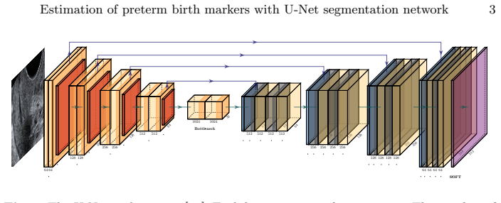
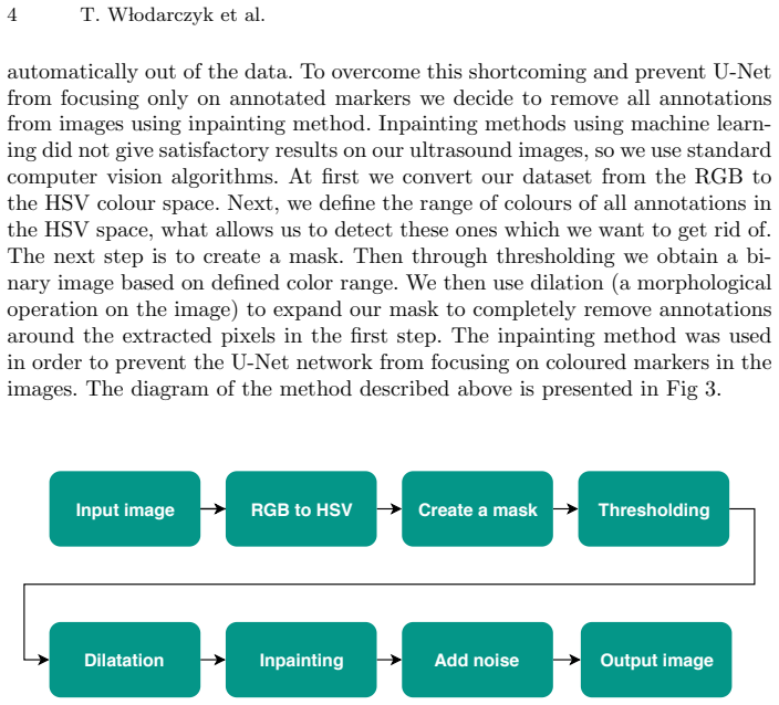
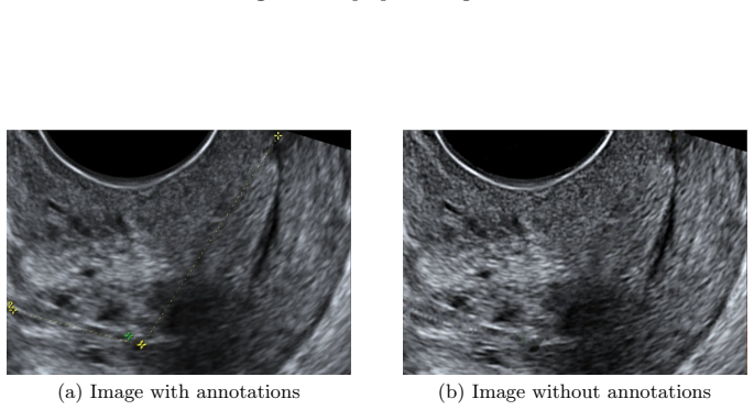
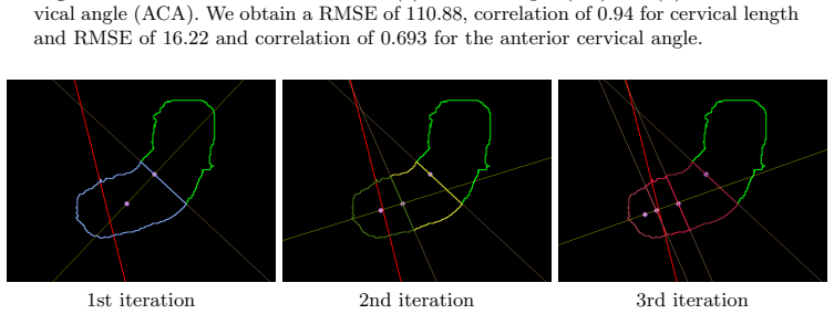
The central machinery is the U-Net, an encoder-decoder convolutional network for biomedical image segmentation, which produces a binary mask of the cervix from each preprocessed ultrasound image. Cervical length is then estimated by a centerline algorithm that builds a Voronoi skeleton of the mask and smooths the selected centerline; anterior cervical angle is estimated by a three-iteration recursive centroid split that divides the mask and measures the angle between the anterior wall and the line through the last two centroids. A naive Bayes classifier maps the resulting (CL, ACA) pairs to preterm/control labels. This chain converts subjective visual features into numerical markers and then into a risk decision.
What would settle it
Apply the trained naive Bayes classifier to the CL and ACA values produced automatically by the U-Net pipeline for the 52 preterm and 62 control pregnancies, and compare the confusion matrix with the one obtained from the 380 precomputed samples. A false-negative count of 21 out of 52 (40%) would show that the 18% figure does not transfer; alternatively, a paired test on both manual and automatic markers would settle whether segmentation accuracy is the limiting factor.
Extended reading notes
Core claim
The central claim is that a complete deep-learning pipeline can extract the two ultrasound markers used in preterm birth screening—cervical length and anterior cervical angle—from raw transvaginal ultrasound images, and that combining these markers predicts spontaneous preterm birth better than current manual screening. On the segmentation task the U-Net achieves an average Jaccard index of 0.91. The cervical-length estimate has correlation 0.94 with ground truth (RMSE 110.88), and the anterior-cervical-angle estimate has correlation 0.693 (RMSE 16.22). Using first- and second-trimester (CL, ACA) pairs, a naive Bayes classifier reaches 77.5% accuracy, 85% precision, 74% recall, and 0.78 AUC, which the paper reports as 18% false negatives versus about 30% for manual screening and 14% false positives. The intended upshot is an objective, repeatable alternative to visual assessment, with the same segmentation masks available for other biophysical markers such as cervical shape and tissue density.
Load-bearing premise
The load-bearing premise is that the classifier's 18% false-negative rate, obtained from a separate 380-sample dataset of expert-measured cervical lengths and angles, will also hold for the automatically segmented and estimated markers, even though the automatic angle estimates correlate only 0.693 with expert measurement.
Editorial extensions
If this is right
- Ultrasound screening for preterm risk could become a fully automatic pipeline: image in, risk score out, removing the subjective step that currently misses about 30% of spontaneous preterm deliveries.
- Because the segmentation masks are reusable geometry, the same network can support additional markers such as cervical shape or tissue density without retraining the segmentation stage.
- Adding anterior cervical angle to cervical length gives a concrete way to improve first-trimester prediction, the period in which current screening is least effective.
- A detection rate near 74% at a 10% false-positive rate would outperform the 54.8% detection rate of the standard maternal-factor-and-placental-perfusion model, making earlier and more reliable risk stratification possible.
Reading between the lines
- The 18% false-negative figure comes from expert-measured markers on a separate 380-sample dataset; if the automatic ACA estimates (correlation 0.693) feed the classifier, the real false-negative rate could be substantially higher, so an end-to-end evaluation is the decisive test the paper leaves open.
- The recursive centroid-split ACA algorithm is sensitive to segmentation boundary noise; evaluating it on masks with artificially degraded Jaccard indices would show how much of the prediction quality depends on the U-Net rather than on the classifier.
- Because the classifier was trained on a balanced 50:50 preterm/control set, the reported 14% false-positive and 18% false-negative rates do not directly reflect clinical prevalence; recalibrating to the observed ~20% preterm rate would change both rates.
Signed reviews
Editorial analysis
A structured set of objections, weighed in public.
Referee Report
Summary. The paper proposes an automatic pipeline for estimating two ultrasound markers of preterm birth risk, cervical length (CL) and anterior cervical angle (ACA), from transvaginal ultrasound images. The pipeline first segments the cervix with a U-Net, then estimates CL via a centerline algorithm and ACA via recursive centroid splitting, and finally uses these markers in binary classifiers (SVM, KNN, Naive Bayes, decision trees) to distinguish preterm from control pregnancies. On a separate dataset of 380 precomputed manual measurements, the authors report that combining CL and ACA decreases the false-negative ratio from 30% to 18%. The abstract presents this as the main result and frames the method as an objective alternative to subjective visual assessment by gynecologists.
Significance. If the end-to-end pipeline were validated, automatic and objective extraction of CL and ACA could have practical value for preterm-birth screening, and the paper would address an important clinical problem. The authors are transparent about the weak correlation of their automatic ACA estimates with ground truth (correlation 0.693, RMSE 16.22 degrees), and they report training details for the U-Net in reasonable detail. However, the central claimed clinical outcome is contradicted by the paper's own tables, and the classifier evaluation is not performed on the automatic marker estimates that the paper advertises. As submitted, the manuscript does not support the abstract's main claim and requires substantial additional experiments and corrections before it could be considered for publication.
major comments (4)
- [Abstract and Section 3.3 (Table 2)] The abstract's central claim that combined CL and ACA markers decrease the false-negative ratio from 30% to 18% is not supported by the results in Table 2. The confusion matrix shows 21 false negatives among 52 actual preterm cases, giving a conditional false-negative rate of 21/52 = 40.4%, not 18%. The value 18% appears only when 21 is divided by all 114 test instances (21/114 = 18.4%), but the baseline of 30% is defined conditionally on spontaneous preterm deliveries, so changing the denominator invalidates the comparison. Table 1 also contradicts the claim: the best Naive Bayes classifier with first- and second-trimester features has recall 74%, implying 26% false negatives, and the first-trimester Naive Bayes classifier has recall 69%, implying 31% false negatives. Therefore the text in Section 3.3 stating that "for the first trimester, we obtained 18% of false negatives" is unsupported by the paper's own data.
- [Section 3.3 (first paragraph) and Abstract] The classifiers are trained and evaluated on a different dataset of 380 balanced numerical samples with precomputed CL and ACA measurements, not on marker values estimated automatically by the U-Net pipeline described in Sections 3.1 and 3.2. Because the automatic ACA estimates have a correlation of only 0.693 with ground truth and an RMSE of 16.22 degrees, classifier performance measured on manual measurements cannot be assumed to transfer to the automatic estimates. The abstract claims that the proposed automatic pipeline allows the false-negative ratio to be reduced to 18%, but no experiment in the paper evaluates the complete pipeline from ultrasound image to classification. An end-to-end evaluation on automatically estimated markers is missing and is load-bearing for the paper's main claim.
- [Section 3.1 (Jaccard Index results)] The reported segmentation performance in Section 3.1 is internally inconsistent. The average Jaccard Index is reported as 0.91 with minimum 0.89, maximum 0.92, and standard deviation 0.1. For any distribution taking values in the interval [0.89, 0.92], the standard deviation cannot exceed 0.015 (half the range width), so a reported standard deviation of 0.1 is mathematically impossible. This indicates an error in either the computation or the reporting of the segmentation statistics and makes the claimed segmentation quality unreliable as stated.
- [Section 3.3 (Table 1 and comparison to [10])] The comparison between the reported results and the baseline from reference [10] is not a fair or meaningful comparison. The paper contrasts its detection rate of 74% with the 54.8% detection rate reported in [10], but reference [10] reports detection at a fixed 10% false-positive rate, whereas the present study uses a balanced dataset and does not report performance at a matched false-positive rate. The authors acknowledge a higher false-negative ratio than [10] but do not provide an operating-point analysis such as ROC curves at comparable false-positive rates. Without this, the claimed superiority over prior screening methods is not established.
minor comments (4)
- [Section 3.2 (CL estimation)] The RMSE of 110.88 for cervical length estimation is reported without units. If the values are in millimeters, such an error would be clinically implausible; if they are in pixels or some other unit, the unit should be stated explicitly.
- [Section 3.1 (Experimental settings)] The text states "We start training a U-Net model" and contains fragments such as "to overcome fact that our ultrasound dataset"; these should be corrected for grammatical clarity.
- [Section 3.3 (Table 1)] The performance metrics in Table 1 are not accompanied by confidence intervals or standard deviations, despite the paper mentioning 5-fold cross-validation. Reporting the variability across folds would help assess whether the differences between classifiers are meaningful.
- [Figure 2] The U-Net architecture diagram is difficult to interpret because the feature-map dimensions are not clearly labeled along both axes and the caption is too brief to explain the visual encoding.
Circularity Check
No circular derivation: the classifier results are independent of the segmentation fit, though the headline 18% false-negative figure is not supported by the paper's own Table 2.
full rationale
The claimed derivation chain is not circular. The U-Net is trained on annotated cervix masks and evaluated by Jaccard index; the CL and ACA estimators are geometric post-processing algorithms (centerline and centroid splitting) validated by linear regression against manually measured ground truth, so these stages do not use the preterm/control labels. The preterm-birth classifier is trained and cross-validated on a separate numerical dataset of 380 precomputed CL and ACA values, not on the U-Net outputs or on the same fitted segmentation parameters; therefore the classification result is not a refit of the marker-estimation parameters. The only same-group reference, [12], motivates the ACA feature and supplies the 30% manual baseline, but the paper computes its own classifier accuracy, sensitivity, and AUC on its own numerical data, so the self-citation is motivational rather than load-bearing. To the extent that the 18% false-negative claim is inconsistent with Table 2's 21 false negatives among 52 preterm cases, and to the extent that the abstract implies an end-to-end automatic pipeline that Section 3.3 does not evaluate, these are correctness and generalizability concerns, not circular reductions: no equation or fitted parameter in the paper is defined in terms of the output it is said to predict.
Assumptions & free parameters
free parameters (1)
- Data augmentation ranges
assumptions (5)
- domain assumption U-Net can learn cervix segmentation from the annotated ultrasound images
- domain assumption The centerline algorithm from [13] provides a valid cervical length measure
- domain assumption The recursive centroid splitting algorithm measures anterior cervical angle
- domain assumption Cervical length and anterior cervical angle are predictive markers for spontaneous preterm birth
- domain assumption The numerical dataset of 380 balanced samples is representative and correctly labeled
Cite this review
Pith. "Pith review of Estimation of preterm birth markers with U-Net segmentation network." pith.science (2026). https://pith.science/paper/B2E3EIG2
@misc{pith2026190809148,
  author       = {Pith},
  title        = {Pith review of: Estimation of preterm birth markers with U-Net segmentation network},
  year         = {2026},
  howpublished = {\url{https://pith.science/paper/B2E3EIG2}},
  note         = {Machine review of arXiv:1908.09148}
}
read the original abstract
Preterm birth is the most common cause of neonatal death. Current diagnostic methods that assess the risk of preterm birth involve the collection of maternal characteristics and transvaginal ultrasound imaging conducted in the first and second trimester of pregnancy. Analysis of the ultrasound data is based on visual inspection of images by gynaecologist, sometimes supported by hand-designed image features such as cervical length. Due to the complexity of this process and its subjective component, approximately 30% of spontaneous preterm deliveries are not correctly predicted. Moreover, 10% of the predicted preterm deliveries are false-positives. In this paper, we address the problem of predicting spontaneous preterm delivery using machine learning. To achieve this goal, we propose to first use a deep neural network architecture for segmenting prenatal ultrasound images and then automatically extract two biophysical ultrasound markers, cervical length (CL) and anterior cervical angle (ACA), from the resulting images. Our method allows to estimate ultrasound markers without human oversight. Furthermore, we show that CL and ACA markers, when combined, allow us to decrease false-negative ratio from 30% to 18%. Finally, contrary to the current approaches to diagnostics methods that rely only on gynaecologist's expertise, our method introduce objectively obtained results.
Figures

Figures from the paper (4 more)


Reference graph
Works this paper leans on
-
[10]
Beta J. et al.: Prediction of spontaneous preterm delivery from maternal factors, obstetric history and placental perfusion and function at 11-13 weeks. Prenatal Diagnosis 31, 75–83 (2011)
work page 2011
-
[1]
Born Too Soon: The Global Action Report on Preterm Birth
Howson, C., Kinney, M., Lawn, J.: March of Dimes, PMNCH, Save the Children, WHO. Born Too Soon: The Global Action Report on Preterm Birth. World Health Organization, Geneva (2012)
work page 2012
-
[2]
Barros F et al.: Epidemiology and causes of preterm birth. Lancet 371, 75–84 (2008)
work page 2008
-
[3]
Celik E. et al.: Cervical length and obstetric history predict spontaneous preterm birth: development and validation of a model to provide individualized risk assess- ment. Ultrasound Obstet Gynecol 31, 549–554 (2008)
work page 2008
-
[4]
et al.: Cervical pessaries for prevention of spontaneous preterm births: Past, present and future
Arabin B. et al.: Cervical pessaries for prevention of spontaneous preterm births: Past, present and future. Ultrasound Obstet Gynecol 44, 390–399 (2013)
work page 2013
-
[5]
Berghella V. et al.: Cerclage for short cervix on ultrasonography: meta-analysis of trials using individual patient-level data. Ultrasound Obstet Gynecol 106, 181–189 (2005)
work page 2005
-
[6]
et al.: Progesterone and the risk of preterm birth among women with a short cervix
Fonseca E. et al.: Progesterone and the risk of preterm birth among women with a short cervix. The New England Journal of Medicine 357, 462–469 (2007)
work page 2007
-
[7]
Goya M. et al.: Cervical pessary in pregnant women with a short cervix (PECEP): an open-label randomised controlled trial. Lancet 379, 1800–1806 (2012)
work page 2012
Show all 13 references
-
[8]
et al.: A Standardized Template for Clinical Studies in Preterm Birth
Myatt L. et al.: A Standardized Template for Clinical Studies in Preterm Birth. Reproductive Sciences 19, 474–482 (2012)
2012
-
[9]
et al.: Cervical cerclage for prevention of preterm delivery in women with short cervix
To M. et al.: Cervical cerclage for prevention of preterm delivery in women with short cervix. Lancet 364, 1849–1853 (2005)
2005
-
[11]
et al.: U-net: Convolutional networks for biomedical image segmen- tation
Ronneberger O. et al.: U-net: Convolutional networks for biomedical image segmen- tation. In: International Conference on Medical Image Computing and Computer- Assisted Intervention, pp. 234–241, Munich (2015) 10 T. W lodarczyk et al
2015
-
[12]
Ultrasound in Obstetrics and Gynecology 46, 377—378 (2015)
Nicole Sochacki-Wojcicka et al.: Anterior cervical angle as a new biophysical ultra- sound marker for prediction of spontaneous preterm birth. Ultrasound in Obstetrics and Gynecology 46, 377—378 (2015)
2015
-
[13]
https://github.com/ungarj/label centerlines
Reviewed August 14, 2026 · model on record in the stance chip above.
Discussion (0). Continue with ORCID to comment.