Learning a Sampling-Free Variational DNN Plugin from Tiny Training Sets to Refine OOD Segmentation With Uncertainty Estimation
Pith reviewed 2026-07-01 07:54 UTC · model grok-4.3
The pith
VarDeepPCA restores OOD medical segmentations by learning anatomical geometry distributions from small in-distribution datasets alone.
A machine-rendered reading of the paper's core claim, the machinery that carries it, and where it could break.
Core claim
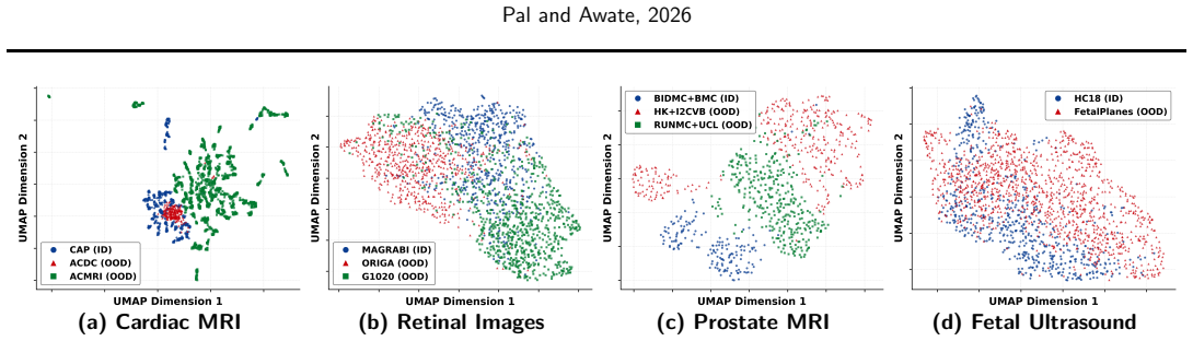
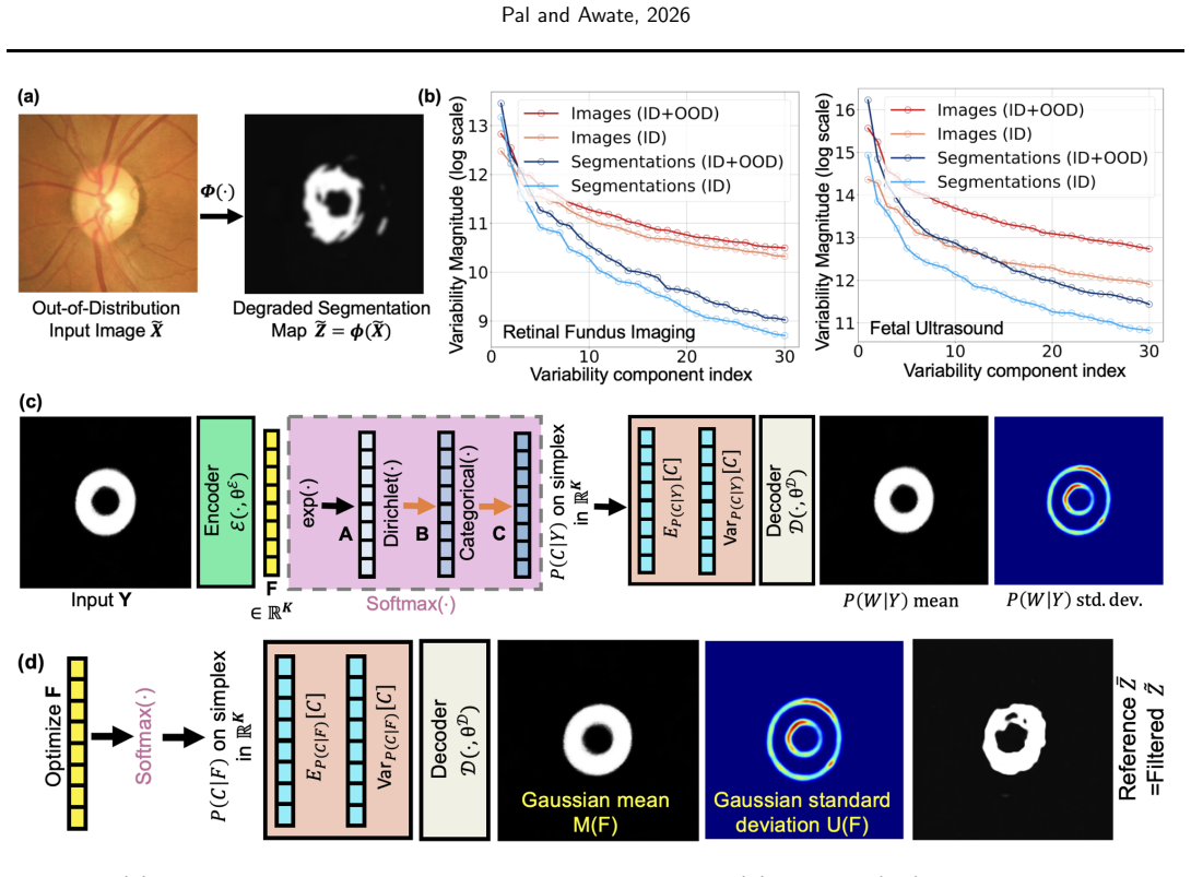
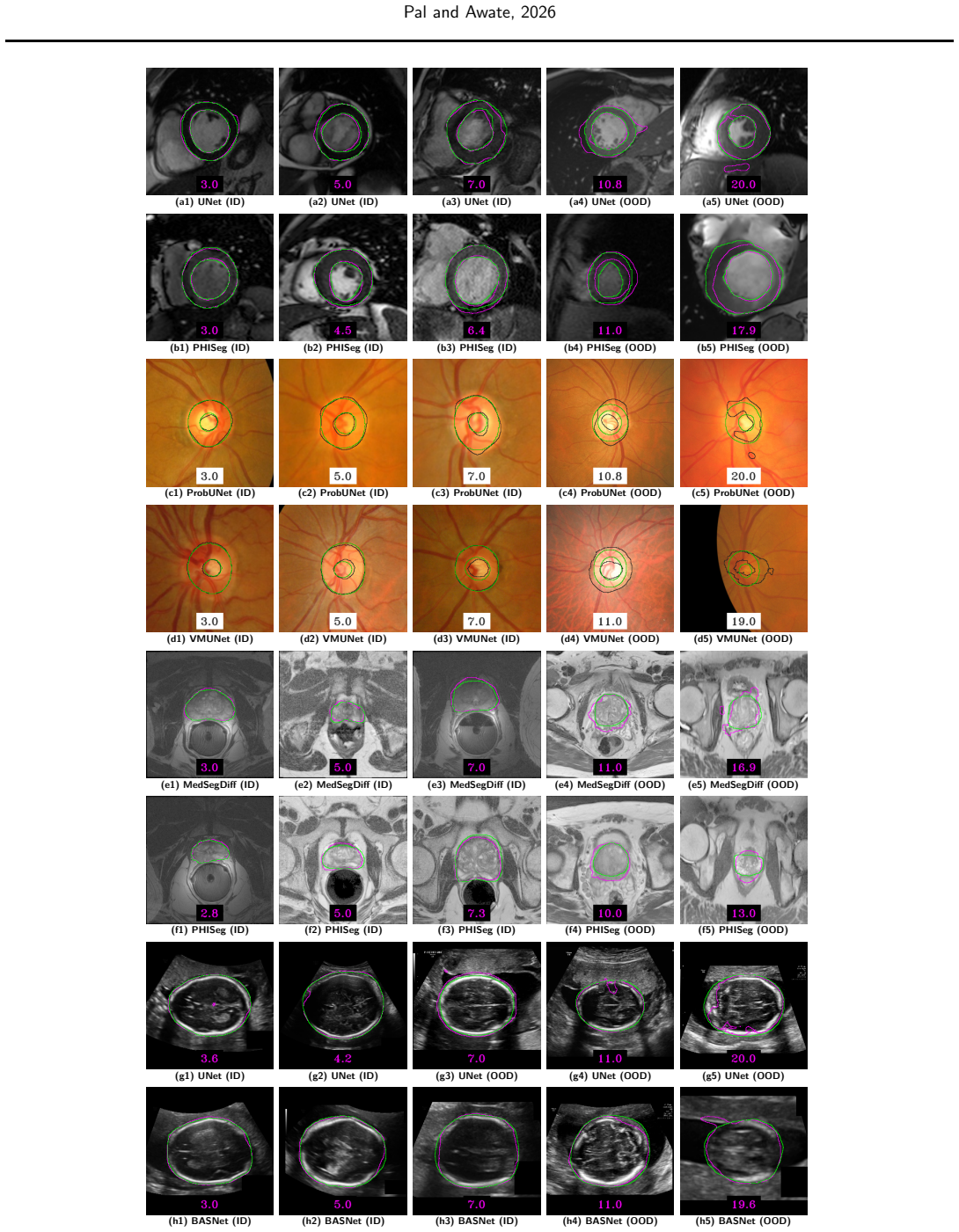
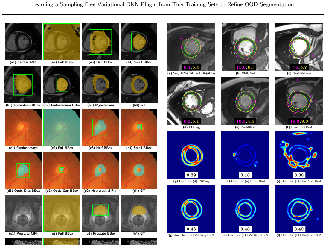
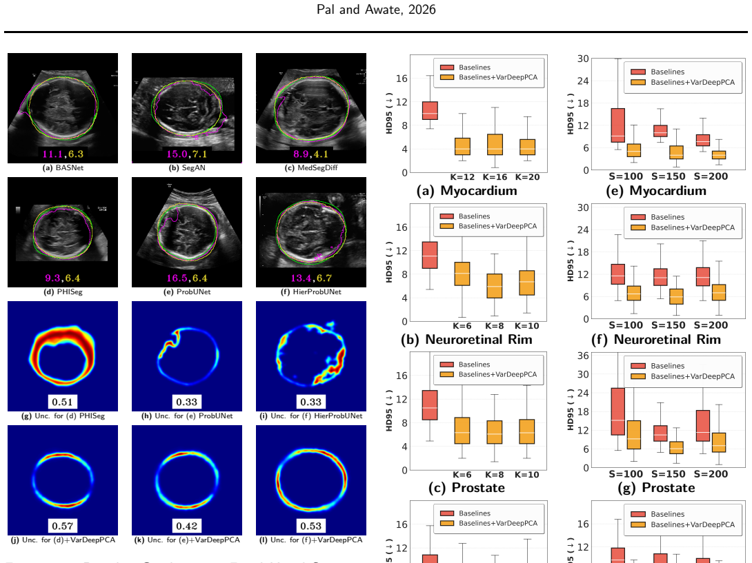
VarDeepPCA explicitly learns a distribution of valid anatomical geometries from small in-distribution datasets. Its novel variational framework reinterprets the softmax mapping to perform exact distribution modeling, which enables computationally efficient sampling-free learning and inference along with associated uncertainty estimates. When used to restore segmentation maps produced by existing methods on out-of-distribution data, the plugin improves anatomical plausibility of the geometries, clinical utility of the segmentations, and reduces errors without needing any more training data than the baselines.
What carries the argument
VarDeepPCA, a variational DNN plugin that models distributions of anatomical geometries via reinterpretation of the softmax mapping for sampling-free exact inference.
If this is right
- Restored segmentations exhibit higher anatomical plausibility and clinical utility on OOD data.
- Segmentation errors decrease across myocardium, neuroretinal rim, prostate, and fetal head tasks.
- Uncertainty estimates accompany each restored map for downstream use.
- Performance gains hold when training data volume matches that of the fifteen comparison methods.
- The same plugin works across fourteen public datasets spanning four distinct applications.
Where Pith is reading between the lines
- The sampling-free property could allow integration into existing segmentation pipelines without added computational overhead at test time.
- Uncertainty maps from the plugin might serve as inputs for selective re-acquisition or human review in clinical settings.
- If the softmax reinterpretation generalizes, similar lightweight plugins could be attached to other softmax-based models for OOD correction.
Load-bearing premise
Reinterpreting the softmax mapping enables exact distribution modeling without sampling.
What would settle it
A controlled test on held-out OOD cases where VarDeepPCA-refined segmentations show no improvement in anatomical plausibility scores or error metrics over the unrefined baselines.
Figures
read the original abstract
Deep neural networks (DNNs) frequently fail to generalize to out-of-distribution (OOD) medical images because of variations in scanners and acquisition protocols. Retraining DNN models to address these distribution shifts is often impractical due to the high cost of acquiring and annotating new medical datasets. To address this, we introduce VarDeepPCA, a novel lightweight variational DNN framework designed to restore/refine degraded segmentation maps by leveraging intrinsic geometric priors. Unlike existing approaches that require target-domain data or extensive pre-training, our VarDeepPCA explicitly learns a distribution of valid anatomical geometries using only small in-distribution (ID) datasets. Theoretically, our novel variational learning framework leverages a reinterpretation of the softmax mapping to implicitly perform exact distribution modeling, thereby enabling computationally efficient, sampling-free learning and inference. This also enables VarDeepPCA to provide uncertainty estimates associated with its restored segmentation maps. We empirically validate our framework across 4 distinct clinical applications, using 14 publicly available datasets, involving segmentation of the myocardium, neuroretinal rim, prostate, and fetal head. Comparisons against 15 existing methods demonstrate that VarDeepPCA consistently restores segmentation maps produced by the existing methods on OOD data to (i) significantly improve anatomical plausibility of geometries and clinical utility of the segmentations, and (ii) significantly reduce errors, without needing any more training data than that used by existing methods.
Editorial analysis
A structured set of objections, weighed in public.
Referee Report
Summary. The manuscript introduces VarDeepPCA, a lightweight variational DNN plugin for restoring OOD segmentation maps in medical imaging. It claims to explicitly learn a distribution of valid anatomical geometries from small ID datasets only, via a novel variational framework that reinterprets the softmax mapping to achieve exact distribution modeling. This enables sampling-free learning/inference and uncertainty estimation. Empirical validation across 4 clinical applications and 14 datasets shows consistent improvements over 15 baselines in anatomical plausibility, clinical utility, and error reduction, without requiring target-domain data.
Significance. If the central theoretical claim holds, the result would be significant: a data-efficient plugin that addresses distribution shifts in medical segmentation without retraining or target annotations, while supplying uncertainty estimates. The multi-application, multi-dataset empirical scope is a strength.
major comments (2)
- [Abstract] Abstract: the central claim that 'a reinterpretation of the softmax mapping to implicitly perform exact distribution modeling' enables sampling-free exact posterior inference over geometries is presented without any derivation, loss function, variational objective, or proof. This is load-bearing for both the sampling-free property and the avoidance of standard VAE gaps.
- [Abstract] Abstract: no equations, training objective, or architectural details are supplied showing how the framework produces an exact (rather than approximate or heuristic) distribution over valid geometries from tiny ID sets, nor how it differs from standard variational PCA or VAE formulations.
minor comments (1)
- [Abstract] Abstract: claims of 'significantly improve' and 'significantly reduce errors' are not accompanied by error bars, statistical tests, or explicit metrics for significance assessment.
Simulated Author's Rebuttal
We thank the referee for their review and the opportunity to clarify the presentation of our theoretical contributions. We respond to each major comment below.
read point-by-point responses
-
Referee: [Abstract] Abstract: the central claim that 'a reinterpretation of the softmax mapping to implicitly perform exact distribution modeling' enables sampling-free exact posterior inference over geometries is presented without any derivation, loss function, variational objective, or proof. This is load-bearing for both the sampling-free property and the avoidance of standard VAE gaps.
Authors: The full manuscript derives the softmax reinterpretation in Section 3.1, presents the exact variational objective in Equation (5), and proves exact posterior inference (no ELBO gap) in Theorem 1. The abstract is intentionally concise, but we agree it should better signal these elements and will revise it to include a brief reference to the key theoretical result. revision: yes
-
Referee: [Abstract] Abstract: no equations, training objective, or architectural details are supplied showing how the framework produces an exact (rather than approximate or heuristic) distribution over valid geometries from tiny ID sets, nor how it differs from standard variational PCA or VAE formulations.
Authors: Section 3.2 details the architecture, Equation (7) gives the training objective, and Section 2.2 explicitly contrasts the approach with standard variational PCA and VAE formulations (exact modeling via softmax reinterpretation vs. approximate sampling). We will revise the abstract to incorporate a short statement highlighting these distinctions and the exact nature of the distribution. revision: yes
Circularity Check
No circularity: derivation self-contained from ID data
full rationale
The paper introduces VarDeepPCA as learning a distribution of valid anatomical geometries from small ID datasets via a novel variational framework that reinterprets softmax for exact distribution modeling. No equations, loss functions, or self-citations are quoted that reduce this claim to fitted inputs, self-definitions, or prior author work by construction. The abstract presents the approach as independent of target-domain data and validated empirically against 15 methods on 14 datasets, with no load-bearing steps that collapse to renaming or tautological fits. This is the normal case of a self-contained method description.
Axiom & Free-Parameter Ledger
axioms (1)
- ad hoc to paper Reinterpretation of the softmax mapping implicitly performs exact distribution modeling
Reference graph
Works this paper leans on
-
[1]
J. B. Pal and S. Welling and H. Saini and S. P. Awate , title =. 2025 , pages =
2025
-
[2]
B. Sch. Nonlinear Component Analysis as a Kernel Eigenvalue Problem , journal = NeuralComput, volume =
-
[3]
Tipping and C
M. Tipping and C. Bishop , title=. 1999 , volume=
1999
-
[4]
Tonin and Q
F. Tonin and Q. Tao and P. Patrinos and J. Suykens , title =
-
[5]
Bishop , booktitle=ICANN, title=
C. Bishop , booktitle=ICANN, title=. 1999 , volume=
1999
-
[6]
Lian , title=
H. Lian , title=. 2008 , volume=
2008
-
[7]
Farquhar and Y
S. Farquhar and Y. Gal , title=. Adv. Neural Inform. Process. Syst
-
[8]
He and X
K. He and X. Zhang and S. Ren and J. Sun , title =
-
[9]
Hu and L
J. Hu and L. Shen and G. Sun , booktitle = CVPR, title=. 2018 , pages=
2018
-
[10]
Pal and S
J. Pal and S. Awate , booktitle = MICCAI, pages=. Convex Segments for Convex Objects Using
-
[11]
Pal and S
J. Pal and S. Awate , booktitle = ICIP, pages=. A Hard Convex-Shape Constraint In
-
[12]
S Awate and S Garg and R Jena , title=. Med. Imag. Analysis , year=
-
[13]
Jena and S
R. Jena and S. P. Awate , booktitle=. A
-
[14]
Varma and A
H. Varma and A. Gaikwad and S. Awate , booktitle=ISBI, title=. 2023 , pages=
2023
-
[15]
Deep Variational Segmentation of Topology-Constrained Object Sets, with Correlated Uncertainty Models, for Robustness to Degradations , author=
-
[16]
Gao and Q
J. Gao and Q. Lao and P. Liu and H. Yi and Q. Kang and Z. Jiang and X. Wu and K. Li and Y. Chen and L. Zhang , journal = JBHI, title=. 2023 , volume=
2023
-
[17]
Kingma and M
D. Kingma and M. Welling , title =
-
[18]
Neural discrete representation learning , author=
-
[19]
Razavi and A
A. Razavi and A. Generating diverse high-fidelity images with
-
[20]
Learning structured output representation using deep conditional generative models , author=
-
[21]
Kohl and B
S. Kohl and B. Romera-Paredes and C. Meyer and J. A probabilistic
-
[23]
C. F. Baumgartner and K. C. Tezcan and K. Chaitanya and A. M. H. 2019 , organization=
2019
-
[24]
Painchaud and Y
N. Painchaud and Y. Skandarani and T. Judge and O. Bernard and A. Lalande and P. Jodoin , journal = TMI, title=. 2020 , volume=
2020
-
[25]
Wang and C
S. Wang and C. Qin and N. Savioli and C. Chen and D. O'Regan and S. Cook and Y. Guo and D. Rueckert and W. Bai , title =
-
[26]
Oktay and E
O. Oktay and E. Ferrante and K. Kamnitsas and M. Heinrich and W. Bai and J. Caballero and S. Cook and A. Marvao and T. Dawes and D. O'Regan and B. Kainz and B. Glocker and D. Rueckert , journal=TMI, year=
-
[27]
Jacob and P
A. Jacob and P. Sharma and D. Rueckert , booktitle=ISBI, title=. 2024 , pages=
2024
-
[28]
Jacob and P
A. Jacob and P. Sharma and D. Rueckert , title =. Statistical Atlases and Computational Models of the Heart. Regular and
-
[29]
American Mathematical Monthly , year=
A Characterization of Star-Shaped Sets , author=. American Mathematical Monthly , year=
-
[30]
Practical uncertainty estimation and out-of-distribution robustness in deep learning , author=. Adv. Neural Inform. Process. Syst. Tutorial , year=
-
[31]
Ronneberger and P
O. Ronneberger and P. Fischer and T. Brox , title =
-
[32]
Oktay and J
O. Oktay and J. Schlemper and L. Folgoc and M. Lee and M. Heinrich and K. Misawa and K. Mori and S. McDonagh and N. Hammerla and B. Kainz and B. Glocker and D. Rueckert , booktitle = MIDL, pages=. Attention
-
[33]
Zhang and Q
Z. Zhang and Q. Liu and Y. Wang , title =. 2018 , volume =
2018
-
[34]
Jha and P
D. Jha and P. Smedsrud and M. Riegler and D. Johansen and T. Lange and P. Halvorsen and H. Johansen , journal=. 2019 , pages=
2019
-
[35]
Encoder-Decoder with Atrous Separable Convolution for Semantic Image Segmentation , author=
-
[36]
Qin and Z
X. Qin and Z. Zhang and C. Huang and C. Gao and M. Dehghan and M. J
-
[37]
Lin and B
A. Lin and B. Chen and J. Xu and Z. Zhang and G. Lu and D. Zhang , journal=TIM, year=
-
[38]
Xue and T
Y. Xue and T. Xu and H. Zhang and L. Long and X. Huang , journal = NeuroInfo, volume=
-
[39]
Goodfellow and J
I. Goodfellow and J. Pouget-Abadie and M. Mirza and B. Xu and D. Warde-Farley and S. Ozair and A. Courville and Y. Bengio , title =. 2014 , pages =
2014
-
[40]
Wu and R
J. Wu and R. Fu and H. Fang and Y. Zhang and Y. Yang and H. Xiong and H. Liu and Y. Xu , title =
-
[41]
Ho and A
J. Ho and A. Jain and P. Abbeel , title =
-
[42]
Kazerouni and E
A. Kazerouni and E. Diffusion Models for Medical Image Analysis:. 2023 , volume=
2023
-
[43]
Li and H
X. Li and H. Ding and H. Yuan and W. Zhang and J. Pang and G. Cheng and K. Chen and Z. Liu and C. Loy , journal=PAMI, year=. Transformer-Based Visual Segmentation:
-
[44]
Chen and J
J. Chen and J. Mei and X. Li and Y. Lu and Q. Yu and Q. Wei and X. Luo and Y. Xie and E. Adeli and Y. Wang and M. P. Lungren and S. Zhang and L. Xing and L. Lu and A. Yuille and Y. Zhou , journal = MIA, volume=
-
[45]
Attention is all you need , author=
-
[46]
An Image is Worth 16x16 Words: Transformers for Image Recognition at Scale , author=
-
[47]
Fine-tuning diffusion models with limited data , author=
-
[48]
Wang and A
Z. Wang and A. C. Bovik and H. R. Sheikh and E. P. Simoncelli , journal = TIP, title=. 2004 , volume=
2004
-
[49]
Zhao and O
H. Zhao and O. Gallo and I. Frosio and J. Kautz , title =
-
[50]
Zhou and J
D. Zhou and J. Fang and X. Song and C. Guan and J. Yin and Y. Dai and R. Yang , journal=. 2019 , pages=
2019
-
[51]
C. H. Sudre and W. Li and T. Vercauteren and S. Ourselin and M. J. Cardoso , title =. Deep Learning in Medical Image Analysis and Multimodal Learning for Clinical Decision Support , series = LNCS, pages =
-
[52]
International Journal of Imaging Systems and Technology , volume=
Image Segmentation Evaluation With the Dice Index: Methodological Issues , author=. International Journal of Imaging Systems and Technology , volume=. 2024 , publisher=
2024
-
[53]
Li and Y
B. Li and Y. Liu and C. Occleshaw and B. Cowan and A. Young , title =. 2010 , volume =
2010
-
[54]
Kadish and D
A. Kadish and D. Bello and J. Finn and R. Bonow and A. Schaechter and H. Subacius and C. Albert and J. Daubert and C. Fonseca and J. Goldberger , title=. Journal of Cardiovascular Electrophysiology , year=
-
[55]
Suinesiaputra and B
A. Suinesiaputra and B. Cowan and A. Al-Agamy and M. Elattar and N. Ayache and A. Fahmy and A. Khalifa and P. Medrano-Gracia and M. Jolly and A. Kadish and D. Lee and J. Margeta and S. Warfield and A. Young , title=. 2014 , volume=
2014
-
[56]
Andreopoulos and J
A. Andreopoulos and J. Tsotsos , title =. 2008 , volume =
2008
-
[57]
Bajwa and G
M. Bajwa and G. Singh and W. Neumeier and M. Malik and A. Dengel and S. Ahmed , title =. 2020 , pages =
2020
-
[58]
Zhang and F
Z. Zhang and F. Yin and J. Liu and W. K. Wong and N. M. Tan and B. H. Lee and J. Cheng and T. Y. Wong , title=. 2010 , pages=
2010
-
[59]
Almazroa and S
A. Almazroa and S. Alodhayb and E. Osman and E. Ramadan and M. Hummadi and M. Dlaim and M. Alkatee and K. Raahemifar and V. Lakshminarayanan , booktitle=. Retinal fundus images for glaucoma analysis: the
-
[60]
2020 , publisher=
Evaluation of deep convolutional neural networks for automatic classification of common maternal fetal ultrasound planes , author=. 2020 , publisher=
2020
-
[61]
T. L. Automated measurement of fetal head circumference using. 2018 , volume=
2018
-
[62]
Shigwan and A
S. Shigwan and A. Gaikwad and S. Awate , booktitle=ISBI, title=. 2020 , pages=
2020
-
[63]
Topology-Preserving Deep Image Segmentation , author=
-
[64]
Lee and K
M. Lee and K. Petersen and N. Pawlowski and B. Glocker and M. Schaap , title =. 2019 , volume =
2019
-
[65]
Kepp and J
T. Kepp and J. Andresen and C. Shape-based segmentation of retinal layers and fluids in. Medical Imaging 2023: Computer-Aided Diagnosis , volume=
2023
-
[66]
Rethinking the Inception Architecture for Computer Vision , author =
-
[67]
Heusel and H
M. Heusel and H. Ramsauer and T. Unterthiner and B. Nessler and S. Hochreiter , booktitle = NIPS, year =
-
[68]
Journal of the American statistical Association , volume=
The Kolmogorov-Smirnov test for goodness of fit , author=. Journal of the American statistical Association , volume=. 1951 , publisher=
1951
-
[69]
2015 , publisher=
Optic disc and optic cup segmentation methodologies for glaucoma image detection: a survey , author=. 2015 , publisher=
2015
-
[70]
Deng and W
J. Deng and W. Dong and R. Socher and L. Li and K. Li and L. Fei-Fei , booktitle=CVPR, title=. 2009 , pages=
2009
-
[71]
Bernard and A
O. Bernard and A. Lalande and C. Zotti and F. Cervenansky and X. Yang and P.-A. Heng and I. Cetin and K. Lekadir and O. Camara and M. A. Gonzalez Ballester and G. Sanroma and S. Napel and S. Petersen and G. Tziritas and E. Grinias and M. Khened and V. A. Kollerathu and G. Krishnamurthi and M.-M. Rohé and X. Pennec and M. Sermesant and F. Isensee and P. Jä...
2018
-
[72]
A review of heart chamber segmentation for structural and functional analysis using cardiac magnetic resonance imaging , author=. Magn. Reson. Mater. Phy. , volume=
-
[73]
J. I. Orlando and H. Fu and J. B. Breda and K
-
[74]
Shen and H
Z. Shen and H. Fu and J. Shen and L. Shao , journal=TMI, title=. 2021 , volume=
2021
-
[75]
Lu , title=
S. Lu , title=. 2011 , volume=
2011
-
[76]
Automated segmentation of the optic disc from fundus images using an asymmetric deep learning network , author=
-
[77]
Wang and X
B. Wang and X. Gu and C. Fan and H. Xie and S. Zhang and X. Tian and L. Gu , title =. 2015 , volume =
2015
-
[78]
Sun and Z
F. Sun and Z. Luo and S. Li , title=. 2023 , pages=
2023
-
[79]
Boundary loss for highly unbalanced segmentation , author=
-
[80]
2022 , publisher=
Efficient fetal ultrasound image segmentation for automatic head circumference measurement using a lightweight deep convolutional neural network , author=. 2022 , publisher=
2022
-
[81]
Challenges with segmenting intraoperative ultrasound for brain tumours , author=
discussion (0)
Sign in with ORCID, Apple, or X to comment. Anyone can read and Pith papers without signing in.