REVIEW 4 major objections 6 minor 82 references
The paper claims that a single foundation model—LPFM—restores blurry, noisy, low-resolution pathology images and performs virtual staining through one prompt-controlled architecture, reporting better performance than specialized methods on
Reviewed by Pith at T0; open to challenge. T0 means a machine referee read the full paper against a public rubric. the ladder, T0–T4 →
A prompt-guided diffusion model pretrained on 190 million pathology patches outperforms task-specific models across most restoration and virtual staining benchmarks.
T0 review reviewed 2026-08-05 challenge →
load-bearing objection Big unified pathology restoration model, but the headline significance claim is undercut by pretraining on internal test patches. the 4 major comments →
A Unified Low-level Foundation Model for Enhancing Pathology Image Quality
The pith
A machine-rendered reading of the paper's core claim, the machinery that carries it, and where it could break.
The reading
Core claim
The paper's central claim is that low-level pathology image enhancement does not need a collection of task-specific networks. LPFM is a unified architecture trained in two stages: a contrastive pre-training stage learns representations that are invariant to stain and degradation type from 190 million unlabeled patches, producing coarse restored images; a prompt-conditioned latent diffusion stage refines those images using a text prompt such as "obtain high-quality H&E pathology image" or "translate the label-free patch to H&E image." Because tasks are selected by prompt rather than by architecture, the same model performs super-resolution (2x, 4x, 8x), deblurring (Gaussian kernels of size 7–
What carries the argument
The load-bearing mechanism is a two-stage generative pipeline. Stage one is a KL-autoencoder pre-trained with a contrastive loss that pulls together latent features of degraded and high-quality views of the same tissue while pushing apart features of different tissues, plus reconstruction, perceptual, and adversarial losses; this yields a coarse restoration and a degradation-robust, stain-invariant latent space. Stage two is a latent conditional diffusion model whose U-Net receives both the coarse restoration and a pretrained text-prompt embedding as conditions, so a single set of weights switches among restoration and staining tasks. The text prompt is the control surface: changing the prom
Load-bearing premise
The load-bearing premise is that the synthetic degradations used to create training and test pairs—Gaussian blur with kernel sizes 7–15, Gaussian and Poisson noise with sigma 21/31/41, and downscaling by 2/4/8 with area, bilinear, or bicubic interpolation—faithfully represent real artifacts found in clinical whole-slide images.
What would settle it
Collect a set of real clinical whole-slide images with paired low-quality and high-quality acquisitions (for example, the same slides scanned twice, once with defocus or added noise and once at full quality, or same-slide autofluorescence/H&E pairs) and compare LPFM against SwinIR and RegGAN on those real pairs using PSNR and SSIM. If LPFM's advantage over these specialized baselines shrinks to near zero or reverses, the claim that its synthetic degradation pipeline captures clinical artifacts—and hence that the unified model transfers—would be falsified.
If this is right
- A hospital could deploy one model instead of separate denoising, deblurring, super-resolution, and virtual-staining systems, with task selection by text prompt.
- The same pretrained weights can be applied to tissue types and scanners represented in the external sets (TIGER, MIDOG, OCELOT) without retraining, as shown by the external validation tasks.
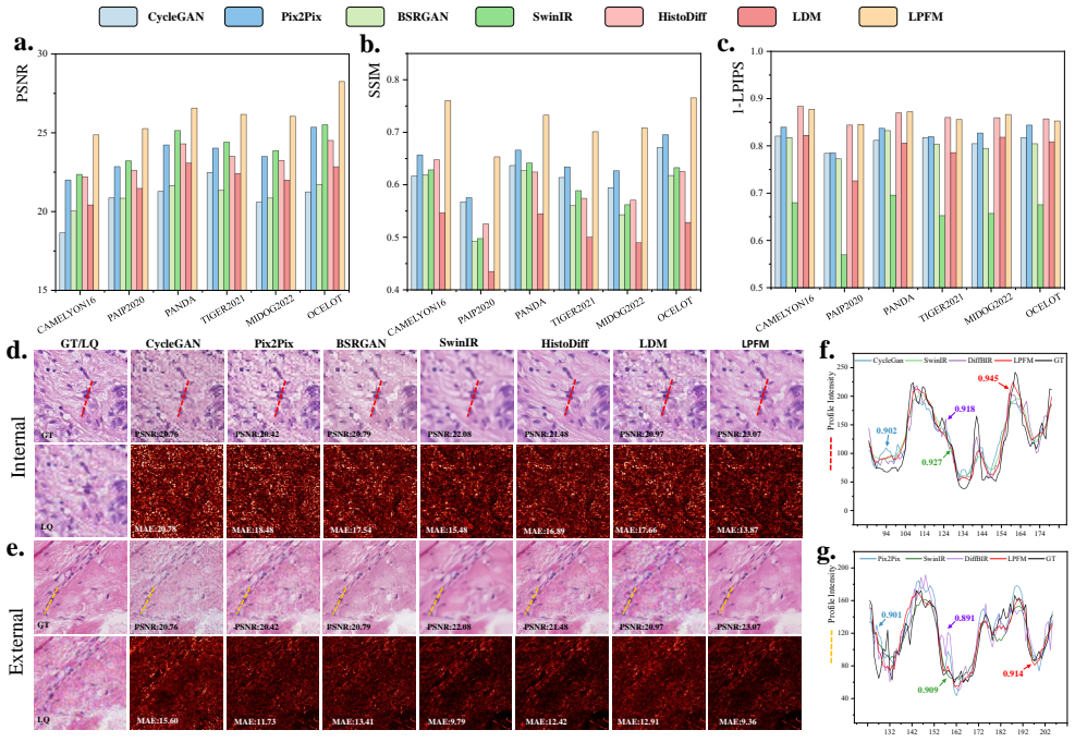
- Coupled-degradation restoration (blur plus noise plus downsampling) outperforms specialized single-task models by 2.10 dB PSNR, suggesting combined artifacts can be corrected jointly rather than sequentially.
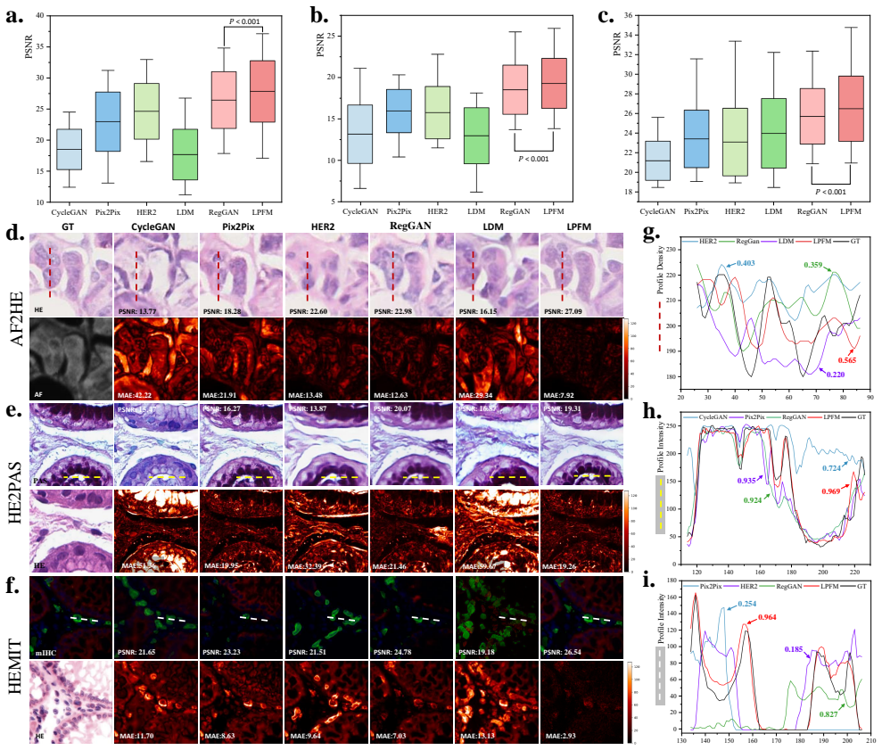
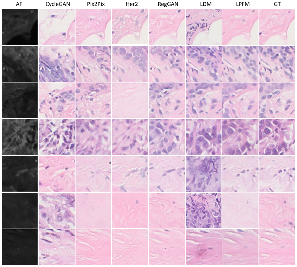
- Virtual staining of H&E into PAS-AB and mIHC, and of autofluorescence into H&E, reaches pixel-level and structural metrics above current GAN baselines, which would support stain-free and retrospective-staining workflows if diagnostic concordance is confirmed.
- Prompt-based control implies output can be steered by user intent (for example, "upscale x2" versus "remove noise"), enabling interactive or prioritized enhancement during review.
Where Pith is reading between the lines
- If the synthetic degradation pipeline mirrors real scanner and preparation artifacts, LPFM could be inserted upstream of existing diagnostic foundation models as a pre-processing step; the paper evaluates image-quality metrics, not downstream diagnostic accuracy, so that benefit is an inference.
- Because task switching happens through prompts, LPFM may adapt to new stain protocols or artifact types zero-shot by editing the prompt; the paper demonstrates prompt-controlled generation but does not test unchanged-weight generalization to unseen stain pairs.
- The finding that LPFM scores higher PSNR on degraded HEMIT inputs than on high-quality ones (26.99 versus 26.49 dB) suggests the model may actively exploit artifact patterns during stain conversion; if confirmed, it argues for jointly optimizing restoration and translation rather than separating them.
- A practical extension would be a controlled clinical study comparing pathologist diagnostic concordance on LPFM-enhanced and physically restained slides; such a study would test whether the reported metric gains translate into decisions.
Editorial analysis
A structured set of objections, weighed in public.
Referee Report
Summary. The paper proposes LPFM, a unified low-level pathology foundation model intended to handle both image restoration (super-resolution, deblurring, denoising, coupled degradations) and image translation (virtual staining) within a single architecture. The method combines contrastive pretraining on roughly 190 million patches from 87,810 WSIs with a prompt-conditioned latent diffusion refinement stage. The authors report results on 66 tasks with 95% confidence intervals and claim statistically significant superiority (p<0.01) over eight baselines in 56/66 tasks. Three of the benchmark datasets (MIDOG2022, TIGER2021, OCELOT) are explicitly excluded from pretraining for external validation. The manuscript is well organized and the scale of the curated dataset is notable, but the experimental section contains a serious internal-validity problem: the internal test sets are also present in the contrastive pretraining corpus. In addition, the statistical test underlying the headline significance claim is never described, and all degraded/restoration benchmarks are generated from the authors' own synthetic degradation pipeline.
Significance. If the reported effects are real, LPFM would be a meaningful step toward unified low-level vision in computational pathology: the combination of contrastive pretraining with prompt-conditioned diffusion is sensible, and the decision to hold out three datasets from pretraining is a genuine strength. The scale of data curation and the breadth of tasks also add value. However, the conclusions currently rest on comparisons whose internal test portions are confounded by pretraining data overlap, and on a significance claim with no described test. The external datasets provide a cleaner test bed, but the aggregate 56/66 claim mixes contaminated and uncontaminated tasks. The paper's significance is therefore conditional on repairing the experimental protocol and reporting external/clean results separately.
major comments (4)
- [Sec. 2.1 / Sec. 4.5] The internal evaluation is confounded by pretraining data overlap. Sec. 4.5 states the contrastive pretraining corpus includes CAMELYON16, PANDA, PAIP2020, AF2HE, PASAB, and HEMIT, while Sec. 2.1 uses the first three as 'internal' benchmarks with train/val/test splits. The test patches from these datasets were therefore seen by the LPFM encoder during contrastive pretraining. The baselines (CycleGAN, SwinIR, etc.) are trained from scratch on the downstream training split only. The headline claim of 56/66 statistically significant improvements includes these contaminated internal tasks. Please re-run with the internal test sets excluded from pretraining, and separately report results for the clean external sets (MIDOG2022, TIGER2021, OCELOT).
- [Abstract / Sec. 2] No statistical test is specified for the p<0.01 / 56/66 claim. The text and figures report p-values (e.g., Fig. 2-5) and mention 'significance testing' and 95% CIs, but no test name, null hypothesis, sample unit, or multiple-comparison correction is given. With 66 tasks and 3 metrics, uncorrected pairwise tests would make many 'significant' findings expected by chance. Please specify the test, the multiplicity adjustment, and provide a per-task significance table with internal/external breakdown.
- [Sec. 4.1.2 / Sec. 3] All restoration and degraded-staining evaluations are based on the authors' synthetic degradation pipeline (Gaussian blur kernels 7-15, Gaussian/Poisson noise σ=21/31/41, downscaling 2/4/8 by area/bilinear/bicubic). The 'external' datasets test tissue/scanner/stain transfer under this same pipeline, not generalization to real scanner artifacts or preparation defects. The Discussion's clinical claims (e.g., 'transform diagnostic workflows') require support from real or independently acquired degraded pathology images. At minimum, validate on a real degraded dataset or substantially soften the generality claims.
- [Tables 1-3, 6, 11-12] The extended-data tables contain implausible repetitions that undermine confidence in the reported rankings. Tables 1-3 list identical Pix2Pix rows (same PSNR/SSIM/LPIPS and CIs) for CAMELYON16, PAIP2020, and PANDA at every super-resolution scale. Table 6 shows the OCELOT ×4 LDM row identical to the BSRGAN row of Table 4. Tables 11-12 have repeated or mislabeled kernel-size rows (15, 11, 15 and 15, 7, 15 instead of 7/11/15). These must be corrected and regenerated from the actual experiments before the extended-data results can support the aggregate rankings.
minor comments (6)
- [Abstract / Fig. 1] Typographical issues: 'whole slied images' in the abstract; 'virutal' in Fig. 1; '0=cs' in Fig. 13 caption. These should be corrected.
- [Eq. (7)] The equation uses x_d but the surrounding text defines 'xp' as the paired degraded/source-stained image. Please define the variable consistently.
- [Eq. (15) / Sec. 4.3] The text states 'A lower MAE indicates better pixel-wise accuracy' immediately after defining MSE. MAE is not defined; this appears to be a typo for MSE or should be a separate metric.
- [Fig. 12 / Fig. 13] These figures contain Chinese-language placeholder text (e.g., '很丑,要换的一个过程') and random filename strings. These should be removed before submission.
- [Sec. 2.6] The sentence 'LPFM showed merely 6.2' is incomplete, breaking the logic of the robustness comparison. Please complete or rephrase.
- [Table 27 / Sec. 1] The paper claims '34 tissue types' but Table 27 lists 40 primary-site rows, including 'Pancancer' and 'Prostate,' which are not tissue types. Please reconcile the count.
Circularity Check
Internal 'held-out' test patches were included in contrastive pretraining, so the 56/66 significance claim is partially circular; external validation remains independent.
specific steps
-
fitted input called prediction
[Sec 4.5 (Datasets) and Sec 2.1 (Super Resolution), also affecting internal tasks in Secs 2.2-2.6 and the abstract's 56/66 claim]
"Our unified low-level pathology foundation model is pretrained on all the datasets excluding MIDOG2022, TIGER2021, and OCELOT which are reserved for external validation. ... For internal validation, we employed three benchmark datasets (CAMELYON16, PANDA, and PAIP2020), which were rigorously partitioned into training (70%), validation (10%), and test (20%) sets with no data overlap to ensure unbiased evaluation."
Section 4.5 places CAMELYON16, PANDA, and PAIP2020 inside the pretraining corpus: they are part of 'all the datasets' from which 190M patches are drawn. Section 2.1 then uses these same three datasets to define internal 'held-out' test sets. Because LPFM's contrastive encoder was pretrained on the exact test patches, the internal evaluation is not held out: the model has already seen the test images. Calling these results 'predictions' on held-out data is therefore incorrect by the paper's own dataset construction. The headline aggregate '56/66 tasks with p<0.01' counts these contaminated internal tasks together with truly external ones, so a portion of the central claim reduces to pretraining exposure rather than independent generalization.
full rationale
The core architecture and losses (contrastive pretraining, conditional diffusion, prompt conditioning) are not self-referential, and no parameter is fitted to test labels. The external datasets (MIDOG2022, TIGER2021, OCELOT) are explicitly excluded from pretraining and provide genuinely independent evidence of generalization. However, the internal benchmark is circular in an evaluation sense: the pretraining corpus includes all slides from CAMELYON16, PANDA, and PAIP2020, which are later 'rigorously partitioned' into train/val/test. LPFM's contrastive encoder has therefore already seen the exact internal test patches, while baselines trained only on the downstream train split have not. This gives LPFM an unfair advantage on the 36 internal tasks and confounds the aggregated 56/66 significance claim. The paper does not provide a per-task internal/external breakdown of the p<0.01 count, so the strength of the headline claim cannot be assessed from the reported numbers. This is a partial circularity in the evaluation protocol, not a derivation-level circularity: the external results and the virtual-staining results on datasets not in the pretraining corpus still carry independent weight. Score 6 reflects that a substantial portion of the central empirical claim is contaminated by the train/test overlap, while acknowledging the existence of independent external support.
Axiom & Free-Parameter Ledger
free parameters (4)
- Degradation simulation parameters =
blur kernel 7-15, sigma1/sigma2 1.5-3.5; noise sigma 21/31/41; SR scales 2/4/8
- Coupled degradation composition =
Randomized combinations, exact distributions not specified
- Prompt templates =
Hand-written prompts such as 'Obtain the high-quality H&E pathology image'
- Training hyperparameters =
Not reported
axioms (4)
- standard math Standard diffusion and latent diffusion model mathematics (Eqs. 11-14)
- domain assumption Synthetic degradation models are representative of clinical artifacts
- domain assumption Contrastive pretraining on public pathology datasets yields stain-invariant features
- ad hoc to paper Hand-crafted prompts sufficiently specify each task
Cite this review
Pith. "Pith review of A Unified Low-level Foundation Model for Enhancing Pathology Image Quality." pith.science (2026). https://pith.science/paper/BZCWZK5T
@misc{pith2026250901071,
  author       = {Pith},
  title        = {Pith review of: A Unified Low-level Foundation Model for Enhancing Pathology Image Quality},
  year         = {2026},
  howpublished = {\url{https://pith.science/paper/BZCWZK5T}},
  note         = {Machine review of arXiv:2509.01071}
}
read the original abstract
Foundation models have revolutionized computational pathology by achieving remarkable success in high-level diagnostic tasks, yet the critical challenge of low-level image enhancement remains largely unaddressed. Real-world pathology images frequently suffer from degradations such as noise, blur, and low resolution due to slide preparation artifacts, staining variability, and imaging constraints, while the reliance on physical staining introduces significant costs, delays, and inconsistency. Although existing methods target individual problems like denoising or super-resolution, their task-specific designs lack the versatility to handle the diverse low-level vision challenges encountered in practice. To bridge this gap, we propose the first unified Low-level Pathology Foundation Model (LPFM), capable of enhancing image quality in restoration tasks, including super-resolution, deblurring, and denoising, as well as facilitating image translation tasks like virtual staining (H&E and special stains), all through a single adaptable architecture. Our approach introduces a contrastive pre-trained encoder that learns transferable, stain-invariant feature representations from 190 million unlabeled pathology images, enabling robust identification of degradation patterns. A unified conditional diffusion process dynamically adapts to specific tasks via textual prompts, ensuring precise control over output quality. Trained on a curated dataset of 87,810 whole slied images (WSIs) across 34 tissue types and 5 staining protocols, LPFM demonstrates statistically significant improvements (p<0.01) over state-of-the-art methods in most tasks (56/66), achieving Peak Signal-to-Noise Ratio (PSNR) gains of 10-15% for image restoration and Structural Similarity Index Measure (SSIM) improvements of 12-18% for virtual staining.
Figures






Reference graph
Works this paper leans on
-
[1]
& Chen, H
Ma, J., Chan, R., Wang, J., Fei, P. & Chen, H. A generalizable pathology foundation model using a unified knowledge distillation pretraining framework. Nat. Biomed. Eng. (2025)
2025
-
[2]
Chen, R. J. et al. Towards a general-purpose foundation model for computational pathology. Nat. Medicine 30, 850–862 (2024)
2024
-
[3]
Song, A. H. et al. Artificial intelligence for digital and computational pathology. Nat. Rev. Bioeng. 1, 930–949 (2023)
work page 2023
-
[4]
Bejnordi, B. E. et al. Diagnostic assessment of deep learning algorithms for detection of lymph node metastases in women with breast cancer. Jama 318, 2199–2210 (2017)
work page 2017
-
[5]
Ma, J., Chan, R. & Chen, H. Pathbench: A comprehensive comparison benchmark for pathology foundation models towards precision oncology. arXiv:2505.20202 (2025)
Pith/arXiv arXiv 2025
-
[6]
Lai, B., Fu, J. et al. Artificial intelligence in cancer pathology: Challenge to meet increasing demands of precision medicine. Int. J. Oncol. 63, 1–30 (2023)
work page 2023
-
[7]
Xu, Y .et al. A multimodal knowledge-enhanced whole-slide pathology foundation model.arXiv preprint arXiv:2407.15362 (2024)
Pith/arXiv arXiv 2024
-
[8]
Srinidhi, C. L., Ciga, O. & Martel, A. L. Deep neural network models for computational histopathology: A survey. Med. image analysis 67, 101813 (2021)
work page 2021
-
[9]
Yan, F., Chen, H., Zhang, S., Wang, Z. et al. Pathorchestra: A comprehensive foundation model for computational pathology with over 100 diverse clinical-grade tasks. arXiv:2503.24345 (2025)
work page internal anchor Pith review Pith/arXiv arXiv 2025
-
[10]
Zhuang, J. et al. Mim: Mask in mask self-supervised pre-training for 3d medical image analysis. IEEE Transactions on Med. Imaging (2025)
work page 2025
-
[11]
Liang, J. et al. Swinir: Image restoration using swin transformer. In Proceedings of the IEEE/CVF international conference on computer vision, 1833–1844 (2021)
work page 2021
-
[12]
Jin, C., Chen, H. et al. Hmil: Hierarchical multi-instance learning for fine-grained whole slide image classification. IEEE Transactions on Med. Imaging 44, 1796–1808 (2025)
work page 2025
-
[13]
Echle, A. et al. Deep learning in cancer pathology: a new generation of clinical biomarkers. Br. journal cancer 124, 686–696 (2021)
work page 2021
-
[14]
Wang, H. et al. Rethinking multiple instance learning for whole slide image classification: A bag-level classifier is a good instance-level teacher. IEEE Transactions on Med. Imaging 43, 3964–3976 (2024)
work page 2024
-
[15]
Xiong, C., Chen, H., King, I. et al. Takt: Target-aware knowledge transfer for whole slide image classification. In International Conference on Medical Image Computing and Computer-Assisted Intervention (2024). 20/49
work page 2024
-
[16]
Zhang, K., Zuo, W., Chen, Y ., Meng, D. & Zhang, L. Beyond a gaussian denoiser: Residual learning of deep cnn for image denoising. IEEE transactions on image processing 26, 3142–3155 (2017)
work page 2017
-
[17]
Chen, L. et al. Next token prediction towards multimodal intelligence: A comprehensive survey. arXiv preprint arXiv:2412.18619 (2024)
Pith/arXiv arXiv 2024
-
[18]
Bulten, W. et al. Automated deep-learning system for gleason grading of prostate cancer using biopsies: a diagnostic study. The Lancet Oncol. 21, 233–241 (2020)
work page 2020
-
[19]
Tolkach, Y ., Dohmgörgen, T., Toma, M. & Kristiansen, G. High-accuracy prostate cancer pathology using deep learning. Nat. Mach. Intell. 2, 411–418 (2020)
work page 2020
-
[20]
Siemion, K. et al. What do we know about inflammatory myofibroblastic tumors?–a systematic review. Adv. Med. Sci. 67, 129–138 (2022)
work page 2022
-
[21]
Coudray, N. et al. Classification and mutation prediction from non–small cell lung cancer histopathology images using deep learning. Nat. medicine 24, 1559–1567 (2018)
work page 2018
-
[22]
Wang, J., Yue, Z., Zhou, S., Chan, K. C. & Loy, C. C. Exploiting diffusion prior for real-world image super-resolution. Int. J. Comput. Vis. 132, 5929–5949 (2024)
work page 2024
-
[23]
Krithiga, R. & Geetha, P. Breast cancer detection, segmentation and classification on histopathology images analysis: a systematic review. Arch. Comput. Methods Eng. 28, 2607–2619 (2021)
work page 2021
-
[24]
Xia, B. et al. Diffir: Efficient diffusion model for image restoration. In Proceedings of the IEEE/CVF International Conference on Computer Vision, 13095–13105 (2023)
work page 2023
-
[25]
Guo, Z., Chen, H. et al. Focus: Knowledge-enhanced adaptive visual compression for few-shot whole slide image classification. In Proceedings of the Computer Vision and Pattern Recognition Conference (2025)
work page 2025
-
[26]
Isola, P., Zhu, J.-Y ., Zhou, T. & Efros, A. A. Image-to-image translation with conditional adversarial networks. In Proceedings of the IEEE conference on computer vision and pattern recognition , 1125–1134 (2017)
work page 2017
-
[27]
Xie, S. et al. Towards unifying understanding and generation in the era of vision foundation models: A survey from the autoregression perspective. arXiv preprint arXiv:2410.22217 (2024)
Pith/arXiv arXiv 2024
-
[28]
Rombach, R., Blattmann, A., Lorenz, D., Esser, P. & Ommer, B. High-resolution image synthesis with latent diffusion models. In Proceedings of the IEEE/CVF conference on computer vision and pattern recognition , 10684–10695 (2022)
work page 2022
-
[29]
Qu, L. et al. Tokenflow: Unified image tokenizer for multimodal understanding and generation. arXiv preprint arXiv:2412.03069 (2024)
Pith/arXiv arXiv 2024
-
[30]
Liang, M. et al. Multi-scale self-attention generative adversarial network for pathology image restoration. The Vis. Comput. 39, 4305–4321 (2023)
work page 2023
-
[31]
Xiong, J. et al. Autoregressive models in vision: A survey. arXiv preprint arXiv:2411.05902 (2024)
Pith/arXiv arXiv 2024
-
[32]
Li, T., Tian, Y ., Li, H., Deng, M. & He, K. Autoregressive image generation without vector quantization.Adv. Neural Inf. Process. Syst. 37, 56424–56445 (2025)
work page 2025
-
[33]
Han, J. et al. Infinity: Scaling bitwise autoregressive modeling for high-resolution image synthesis. arXiv preprint arXiv:2412.04431 (2024)
Pith/arXiv arXiv 2024
-
[34]
Fan, L. et al. Fluid: Scaling autoregressive text-to-image generative models with continuous tokens. arXiv preprint arXiv:2410.13863 (2024)
Pith/arXiv arXiv 2024
-
[35]
Huynh-Thu, Q. & Ghanbari, M. Scope of validity of psnr in image/video quality assessment. Electron. letters 44, 800–801 (2008)
work page 2008
-
[36]
Wang, Z., Bovik, A. C., Sheikh, H. R. & Simoncelli, E. P. Image quality assessment: from error visibility to structural similarity. IEEE transactions on image processing 13, 600–612 (2004)
work page 2004
-
[37]
Johnson, J., Alahi, A. & Fei-Fei, L. Perceptual losses for real-time style transfer and super-resolution. In Computer Vision–ECCV 2016: 14th European Conference, Amsterdam, The Netherlands, October 11-14, 2016, Proceedings, Part II 14, 694–711 (Springer, 2016)
work page 2016
-
[38]
Litjens, G. et al. 1399 h&e-stained sentinel lymph node sections of breast cancer patients: the camelyon dataset. GigaScience 7, giy065 (2018)
work page 2018
-
[39]
Bulten, W. et al. Artificial intelligence for diagnosis and gleason grading of prostate cancer: the panda challenge. Nat. medicine 28, 154–163 (2022). 21/49
work page 2022
-
[40]
Kim, K. et al. Paip 2020: Microsatellite instability prediction in colorectal cancer. Med. Image Analysis 89, 102886 (2023)
work page 2020
-
[41]
Ryu, J. et al. Ocelot: Overlapped cell on tissue dataset for histopathology. In Proceedings of the IEEE/CVF Conference on Computer Vision and Pattern Recognition (CVPR), 23902–23912 (2023)
work page 2023
-
[42]
Benesty, J., Chen, J., Huang, Y . & Cohen, I. Pearson correlation coefficient. InNoise reduction in speech processing, 1–4 (Springer, 2009)
work page 2009
-
[43]
Dai, W., Wong, I. H. & Wong, T. T. A weakly supervised deep generative model for complex image restoration and style transformation. Authorea Prepr. (2022)
work page 2022
-
[44]
Bian, C. et al. Immunoaizer: A deep learning-based computational framework to characterize cell distribution and gene mutation in tumor microenvironment. Cancers 13, 1659 (2021)
work page 2021
-
[45]
Radford, A. et al. Learning transferable visual models from natural language supervision. In International conference on machine learning, 8748–8763 (PmLR, 2021)
work page 2021
-
[46]
Simonyan, K. & Zisserman, A. Very deep convolutional networks for large-scale image recognition. arXiv preprint arXiv:1409.1556 (2014)
Pith/arXiv arXiv 2014
-
[47]
Zhang, L., Rao, A. & Agrawala, M. Adding conditional control to text-to-image diffusion models
- [48]
-
[49]
Zhu, J.-Y ., Park, T., Isola, P. & Efros, A. A. Unpaired image-to-image translation using cycle-consistent adversarial networks. In Computer Vision (ICCV), 2017 IEEE International Conference on (2017)
work page 2017
-
[50]
Zhang, K., Liang, J., Van Gool, L. & Timofte, R. Designing a practical degradation model for deep blind image super-resolution. In IEEE International Conference on Computer Vision , 4791–4800 (2021)
work page 2021
-
[51]
Xu, X., Kapse, S. & Prasanna, P. Histo-diffusion: A diffusion super-resolution method for digital pathology with comprehensive quality assessment. arXiv preprint arXiv:2408.15218 (2024)
work page internal anchor Pith review Pith/arXiv arXiv 2024
-
[52]
DoanNgan, B., Angus, D., Sung, L. et al. Label-free virtual her2 immunohistochemical staining of breast tissue using deep learning. BME frontiers (2022)
work page 2022
-
[53]
Rong, R. et al. Enhanced pathology image quality with restore–generative adversarial network. The Am. J. Pathol. 193, 404–416 (2023)
work page 2023
-
[54]
Aubreville, M. et al. Domain generalization across tumor types, laboratories, and species—insights from the 2022 edition of the mitosis domain generalization challenge. Med. Image Analysis 94, 103155 (2024)
work page 2022
-
[55]
Shephard, A. et al. Tiager: Tumor-infiltrating lymphocyte scoring in breast cancer for the tiger challenge. arXiv preprint arXiv:2206.11943 (2022)
Pith/arXiv arXiv 2022
-
[56]
Ryu, J. et al. Ocelot: Overlapped cell on tissue dataset for histopathology. In Proceedings of the IEEE/CVF Conference on Computer Vision and Pattern Recognition, 23902–23912 (2023)
work page 2023
-
[57]
Weinstein, J. N. et al. The cancer genome atlas pan-cancer analysis project. Nat. genetics 45, 1113–1120 (2013)
work page 2013
-
[58]
Carithers, L. J. et al. A novel approach to high-quality postmortem tissue procurement: the gtex project. Biopreservation biobanking 13, 311–319 (2015)
work page 2015
-
[59]
Edwards, N. J. et al. The cptac data portal: a resource for cancer proteomics research. J. proteome research 14, 2707–2713 (2015)
work page 2015
-
[60]
Bandi, P. et al. From detection of individual metastases to classification of lymph node status at the patient level: the camelyon17 challenge. IEEE transactions on medical imaging 38, 550–560 (2018)
work page 2018
-
[61]
Matek, S. S. M. C., C. & Spiekermann, K. A single-cell morphological dataset of leukocytes from aml patients and non-malignant controls. The Cancer Imaging Arch. (2019)
work page 2019
-
[62]
Osteosarcoma data from ut southwestern/ut dallas for viable and necrotic tumor assessment [data set]
Leavey, P.et al. Osteosarcoma data from ut southwestern/ut dallas for viable and necrotic tumor assessment [data set]. Cancer Imaging Arch 14 (2019)
work page 2019
-
[63]
Kim, Y . J.et al. Paip 2019: Liver cancer segmentation challenge. Med. image analysis 67, 101854 (2021)
work page 2019
-
[64]
A., Shvetsov, N., Busund, L.-T
Tafavvoghi, M., Bongo, L. A., Shvetsov, N., Busund, L.-T. R. & Møllersen, K. Publicly available datasets of breast histopathology h&e whole-slide images: a scoping review. J. Pathol. Informatics 15, 100363 (2024)
work page 2024
-
[65]
A multi-stain breast cancer histological whole-slide-image data set from routine diagnostics
Weitz, P.et al. A multi-stain breast cancer histological whole-slide-image data set from routine diagnostics. Sci. Data 10, 562 (2023). 22/49
work page 2023
-
[66]
Petrick, N. et al. Spie-aapm-nci breastpathq challenge: an image analysis challenge for quantitative tumor cellularity assessment in breast cancer histology images following neoadjuvant treatment. J. Med. Imaging 8, 034501–034501 (2021)
work page 2021
-
[67]
Koziarski, M. et al. Diagset: a dataset for prostate cancer histopathological image classification. Sci. Reports 14, 6780 (2024)
work page 2024
-
[68]
Xu, F. et al. Predicting axillary lymph node metastasis in early breast cancer using deep learning on primary tumor biopsy slides. Front. oncology 11, 759007 (2021)
work page 2021
-
[69]
Wilkinson, S. et al. Nascent prostate cancer heterogeneity drives evolution and resistance to intense hormonal therapy. Eur. urology 80, 746–757 (2021)
work page 2021
-
[70]
Huo, X. et al. A comprehensive ai model development framework for consistent gleason grading. Commun. Medicine 4, 84 (2024)
work page 2024
-
[71]
Wang, C.-W. et al. Weakly supervised deep learning for prediction of treatment effectiveness on ovarian cancer from histopathology images. Comput. Med. Imaging Graph. 99, 102093 (2022)
work page 2022
-
[72]
Wang, C.-W. et al. Histopathological whole slide image dataset for classification of treatment effectiveness to ovarian cancer. Sci. Data 9, 25 (2022)
work page 2022
-
[73]
Vrabac, D. et al. Dlbcl-morph: Morphological features computed using deep learning for an annotated digital dlbcl image set. Sci. Data 8, 135 (2021)
work page 2021
-
[74]
Pataki, B. Á. et al. Huncrc: annotated pathological slides to enhance deep learning applications in colorectal cancer screening. Sci. Data 9, 370 (2022)
work page 2022
-
[75]
Kemalo˘glu, N., Aydo˘gan, T. & Küçüksille, E. U. Deep learning approaches in metastatic breast cancer detection. Artif. Intell. for Data-Driven Med. Diagn. 3, 55 (3)
-
[76]
Aresta, G. et al. Bach: Grand challenge on breast cancer histology images. Med. image analysis 56, 122–139 (2019)
work page 2019
-
[77]
Gamper, J. & Rajpoot, N. Multiple instance captioning: Learning representations from histopathology textbooks and articles. In Proceedings of the IEEE/CVF conference on computer vision and pattern recognition , 16549–16559 (2021)
work page 2021
-
[78]
Liu, S. et al. Bci: Breast cancer immunohistochemical image generation through pyramid pix2pix. In Proceedings of the IEEE/CVF conference on computer vision and pattern recognition , 1815–1824 (2022)
work page 2022
-
[79]
Xu, G. et al. Camel: A weakly supervised learning framework for histopathology image segmentation. In Proceedings of the IEEE/CVF International Conference on computer vision , 10682–10691 (2019)
work page 2019
-
[80]
Janowczyk, A. & Madabhushi, A. Deep learning for digital pathology image analysis: A comprehensive tutorial with selected use cases. J. pathology informatics 7, 29 (2016)
work page 2016
This paper was first reviewed by deepseek-v4-flash on August 5, 2026.
discussion (0)
Sign in with ORCID, Apple, or X to comment. Anyone can read and Pith papers without signing in.