REVIEW 3 major objections 4 minor 39 references
Programmable Beam Control for Electron Energy-Loss Spectroscopy and Ptychography
T0 review · 3 major / 4 minor · reviewed 2026-08-04 · deepseek-v4-flash
Pith's one-line read Non-raster beam paths spread dose unevenly, but ptychography keeps atomic resolution from jumpy scans that leave EELS elemental maps patchy.
desk verdict Useful direct multimodal comparison of scan trajectories for EELS vs ptychography; the central claim is plausible, but residual thickness/defocus variation is a real confound and the support is qualitative. read the letter →
The pith
A machine-rendered reading of the paper's core claim, the machinery that carries it, and where it could break.
The reading
What carries the argument
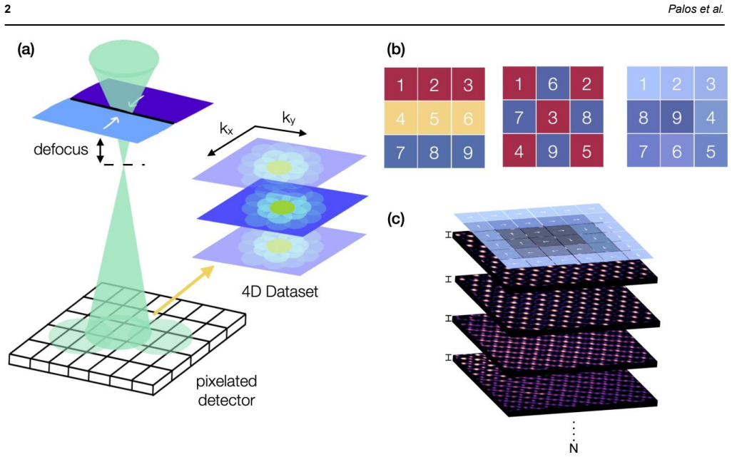
The central object is the programmable scan trajectory—raster line-by-line, inward/outward spiral, and N×N block-sequential paths—and how each one distributes probe position errors and dose over time. The argument's load-bearing contrast is between direct imaging (ADF/EELS), where each pixel's intensity is assigned to a commanded position so positional errors become visible artifacts, and iterative ptychography, which reconstructs the object from overlapping, redundant diffraction patterns and can absorb or correct moderate scan-path irregularities. The paper also uses Fourier transforms of ADF images as a diagnostic: replicated Bragg peaks mark large-jump settling errors, vertical streaks m
What would settle it
Re-acquire the same DyScO3 region with raster, spiral, and N=8 sequential scans, interleaved under fixed thickness and defocus, and check whether EELS patchiness and ptychography robustness persist; also lower the dose for N=8 to see whether ptychography still reaches ~0.7 Å resolution.
Extended reading notes
Core claim
The paper's central claim is that the scan trajectory itself, not just the total electron dose, determines data quality in atomic-resolution STEM, and that the two measurement modalities sit at opposite ends of sensitivity. For ptychography, iterative recovery from overlapping diffraction patterns absorbs large probe-position jumps: N=2, 4, and 8 sequential scans all give phase reconstructions with Bragg peaks visible to about 0.7 Å, even though ADF images from the same datasets show discontinuities and replicated Bragg reflections. For EELS, the elemental maps depend on precise spatial registration and uniform temporal illumination, so high-N sequential and some spiral scans produce non-uni
Load-bearing premise
The load-bearing premise is that differences in sample thickness and defocus between datasets were small enough not to cause the observed scan-mode differences; the paper says these variations were 'minimized, but not eliminated'.
Editorial extensions
If this is right
- Ptychographic reconstructions can be paired with non-raster trajectories that spread dose, without sacrificing the ~0.7 Å resolution demonstrated in DyScO3.
- EELS elemental maps from large-jump scans (N=8 sequential) show non-uniform contrast and replicated Bragg artifacts; users should avoid large jumps when quantitative spectra are needed.
- Spiral scans suppress flyback streaks and produce cleaner Fourier-space data than raster; the mild radial dose gradient is a secondary trade-off.
- At cryogenic temperature, outward spiral scans recover oxygen-column contrast in BaTiO3 that raster scans miss, enabling detection of subtle displacements.
- FFT inspection of ADF frames is a useful diagnostic: replicated peaks indicate probe-settling or large jumps; vertical streaks indicate flyback.
Reading between the lines
- An untested extension: random or interleaved trajectories may inherit the same ptychographic robustness, but only if positional metadata remains reliable; at very low dose, ptychographic convergence could fail.
- The EELS sensitivity implies that quantitative atomic-scale spectroscopy with programmable scans should be validated with dose-uniformity metrics, such as per-pixel exposure histograms, before relying on elemental ratios.
- The cryo-BaTiO3 oxygen shifts are consistent with a polar phase, but since ptychography is a 2D projection, full 3D displacement vectors would need multislice or tilt-series ptychography.
- If the temporal-dose argument generalizes, spiral or block-sequential scans may delay radiolysis in organic or biological materials; this is a direct, testable prediction beyond the oxides studied here.
Editorial analysis
A structured set of objections, weighed in public.
Referee Report
Summary. The paper reports a systematic comparison of programmable STEM scan trajectories (raster, inward/outward spiral, and N-sequential) applied to electron energy-loss spectroscopy (EELS), annular dark-field (ADF) imaging, and ptychography. Using DyScO3 as a model perovskite, the authors evaluate atomic-resolution EELS maps, ADF-derived FFTs, and ptychographic phase reconstructions. They conclude that ptychography is largely robust to large probe jumps and non-raster trajectories, whereas EELS and ADF are sensitive to trajectory-induced artifacts such as probe-settling delays, flyback streaks, and non-uniform dose delivery. A cryogenic BaTiO3 experiment is used to argue that outward spiral scanning improves oxygen-column visibility and reveals subtle oxygen displacements consistent with a low-temperature ferroelectric phase. The paper proposes practical guidelines for choosing scan strategies in low-dose 4D-STEM.
Significance. If the conclusions hold, the paper provides useful practical guidance for the growing use of programmable scan trajectories in dose-sensitive electron microscopy. The claim that ptychography tolerates large scan jumps while EELS requires precise, temporally uniform illumination is of broad interest and would help experimentalists allocate dose and choose scan patterns. The use of py4DSTEM for reconstructions and the focus on cryogenic low-dose conditions are strengths. However, the support is primarily qualitative: resolution is judged from visual inspection of FFT peaks, EELS maps come from single acquisitions without error bars, and the Methods explicitly acknowledge residual sample-thickness and defocus variations across scan-pattern datasets. These limitations are load-bearing for the central attribution of observed differences to scan trajectory. The paper also gives limited side-by-side ptychographic comparisons for raster and spiral scans in the same material system, so the strongest claim of general ptychographic robustness is only partially supported.
major comments (3)
- [Materials and Methods, 4D-STEM Data Acquisition] The Methods state: 'Variations in sample thickness and defocus were minimized, but not eliminated, across the different scan pattern datasets.' This residual variation is a direct confound for the central claim that observed differences in EELS, ADF, and ptychography arise from the scan trajectory. Since each scan-pattern dataset is acquired at a different location with manually optimized defocus (10–20 nm), systematic thickness or defocus differences could produce exactly the reported pattern: ptychography, which reconstructs the probe via mixed-state multislice, would appear robust, while EELS/ADF would show non-uniform contrast or replicated features. The authors need to provide per-dataset thickness/defocus estimates, repeat acquisitions, or otherwise demonstrate that these variations are uncorrelated with scan mode.
- [Fig. 4 and discussion of ptychographic resolution] The claim that 'Bragg peaks remained visible to ~0.7 Å' and that ptychographic reconstructions show 'minimal differences in achievable resolution' is based solely on visual inspection of FFTs. No quantitative resolution metric (e.g., Fourier ring correlation, peak width, or signal-to-noise threshold) is provided, and no uncertainty estimate is given. Furthermore, Fig. 4 compares only N=2, N=4, and N=8 sequential scans; raster and spiral ptychography of DyScO3 are not shown in the main text. Thus the paper's headline conclusion that 'ptychography tolerates large pixel jumps' and is robust across trajectory types is only directly demonstrated for one family of trajectories (N-sequential) in the primary benchmarking material. The cryogenic BaTiO3 comparison (Fig. 5) is a single raster vs. spiral pair and is also not quantitatively assessed.
- [EELS Data Acquisition and Fig. 3] The EELS elemental maps are presented from single acquisitions (15 passes with drift correction between passes) with no repeat measurements, no error bars, and no quantitative measure of contrast non-uniformity. The conclusion that sequential and spiral scans 'introduce non-uniform elemental contrast' is inferred visually. Drift correction between passes, which may itself vary with scan pattern, could generate patchiness or discontinuities that are then attributed to the trajectory. The authors should provide either repeated acquisitions with variance estimates or a quantitative metric of map uniformity across the different scan modes.
minor comments (4)
- [Fig. 3 caption] The caption lists 'raster, random, N=8, N=4, N=2 sequential, and spiral' scan modes, but the Materials and Methods state that programmable scan patterns scripted were 'raster, spiral (inward and outward), and N-sequential scans (N = 2, 4, 8)'. There is no description of a 'random' scan mode in the methods. Please clarify whether a random trajectory was used and, if so, provide details; if not, correct the caption.
- [Materials and Methods, EELS Data Acquisition and Fig. 3] The ADF images used for FFT analysis were processed with 'Hanning filtering, quartile-based thresholding, and cropping' before the FFTs were computed. These operations can alter periodic features and introduce or suppress artifacts. Showing the raw or minimally processed FFTs, or discussing the impact of these filters, would strengthen the interpretation of the FFT features.
- [Fig. 4 caption vs. Materials and Methods, Ptychographic Reconstructions] The Fig. 4 caption states '2 probe modes, 18 slices, 2 nm thickness, 20 nm defocus', whereas the Methods state '10–20 slices (2 nm per slice)' and 'defocus was varied between 10 and 20 nm, optimized manually for each dataset'. Please reconcile these numbers and report the actual parameters for each reconstruction, including the number of slices and defocus used for the N=2, N=4, and N=8 datasets.
- [General] The paper would benefit from a data availability statement and a list of the exact code and versions used (e.g., py4DSTEM version) to facilitate reproducibility. The current text only names the software package.
Circularity Check
No significant circularity; experimental benchmarking is self-contained.
full rationale
This paper is an experimental benchmark rather than a derivation-based claim. It contains no fitted parameters that are later called predictions, no equations whose outputs equal their inputs, and no uniqueness theorems invoked from self-citations. The central claim—that ptychography tolerates non-raster trajectories while EELS/ADF do not—is supported directly by the reconstructions, ADF FFTs, and EELS maps in Figures 3–5. The py4DSTEM package and related algorithm papers by co-authors are cited as tools and background, but the resilience of ptychography is not deduced from those citations; it is evidenced by the N=2, 4, and 8 sequential-scan reconstructions retaining Bragg peaks to ~0.7 Å. The acknowledged limitation in Materials and Methods ('Variations in sample thickness and defocus were minimized, but not eliminated, across the different scan pattern datasets.') is a possible confound for attributing all observed differences to scan trajectory, but that is an experimental validity concern, not circularity: no step reduces by definition to its own input. No self-definitional, fitted-input-as-prediction, self-citation-load-bearing, uniqueness-imported, ansatz-smuggled, or renaming pattern is present.
Assumptions & free parameters
free parameters (3)
- Defocus =
10 to 20 nm, manually optimized per dataset
- Number of probe modes =
2 modes (DyScO3), 6 modes (BaTiO3)
- Slice thickness =
2 nm per slice, 10 to 20 slices
assumptions (4)
- domain assumption The mixed-state multislice ptychography algorithm as implemented in py4DSTEM produces an accurate real-space potential at the claimed resolution.
- domain assumption Replicated Bragg peaks in ADF FFTs arise from probe-settling delays during large jumps.
- domain assumption Residual thickness and defocus variations between scan-pattern datasets are small enough not to affect the cross-pattern comparisons.
- domain assumption FFT Bragg-peak visibility is a valid proxy for spatial resolution.
Cite this review
Pith. "Pith review of Programmable Beam Control for Electron Energy-Loss Spectroscopy and Ptychography." pith.science (2026). https://pith.science/paper/CQ3Z2254
@misc{pith2026250910726,
  author       = {Pith},
  title        = {Pith review of: Programmable Beam Control for Electron Energy-Loss Spectroscopy and Ptychography},
  year         = {2026},
  howpublished = {\url{https://pith.science/paper/CQ3Z2254}},
  note         = {Machine review of arXiv:2509.10726}
}
read the original abstract
Programmable electron-beam scanning offers new opportunities to improve dose efficiency and suppress scan-induced artifacts in scanning transmission electron microscopy. Here, we systematically benchmark the impact of non-raster trajectories, including spiral and multi-pass sequential patterns, on two dose sensitive techniques: electron energy-loss spectroscopy (EELS) and ptychography. Using DyScO3 as a model perovskite, we compare spatial resolution, spectral fidelity, and artifact suppression across scan modes. Ptychographic phase reconstructions consistently achieve atomic resolution and remain robust to large jumps in probe position. In contrast, atomic-resolution EELS maps show pronounced sensitivity to probe motion, with sequential and spiral scans introducing non-uniform elemental contrast. Finally, spiral scanning applied under cryogenic conditions in BaTiO3 thin films improves dose uniformity and mitigates drift related distortions. These results establish practical guidelines for the implementation of programmable scan strategies in low-dose 4D-STEM and highlight the inherent resilience of ptychography to trajectory-induced artifacts.
Figures


Figures from the paper (1 more)

Reference graph
Works this paper leans on
-
[1]
R.F. Egerton. New techniques in electron energy - loss spectroscopy and energy -filtered imaging. Micron, 34(3–5):127–139, Apr 2003
2003
-
[2]
Egoavil, N
R. Egoavil, N. Gauquelin, G.T. Martinez, S. Van Aert, G. Van Tendeloo, and J. Verbeeck. Atomic resolution mapping of phonon excitations in stem -eels experiments. Ultramicroscopy , 147:1–7, Dec 2014
2014
-
[3]
Verbeeck and S
J. Verbeeck and S. Van Aert. Model based quantification of eels spectra. Ultramicroscopy , 101(2):207 –224, 2004
2004
-
[4]
Four -dimensional scanning transmission electron microscopy (4d -stem): From scanning nanodiffraction to ptychography and beyond
Colin Ophus. Four -dimensional scanning transmission electron microscopy (4d -stem): From scanning nanodiffraction to ptychography and beyond. Microscopy and Microanalysis , 25(3):563 –582, 06 2019
2019
-
[5]
Quantitative scanning transmission electron microscopy for materials science: Imaging, diffraction, spectroscopy, and tomography
Colin Ophus. Quantitative scanning transmission electron microscopy for materials science: Imaging, diffraction, spectroscopy, and tomography. Annual Review of Materials Research , 53(1):105 –141, 2023
2023
-
[6]
py4dstem: A software package for four - dimensional scanning transmission electron microscopy data analysis
Benjamin H Savitzky, Steven E Zeltmann, Lauren A Hughes, Hamish G Brown, Shiteng Zhao, Philipp M Pelz, Thomas C Pekin, Edward S Barnard, Jennifer Donohue, Luis Rangel DaCosta, Ellis Kennedy, Yujun Xie, Matthew T Janish, Matthew M Schneider, Patrick Herring, Chirranjeevi Gopal, Abraham Anapolsky, Rohan Dhall, Karen C Bustillo, Peter Ercius, Mary C Scott, J...
2021
-
[7]
Iterative phase retrieval algorithms for scanning transmission electron microscopy
Georgios Varnavides, Stephanie M Ribet, Steven E Zeltmann, Yue Yu, Benjamin H Savitzky, Dana O Byrne, Frances I Allen, Vinayak P Dravid, Mary C Scott, and Colin Ophus. Iterative phase retrieval algorithms for scanning transmission electron microscopy. arXiv preprint arXiv:2309.05250 , 2023
arXiv 2023
-
[8]
Velazco, A
A. Velazco, A. B´ech´e, D. Jannis, and J. Verbeeck. Reducing electron beam damage through alternative stem scanning strategies, part i: Experimental findings. Ultramicroscopy , 232:113398, Jan 2022
2022
Show all 39 references
-
[9]
Jannis, A
D. Jannis, A. Velazco, A. B´ech´e, and J. Verbeeck. Reducing electron beam damage through alternative stem scanning strategies, part ii: Attempt towards an empirical model describing the damage process. Ultramicroscopy , 240:113568, 2022
2022
-
[10]
Velazco, M
A. Velazco, M. Nord, A. B´ech´e, and J. Verbeeck. Evaluation of different rectangular scan strategies for stem imaging. Ultramicroscopy, 215:113021, Aug 2020
2020
-
[11]
Goodge, Elisabeth Bianco, Noah Schnitzer, Henny W
Berit H. Goodge, Elisabeth Bianco, Noah Schnitzer, Henny W. Zandbergen, and Lena F. Kourkoutis. Atomic -resolution cryo - stem across continuously variable temperatures. Microscopy and Microanalysis , 26(3):439 –446, 2021
2021
-
[12]
Kim, Emanuela Liberti, Yiming Hu, Takeo Sasaki, Peter D
Xudong Pei, Liqi Zhou, Chen Huang, Mark Boyce, Judy S. Kim, Emanuela Liberti, Yiming Hu, Takeo Sasaki, Peter D. Nellist, Peijun Zhang, David I. Stuart, Angus I. Kirkland, and Peng Wang. Cryogenic electron ptychographic single particle analysis with wide bandwidth information t...
2023
-
[13]
Savitzky, Ismail El Baggari, Colin B
Benjamin H. Savitzky, Ismail El Baggari, Colin B. Clement, Emily Waite, Berit H. Goodge, David J. Baek, John P. Sheckelton, Christopher Pasco, Hari Nair, Nathaniel J. Schreiber, Jason Hoffman, Alemayehu S. Admasu, Jaewook Kim, Sang -Wook Cheong, Anand Bhattacharya, Darrell G. ...
2018
-
[14]
Lupini, Raymond R
Xiahan Sang, Andrew R. Lupini, Raymond R. Unocic, Miaofang Chi, Albina Y. Borisevich, Sergei V. Kalinin, Eirik Endeve, Richard K. Archibald, and Stephen Jesse. Dynamic scan control in stem: Spiral scans. Advanced Structural and Chemical Imaging , 2(1), Jun 2016
2016
-
[15]
Lupini, Jilai Ding, Sergei V
Xiahan Sang, Andrew R. Lupini, Jilai Ding, Sergei V. Kalinin, Stephen Jesse, and Raymond R. Unocic. Precision controlled atomic resolution scanning transmission electron microscopy using spiral scan pathways. Scientific Reports, 7(1), Mar 2017
2017
-
[16]
Ede and Richard Beanland
Jeffrey M. Ede and Richard Beanland. Partial scanning transmission electron microscopy with deep learning. Scientific Reports, 10(1), May 2020
2020
-
[17]
Ortega, D
E. Ortega, D. Nicholls, N. D. Browning, and N. de Jonge. High temporal -resolution scanning transmission electron microscopy using sparse -serpentine scan pathways. Scientific Reports , 11:24301, 2021
2021
-
[18]
Woo, Anna Tararan, Luiz H.G
Alberto Zobelli, Steffi Y. Woo, Anna Tararan, Luiz H.G. Tizei, Nathalie Brun, Xiaoyan Li, Odile St´ephan, Mathieu Kociak, and Marcel Tenc´e. Spatial and spectral dynamics in stem hyperspectral imaging using random scan patterns. Ultramicroscopy, 212:112912, 2020
2020
-
[19]
B´ech´e, B
A. B´ech´e, B. Goris, B. Freitag, and J. Verbeeck. Development of a fast electromagnetic beam blanker for compressed sensing in scanning transmission electron microscopy. Applied Physics Letters, 108(9), Feb 2016
2016
-
[20]
Turner, M
R. Turner, M. Nord, A. Velazco, and J. Verbeeck. Optimal scanning strategies for high -fidelity stem imaging. Ultramicroscopy, 218:113085, 2020
2020
-
[21]
Using your beam efficiently: Reducing electron dose in the stem via flyback compensation
Tiarnan Mullarkey, Jonathan J P Peters, Clive Downing, and Lewys Jones. Using your beam efficiently: Reducing electron dose in the stem via flyback compensation. Microscopy and Microanalysis , 28(4):1428 –1436, 08 2022
2022
-
[22]
Peters, Bryan W
Jonathan J. Peters, Bryan W. Reed, Yu Jimbo, Kanako Noguchi, Karin H. Mu¨ller, Alexandra Porter, Daniel J. Masiel, and Lewys Jones. Event-responsive scanning transmission 9 electron microscopy. Science , 385(6708):549 –553, Aug 2024
2024
-
[23]
Jannis, C
D. Jannis, C. Hofer, C. Gao, X. Xie, A. B´ech´e, T.J. Pennycook, and J. Verbeeck. Event driven 4d stem acquisition with a timepix3 detector: Microsecond dwell time and faster scans for high precision and low dose applications. Ultramicroscopy , 233:113423, 2022
2022
-
[24]
T. Susi, J. C. Meyer, and J. Kotakoski. Manipulating low - dimensional materials down to the level of single atoms with electron irradiation. Nature Reviews Materials
-
[25]
Flexible metallic nanowires with self -adaptive contacts to semiconducting transition-metal dichalcogenide monolayers
Junhao Lin, Ovidiu Cretu, Wu Zhou, Kazu Suenaga, Dhiraj Prasai, Kirill I Bolotin, Nguyen Thanh Cuong, Minoru Otani, Susumu Okada, Andrew R Lupini, et al. Flexible metallic nanowires with self -adaptive contacts to semiconducting transition-metal dichalcogenide monolayers. Natu...
2014
-
[26]
Atom-by-atom fabrication with electron beams
Ondrej Dyck, Maxim Ziatdinov, David B Lingerfelt, Raymond R Unocic, Bethany M Hudak, Andrew R Lupini, Stephen Jesse, and Sergei V Kalinin. Atom-by-atom fabrication with electron beams. Nature Reviews Materials , 4(7):497–507, 2019
2019
-
[27]
Probing the dynamics of topologically protected charged ferroelectric domain walls with the electron beam at the atomic scale
Michele Conroy, Kalani Moore, Eoghan O’Connell, Lewys Jones, Clive Downing, Roger Whamore, Alexei Gruverman, Marty Gregg, and Ursel Bangert. Probing the dynamics of topologically protected charged ferroelectric domain walls with the electron beam at the atomic scale. Microscop...
2020
-
[28]
Contact-free reversible switching of improper ferroelectric domains by electron and ion irradiation
Erik D Roede, Aleksander B Mosberg, Donald M Evans, Edith Bourret, Zewu Yan, Antonius TJ van Helvoort, and Dennis Meier. Contact-free reversible switching of improper ferroelectric domains by electron and ion irradiation. APL Materials, 9(2), 2021
2021
-
[29]
Electron-beam-induced ferroelectric domain behavior in the transmission electron microscope: Toward deterministic domain patterning
James L Hart, Shi Liu, Andrew C Lang, Alexander Hubert, Andrius Zukauskas, Carlota Canalias, Richard Beanland, Andrew M Rappe, Miryam Arredondo, and Mitra L Taheri. Electron-beam-induced ferroelectric domain behavior in the transmission electron microscope: Toward deterministi...
2016
-
[30]
Formation and manipulation of domain walls with 2 nm domain periodicity in batio 3 without contact electrodes
Maya Barzilay and Yachin Ivry. Formation and manipulation of domain walls with 2 nm domain periodicity in batio 3 without contact electrodes. Nanoscale , 12(20):11136 –11142, 2020
2020
-
[31]
Domain structure formation by electron beam irradiation in lithium niobate crystals at elevated temperatures
DS Chezganov, EO Vlasov, EA Pashnina, MA Chuvakova, AA Esin, ED Greshnyakov, and V Ya Shur. Domain structure formation by electron beam irradiation in lithium niobate crystals at elevated temperatures. Applied Physics Letters, 115(9), 2019
2019
-
[32]
Electron-beam domain patterning in sr0
Tatyana R Volk, Lyudmila S Kokhanchik, Yadviga V Bodnarchuk, Radmir V Gainutdinov, Eugene B Yakimov, and Lyudmila I Ivleva. Electron-beam domain patterning in sr0. 61ba0. 39nb2o6 crystals. Coatings, 10(3):299, 2020
2020
-
[33]
Correcting nonlinear drift distortion of scanning probe and scanning transmission electron microscopies from image pairs with orthogonal scan directions
Colin Ophus, Jim Ciston, and Chris T Nelson. Correcting nonlinear drift distortion of scanning probe and scanning transmission electron microscopies from image pairs with orthogonal scan directions. Ultramicroscopy , 162:1–9, 2016
2016
-
[34]
Scanning distortion correction in stem images
Shoucong Ning, Takeshi Fujita, Anmin Nie, Ziqian Wang, Xiandong Xu, Jianghua Chen, Mingwei Chen, Shuhuai Yao, and Tong -Yi Zhang. Scanning distortion correction in stem images. Ultramicroscopy , 184:274–283, 2018
2018
-
[35]
Increasing spatial fidelity and snr of 4d-stem using multi-frame data fusion
Colum M O’Leary, Benedikt Haas, Christoph T Koch, Peter D Nellist, and Lewys Jones. Increasing spatial fidelity and snr of 4d-stem using multi-frame data fusion. Microscopy and Microanalysis , 28(4):1417 –1427, 2022
2022
-
[36]
Atomic resolution cryogenic 4d - stem imaging via robust distortion correction
Jacob Smith, Zhennan Huang, Wenpei Gao, Guannan Zhang, and Miaofang Chi. Atomic resolution cryogenic 4d - stem imaging via robust distortion correction. ACS nano , 17(12):11327 –11334, 2023
2023
-
[37]
R.F. Egerton. Control of radiation damage in the tem. Ultramicroscopy , 127:100 –108, 2013. Frontiers of Electron Microscopy in Materials Science
2013
-
[38]
Konijnenberg, S.F
Priya Dwivedi, A.P. Konijnenberg, S.F. Pereira, and H.P. Urbach. Lateral position correction in ptychography using the gradient of intensity patterns. Ultramicroscopy , 192:29 –36, 2018
2018
-
[39]
Bull, Christopher J
Craig L. Bull, Christopher J. Ridley, Kevin S. Knight, Nicholas P. Funnell, and Alexandra S. Gibbs. Comprehensive determination of the high -pressure structural behaviour of batio3. Mater. Adv. , 2:6094–6103, 2021. 10 Palos et al. Supplementary Information Fig. S1: Elemental E...
2021
Reviewed August 4, 2026 · model on record in the stance chip above.
Discussion (0). Continue with ORCID to comment.