REVIEW 3 major objections 2 minor 101 references
RIPPLE turns point tracking annotation into sparse corrections on motion-guided trajectories, matching exhaustive manual quality with 3-25 times fewer clicks.
Reviewed by Pith at T0; open to challenge. T0 means a machine referee read the full paper against a public rubric. the ladder, T0–T4 →
RIPPLE recasts annotation as sparse correction on motion-guided trajectories, matching exhaustive manual quality while cutting clicks by 3-25x on five diverse microscopy datasets from jellyfish and sperm.
T0 review reviewed 2026-06-29 challenge →
load-bearing objection RIPPLE gives a workable sparse-correction workflow for hard microscopy tracking cases and reports useful click reductions on real data, but the quality-equivalence claim lacks the metrics needed to evaluate it. the 3 major comments →
Motion-guided sparse correction enables expert-quality point tracking across diverse microscopy regimes
The pith
A machine-rendered reading of the paper's core claim, the machinery that carries it, and where it could break.
The reading
Core claim
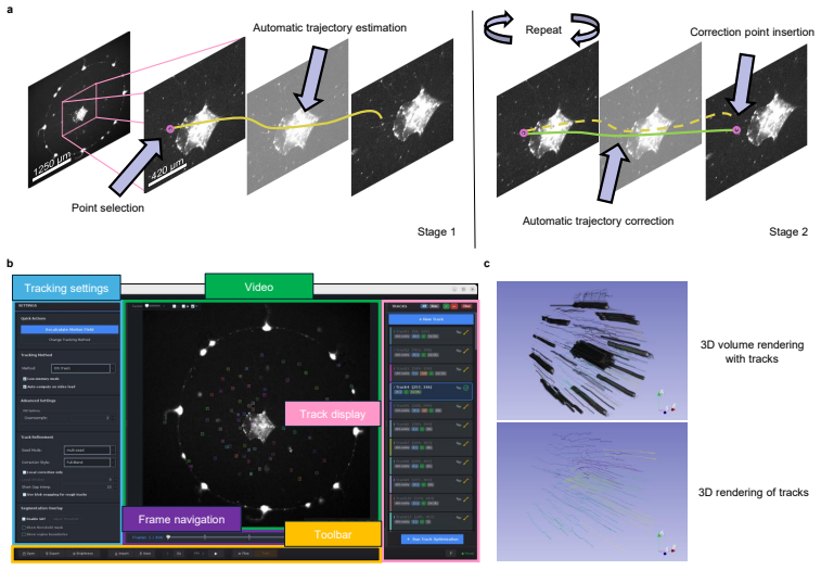
RIPPLE recasts annotation as sparse correction: a user clicks a starting point, the system proposes a full trajectory via motion guidance, and the user intervenes only where the trajectory drifts. Across the five tested microscopy datasets this produced trajectories equivalent in quality to exhaustive manual annotation while reducing the number of manual clicks by a factor of 3 to 25.
What carries the argument
Motion-guided trajectory proposals that support sparse rather than exhaustive user corrections.
Load-bearing premise
The motion-guided proposals remain accurate enough on the tested regimes that corrections stay sparse instead of requiring near-exhaustive intervention.
What would settle it
A new microscopy sequence in which the proposed trajectories require corrections in nearly every frame would demonstrate that the reported reduction in manual effort does not hold.
If this is right
- Enables immediate quantification of biological dynamics in videos previously too costly to annotate exhaustively.
- Supports direct benchmarking of automated trackers against the produced trajectories.
- Generates the gold-standard labeled data required to train and adapt future fully automatic microscopy trackers.
Where Pith is reading between the lines
- The same sparse-correction pattern could be tested on non-microscopy video domains if motion guidance can be adapted.
- Combining RIPPLE proposals with existing automated trackers might further lower the initial click count.
- Longer sequences or higher motion complexity would provide a direct test of whether proposal accuracy holds beyond the five evaluated datasets.
Editorial analysis
A structured set of objections, weighed in public.
Referee Report
Summary. The manuscript introduces RIPPLE (Refinement Interpolation Platform for Point Location Estimation), which reframes point tracking in challenging microscopy videos as sparse user correction of motion-guided trajectory proposals. A user provides an initial click, the system generates a full trajectory, and the user corrects only at drift points. The central claim is that, across five datasets (four from Clytia hemisphaerica and one from sperm landmark tracking), the resulting trajectories match the quality of exhaustive manual annotation while reducing manual clicks by a factor of 3–25×.
Significance. If the equivalence claim holds under rigorous metrics, RIPPLE would supply a scalable intermediate layer between exhaustive manual annotation and fully automatic trackers, directly addressing the annotation bottleneck for non-canonical biological systems. The use of real laboratory datasets from multiple regimes is a concrete strength that supports immediate utility for producing gold-standard data and benchmarking automated methods.
major comments (3)
- [Abstract / Results] Abstract and Results section: the claim that RIPPLE 'matched the quality of exhaustive manual annotation' is presented without any stated error metric (endpoint error, tracklet RMSE, or similar), without reporting numerical values against held-out labels, and without statistical tests or inter-annotator agreement baselines. This directly undermines evaluation of the central equivalence result.
- [Results] Results section: the reported 3–25× click reduction presupposes that motion-guided proposals remain sufficiently close to ground truth that interventions stay sparse; no quantitative analysis of proposal accuracy (e.g., drift statistics before correction) or cases where corrections become dense is supplied, leaving the sparsity assumption untested.
- [Methods / Experiments] Methods / Experiments: no description is given of how the five datasets were split for evaluation, whether any held-out ground truth was used, or how equivalence to exhaustive manual annotation was operationally defined and tested.
minor comments (2)
- [Abstract] The range '3 to 25 times' is given without per-dataset breakdown; a table listing clicks per method and dataset would improve transparency.
- [Abstract] Dataset descriptions in the abstract are brief; adding one-sentence characterizations of imaging conditions (frame rate, resolution, motion type) would aid readers.
Simulated Author's Rebuttal
We thank the referee for the constructive comments. We agree that the current manuscript lacks the quantitative details needed to rigorously support the central claims and will revise the text, figures, and methods to address each point.
read point-by-point responses
-
Referee: [Abstract / Results] Abstract and Results section: the claim that RIPPLE 'matched the quality of exhaustive manual annotation' is presented without any stated error metric (endpoint error, tracklet RMSE, or similar), without reporting numerical values against held-out labels, and without statistical tests or inter-annotator agreement baselines. This directly undermines evaluation of the central equivalence result.
Authors: We acknowledge that the manuscript presents the equivalence claim without the requested quantitative metrics or statistical comparisons. The original evaluation relied on expert visual assessment across the five laboratory datasets, but this is insufficient for the central result. In revision we will add endpoint error and tracklet RMSE values computed against held-out manual annotations, report numerical results per dataset, include inter-annotator agreement baselines from multiple experts, and apply appropriate statistical tests. revision: yes
-
Referee: [Results] Results section: the reported 3–25× click reduction presupposes that motion-guided proposals remain sufficiently close to ground truth that interventions stay sparse; no quantitative analysis of proposal accuracy (e.g., drift statistics before correction) or cases where corrections become dense is supplied, leaving the sparsity assumption untested.
Authors: We agree that the sparsity assumption underlying the click-reduction claim requires explicit quantitative support. The manuscript currently reports only the final click counts without characterizing proposal drift. We will add per-dataset statistics on proposal accuracy (mean and distribution of drift distances before correction), identify any sequences where corrections become dense, and discuss the conditions under which the 3–25× reduction holds. revision: yes
-
Referee: [Methods / Experiments] Methods / Experiments: no description is given of how the five datasets were split for evaluation, whether any held-out ground truth was used, or how equivalence to exhaustive manual annotation was operationally defined and tested.
Authors: We will expand the Methods and Experiments sections to specify the evaluation protocol in full. This will include the splitting strategy for the five datasets, confirmation that held-out ground-truth annotations were used for quantitative comparison, and the precise operational definition of equivalence (error thresholds, statistical criteria, and comparison to inter-annotator variability). revision: yes
Circularity Check
No circularity; empirical tool description with no derivation chain
full rationale
The paper describes RIPPLE as a sparse-correction annotation platform and reports empirical results on five microscopy datasets, claiming reduced clicks while matching manual annotation quality. No equations, fitted parameters, self-citations, or uniqueness theorems are invoked in any load-bearing way. The central claims rest on direct testing against exhaustive manual labels rather than any reduction to inputs by construction. This is the expected non-finding for a methods/empirical paper without a mathematical derivation.
Axiom & Free-Parameter Ledger
Cite this review
Pith. "Pith review of Motion-guided sparse correction enables expert-quality point tracking across diverse microscopy regimes." pith.science (2026). https://pith.science/paper/CSELG3JL
@misc{pith2026260529220,
  author       = {Pith},
  title        = {Pith review of: Motion-guided sparse correction enables expert-quality point tracking across diverse microscopy regimes},
  year         = {2026},
  howpublished = {\url{https://pith.science/paper/CSELG3JL}},
  note         = {Machine review of arXiv:2605.29220}
}
read the original abstract
Tracking the dynamics of non-canonical biological systems in microscopy videos remains a persistent challenge. Both classical and learning-based trackers depend on expert-reviewed data to be evaluated and adapted, yet exhaustive manual annotation rarely scales to the videos where these tools are needed most. We developed RIPPLE (Refinement Interpolation Platform for Point Location Estimation), which recasts annotation as sparse correction: a user clicks a starting point, RIPPLE proposes a full trajectory, and the user intervenes only where the trajectory drifts. We tested RIPPLE on five challenging microscopy datasets from our laboratories, four from the transparent jellyfish Clytia hemisphaerica and one tracking landmarks on rapidly moving sperm. Across these, RIPPLE matched the quality of exhaustive manual annotation while reducing manual clicks by 3 to 25 times across datasets. RIPPLE thereby fills a missing layer between manual annotation and fully automated tracking, enabling immediate quantification of biological dynamics, method benchmarking, and the production of the gold-standard data needed to adapt future automated microscopy trackers.
Figures


Reference graph
Works this paper leans on
-
[1]
Multiscale fluorescence imaging of living samples,
Y. Wu and H. Shroff, “Multiscale fluorescence imaging of living samples,” Histochem. cell biology158, 301–323 (2022)
2022
-
[2]
Live-cell imaging in the deep learning era,
J. W. Pylvänäinen, E. Gómez-de Mariscal, R. Henriques, and G. Jacquemet, “Live-cell imaging in the deep learning era,” Curr. Opin. Cell Biol.85, 102271 (2023)
2023
-
[3]
Robust single-particle tracking in live-cell time-lapse sequences,
K. Jaqaman, D. Loerke, M. Mettlen,et al., “Robust single-particle tracking in live-cell time-lapse sequences,” Nat. methods5, 695–702 (2008)
2008
-
[4]
Objective comparison of particle tracking methods,
N. Chenouard, I. Smal, F. De Chaumont,et al., “Objective comparison of particle tracking methods,” Nat. methods 11, 281–289 (2014)
2014
-
[5]
Featurepointtrackingandtrajectoryanalysisforvideoimagingincellbiology,
I.F.SbalzariniandP.Koumoutsakos,“Featurepointtrackingandtrajectoryanalysisforvideoimagingincellbiology,” J. structural biology151, 182–195 (2005)
2005
-
[6]
A review for cell and particle tracking on microscopy images using algorithms and deep learning technologies,
H.-J. Cheng, C.-H. Hsu, C.-L. Hung, and C.-Y. Lin, “A review for cell and particle tracking on microscopy images using algorithms and deep learning technologies,” biomedical journal45, 465–471 (2022)
2022
-
[7]
A review of progress in single particle tracking: from methods to biophysical insights,
C. Manzo and M. F. Garcia-Parajo, “A review of progress in single particle tracking: from methods to biophysical insights,” Reports on progress physics78, 124601 (2015)
2015
-
[8]
Live cell imaging analysis of receptor function,
D. C. Worth and M. Parsons, “Live cell imaging analysis of receptor function,” inLive Cell Imaging: Methods and Protocols,(Springer, 2009), pp. 311–323
2009
-
[9]
Intracellular transport dynamics revealed by single-particle tracking,
M.-L. Zhang, H.-Y. Ti, P.-Y. Wang, and H. Li, “Intracellular transport dynamics revealed by single-particle tracking,” Biophys. Reports7, 413 (2021)
2021
-
[10]
Phase contrast time-lapse microscopy datasets with automated and manual cell tracking annotations,
D. F. E. Ker, S. Eom, S. Sanami,et al., “Phase contrast time-lapse microscopy datasets with automated and manual cell tracking annotations,” Sci. data5, 180237 (2018)
2018
-
[11]
Delta: Automated cell segmentation, tracking, and lineage reconstruction using deep learning,
J.-B. Lugagne, H. Lin, and M. J. Dunlop, “Delta: Automated cell segmentation, tracking, and lineage reconstruction using deep learning,” PLoS computational biology16, e1007673 (2020)
2020
-
[12]
Long-term tracking of budding yeast cells in brightfield microscopy: Cellstar and the evaluation platform,
C. Versari, S. Stoma, K. Batmanov,et al., “Long-term tracking of budding yeast cells in brightfield microscopy: Cellstar and the evaluation platform,” J. The Royal Soc. Interface14(2017)
2017
-
[13]
Cell population tracking and lineage construction with spatiotemporal context,
K. Li, E. D. Miller, M. Chen,et al., “Cell population tracking and lineage construction with spatiotemporal context,” Med. image analysis12, 546–566 (2008)
2008
-
[14]
Automated reconstruction of whole-embryo cell lineages by learning from sparse annotations,
C. Malin-Mayor, P. Hirsch, L. Guignard,et al., “Automated reconstruction of whole-embryo cell lineages by learning from sparse annotations,” Nat. biotechnology41, 44–49 (2023)
2023
-
[15]
Deep neural networks to register and annotate cells in moving and deforming nervous systems,
A. A. Atanas, A. K.-Y. Lu, B. Goodell,et al., “Deep neural networks to register and annotate cells in moving and deforming nervous systems,” eLife14, RP108159 (2026)
2026
-
[16]
Rethinking deep learning in bioimaging through a data centric lens,
J. Cao, J. Wenzel, S. Zhang,et al., “Rethinking deep learning in bioimaging through a data centric lens,” npj Imaging 3, 29 (2025)
2025
-
[17]
Trackmate: Anopenandextensibleplatformforsingle-particletracking,
J.-Y.Tinevez,N.Perry,J.Schindelin,etal.,“Trackmate: Anopenandextensibleplatformforsingle-particletracking,” Methods115, 80–90 (2017)
2017
-
[18]
Trackmate 7: integrating state-of-the-art segmentation algorithms into tracking pipelines,
D. Ershov, M.-S. Phan, J. W. Pylvänäinen,et al., “Trackmate 7: integrating state-of-the-art segmentation algorithms into tracking pipelines,” Nat. methods19, 829–832 (2022)
2022
-
[19]
u-track3d: Measuring, navigating, and validating dense particle trajectories in three dimensions,
P. Roudot, W. R. Legant, Q. Zou,et al., “u-track3d: Measuring, navigating, and validating dense particle trajectories in three dimensions,” Cell Reports Methods3(2023)
2023
-
[20]
Organoidtracker: Efficient cell tracking using machine learning and manual error correction,
R. N. Kok, L. Hebert, G. Huelsz-Prince,et al., “Organoidtracker: Efficient cell tracking using machine learning and manual error correction,” PLoS One15, e0240802 (2020)
2020
-
[21]
An objective comparison of cell-tracking algorithms,
V. Ulman, M. Maška, K. E. G. Magnusson,et al., “An objective comparison of cell-tracking algorithms,” Nat. methods14, 1141–1152 (2017)
2017
-
[22]
Cellpose: a generalist algorithm for cellular segmentation,
C. Stringer, T. Wang, M. Michaelos, and M. Pachitariu, “Cellpose: a generalist algorithm for cellular segmentation,” Nat. methods18, 100–106 (2021)
2021
-
[23]
Cell detection with star-convex polygons,
U. Schmidt, M. Weigert, C. Broaddus, and G. Myers, “Cell detection with star-convex polygons,” inInternational conference on medical image computing and computer-assisted intervention,(Springer, 2018), pp. 265–273
2018
-
[24]
Sleap: A deep learning system for multi-animal pose tracking,
T. D. Pereira, N. Tabris, A. Matsliah,et al., “Sleap: A deep learning system for multi-animal pose tracking,” Nat. methods19, 486–495 (2022)
2022
-
[25]
Using deeplabcut for 3d markerless pose estimation across species and behaviors,
T. Nath, A. Mathis, A. C. Chen,et al., “Using deeplabcut for 3d markerless pose estimation across species and behaviors,” Nat. protocols14, 2152–2176 (2019)
2019
-
[26]
TAPIR: Tracking any point with per-frame initialization and temporal refinement,
C. Doersch, Y. Yang, M. Vecerik,et al., “TAPIR: Tracking any point with per-frame initialization and temporal refinement,” inProceedings of the IEEE/CVF International Conference on Computer Vision (ICCV),(2023), pp. 10061–10072
2023
-
[27]
Cotracker: Itisbettertotracktogether,
N.Karaev,I.Rocco,B.Graham,etal.,“Cotracker: Itisbettertotracktogether,”inEuropeanconferenceoncomputer vision,(Springer, 2024), pp. 18–35
2024
-
[28]
Local all-pair correspondence for point tracking,
S. Cho, J. Huang, J. Nam,et al., “Local all-pair correspondence for point tracking,” inEuropean Conference on Computer Vision (ECCV),(2024)
2024
-
[29]
Particle video revisited: Tracking through occlusions using point trajectories,
A. W. Harley, Z. Fang, and K. Fragkiadaki, “Particle video revisited: Tracking through occlusions using point trajectories,” inECCV,(2022)
2022
-
[30]
Cotracker3: Simpler and better point tracking by pseudo-labelling real videos,
N. Karaev, Y. Makarov, J. Wang,et al., “Cotracker3: Simpler and better point tracking by pseudo-labelling real videos,” inProceedings of the IEEE/CVF International Conference on Computer Vision,(2025), pp. 6013–6022
2025
-
[31]
Deepprobabilistictrackingofparticlesinfluorescencemicroscopyimages,
R.Spilger,J.-Y.Lee,V.O.Chagin,etal.,“Deepprobabilistictrackingofparticlesinfluorescencemicroscopyimages,” Med. image analysis72, 102128 (2021)
2021
-
[32]
Robustmodel-basedanalysisofsingle-particletrackingexperiments with spot-on,
A.S.Hansen, M.Woringer, J.B.Grimm,etal., “Robustmodel-basedanalysisofsingle-particletrackingexperiments with spot-on,” Elife7, e33125 (2018)
2018
-
[33]
Thecelltrackingchallenge: 10yearsofobjectivebenchmarking,
M.Maška,V.Ulman,P.Delgado-Rodriguez,etal.,“Thecelltrackingchallenge: 10yearsofobjectivebenchmarking,” Nat. Methods20, 1010–1020 (2023)
2023
-
[34]
Pointodyssey: A large-scale synthetic dataset for long-term point tracking,
Y. Zheng, A. W. Harley, B. Shen,et al., “Pointodyssey: A large-scale synthetic dataset for long-term point tracking,” inProceedings of the IEEE/CVF International Conference on Computer Vision,(2023), pp. 19855–19865
2023
-
[35]
Biological data annotation via a human-augmenting ai-based labeling system,
D. van der Wal, I. Jhun, I. Laklouk,et al., “Biological data annotation via a human-augmenting ai-based labeling system,” NPJ digital medicine4, 145 (2021)
2021
-
[36]
A benchmark for comparison of cell tracking algorithms,
M. Maška, V. Ulman, D. Svoboda,et al., “A benchmark for comparison of cell tracking algorithms,” Bioinformatics 30, 1609–1617 (2014)
2014
-
[37]
Tracking cell lineages in 3d by incremental deep learning,
K. Sugawara, Ç. Çevrim, and M. Averof, “Tracking cell lineages in 3d by incremental deep learning,” Elife11, e69380 (2022)
2022
-
[38]
edetect: afasterrordetectionandcorrectiontoolforlivecellimagingdataanalysis,
H.Han,G.Wu,Y.Li,andZ.Zi,“edetect: afasterrordetectionandcorrectiontoolforlivecellimagingdataanalysis,” Iscience13, 1–8 (2019)
2019
-
[39]
Tracurate: efficiently curating cell tracks,
S. Wagner, K. Thierbach, T. Zerjatke,et al., “Tracurate: efficiently curating cell tracks,” SoftwareX13, 100656 (2021)
2021
-
[40]
Segmentation, tracking and cell cycle analysis of live-cell imaging data with cell-acdc,
F. Padovani, B. Mairhörmann, P. Falter-Braun,et al., “Segmentation, tracking and cell cycle analysis of live-cell imaging data with cell-acdc,” BMC biology20, 174 (2022)
2022
-
[41]
Laptrack: linear assignment particle tracking with tunable metrics,
Y. T. Fukai and K. Kawaguchi, “Laptrack: linear assignment particle tracking with tunable metrics,” Bioinformatics 39, btac799 (2023)
2023
-
[42]
Automated cell tracking using stardist and trackmate,
E. Fazeli, N. H. Roy, G. Follain,et al., “Automated cell tracking using stardist and trackmate,” F1000Research9, 1279 (2020)
2020
-
[43]
A genetically tractable jellyfish model for systems and evolutionary neuroscience,
B. Weissbourd, T. Momose, A. Nair,et al., “A genetically tractable jellyfish model for systems and evolutionary neuroscience,” Cell184, 5854–5868 (2021)
2021
-
[44]
Whole-animal multiplexed single-cell rna-seq reveals transcriptional shifts across clytia medusa cell types,
T. Chari, B. Weissbourd, J. Gehring,et al., “Whole-animal multiplexed single-cell rna-seq reveals transcriptional shifts across clytia medusa cell types,” Sci. Adv.7, eabh1683 (2021)
2021
-
[45]
Past, present and future of clytia hemisphaerica as a laboratory jellyfish,
E. Houliston, L. Leclère, C. Munro,et al., “Past, present and future of clytia hemisphaerica as a laboratory jellyfish,” inCurrent topics in developmental biology,vol. 147 (Elsevier, 2022), pp. 121–151
2022
-
[46]
Tap-vid: A benchmark for tracking any point in a video,
C. Doersch, A. Gupta, L. Markeeva,et al., “Tap-vid: A benchmark for tracking any point in a video,” Adv. Neural Inf. Process. Syst.35, 13610–13626 (2022)
2022
-
[48]
Quantitative phase imaging in biomedicine,
Y. Park, C. Depeursinge, and G. Popescu, “Quantitative phase imaging in biomedicine,” Nat. photonics12, 578–589 (2018)
2018
-
[49]
Episodic rolling and transient attachments create diversity in sperm swimming behavior,
D. F. Babcock, P. M. Wandernoth, and G. Wennemuth, “Episodic rolling and transient attachments create diversity in sperm swimming behavior,” BMC biology12, 67 (2014)
2014
-
[50]
Bimodal rheotactic behavior reflects flagellar beat asymmetry in human sperm cells,
A. Bukatin, I. Kukhtevich, N. Stoop,et al., “Bimodal rheotactic behavior reflects flagellar beat asymmetry in human sperm cells,” Proc. National Acad. Sci.112, 15904–15909 (2015)
2015
-
[51]
Chirality and frequency measurement of longitudinal rolling of human sperm using optical trap,
Z. Zhong, C. Zhang, R. Liu,et al., “Chirality and frequency measurement of longitudinal rolling of human sperm using optical trap,” Front. Bioeng. Biotechnol.10, 1028857 (2022)
2022
-
[52]
Imagej for microscopy,
T. J. Collins, “Imagej for microscopy,” Biotechniques43, S25–S30 (2007)
2007
-
[54]
Clytia hemisphaerica: a jellyfish cousin joins the laboratory,
E. Houliston, T. Momose, and M. Manuel, “Clytia hemisphaerica: a jellyfish cousin joins the laboratory,” Trends Genet.26, 159–167 (2010)
2010
-
[55]
High doses of crispr/cas9 ribonucleoprotein efficiently induce gene knockout with low mosaicism in the hydrozoan clytia hemisphaerica through microhomology-mediated deletion,
T. Momose, A. De Cian, K. Shiba,et al., “High doses of crispr/cas9 ribonucleoprotein efficiently induce gene knockout with low mosaicism in the hydrozoan clytia hemisphaerica through microhomology-mediated deletion,” Sci. Reports8, 11734 (2018)
2018
-
[56]
The genome of the jellyfish clytia hemisphaerica and the evolution of the cnidarian life-cycle,
L. Leclère, C. Horin, S. Chevalier,et al., “The genome of the jellyfish clytia hemisphaerica and the evolution of the cnidarian life-cycle,” Nat. ecology & evolution3, 801–810 (2019)
2019
-
[57]
3d slicer: a platform for subject-specific image analysis, visualization, and clinical support,
R. Kikinis, S. D. Pieper, and K. G. Vosburgh, “3d slicer: a platform for subject-specific image analysis, visualization, and clinical support,” inIntraoperative imaging and image-guided therapy,(Springer, 2013), pp. 277–289
2013
-
[58]
Demo for Annotating a Point Track with Optical Flow,
Google DeepMind, “Demo for Annotating a Point Track with Optical Flow,” https://github.com/google-deepmind/ tapnet/blob/main/colabs/optical_flow_track_assist.ipynb (2026). Official TAPNet repository. Accessed: 2026-04-26. Motion-guided sparse correction enables expert-quality point tracking across diverse microscopy regimes LEONIDASZIMIANITIS, 1 PASINDUTH...
2026
-
[59]
Fast optical flow using dense inverse search,
T. Kroeger, R. Timofte, D. Dai, and L. Van Gool, “Fast optical flow using dense inverse search,” inEuropean conference on computer vision,(Springer, 2016), pp. 471–488
2016
-
[60]
Two-frame motion estimation based on polynomial expansion,
G. Farnebäck, “Two-frame motion estimation based on polynomial expansion,” inScandinavian conference on Image analysis,(Springer, 2003), pp. 363–370
2003
-
[61]
A duality based approach for realtime tv-l 1 optical flow,
C. Zach, T. Pock, and H. Bischof, “A duality based approach for realtime tv-l 1 optical flow,” inJoint pattern recognition symposium,(Springer, 2007), pp. 214–223
2007
-
[62]
Robust local optical flow for feature tracking,
T. Senst, V. Eiselein, and T. Sikora, “Robust local optical flow for feature tracking,” IEEE Trans. on Circuits Syst. for Video Technol.22, 1377–1387 (2012)
2012
-
[63]
Deepflow: Large displacement optical flow with deep matching,
P. Weinzaepfel, J. Revaud, Z. Harchaoui, and C. Schmid, “Deepflow: Large displacement optical flow with deep matching,” inProceedings of the IEEE international conference on computer vision,(2013), pp. 1385–1392
2013
-
[64]
Simpleflow: A non-iterative, sublinear optical flow algorithm,
M. Tao, J. Bai, P. Kohli, and S. Paris, “Simpleflow: A non-iterative, sublinear optical flow algorithm,” Comput. Graph. Forum31, 345–353 (2012)
2012
-
[65]
Efficient sparse-to-dense optical flow estimation using a learned basis and layers,
J. Wulff and M. J. Black, “Efficient sparse-to-dense optical flow estimation using a learned basis and layers,” in Proceedings of the IEEE Conference on Computer Vision and Pattern Recognition,(2015), pp. 120–130
2015
-
[66]
Bradski, A
G. Bradski, A. Kaehleret al., “Opencv,” Dr. Dobb’s journal software tools3, 1–81 (2000)
2000
-
[67]
Denseopticalflowbyiterativelocalwindowregistration,
G.LeBesneraisandF.Champagnat,“Denseopticalflowbyiterativelocalwindowregistration,”inIEEEInternational Conference on Image Processing 2005,vol. 1 (IEEE, 2005), pp. I–137
2005
-
[68]
Determining optical flow,
B. K. Horn and B. G. Schunck, “Determining optical flow,” Artif. intelligence17, 185–203 (1981)
1981
-
[69]
RAFT: Recurrent all-pairs field transforms for optical flow,
Z. Teed and J. Deng, “RAFT: Recurrent all-pairs field transforms for optical flow,” inEuropean Conference on Computer Vision (ECCV),(Springer, 2020), pp. 402–419
2020
-
[70]
Learning to estimate hidden motions with global motion aggregation,
S. Jiang, D. Campbell, Y. Lu,et al., “Learning to estimate hidden motions with global motion aggregation,” in Proceedings of the IEEE/CVF international conference on computer vision,(2021), pp. 9772–9781
2021
-
[71]
Flowformer: Atransformerarchitectureforopticalflow,
Z.Huang,X.Shi,C.Zhang,etal.,“Flowformer: Atransformerarchitectureforopticalflow,”inEuropeanconference on computer vision,(Springer, 2022), pp. 668–685
2022
-
[72]
Flowformer++: Masked cost volume autoencoding for pretraining optical flow estimation,
X. Shi, Z. Huang, D. Li,et al., “Flowformer++: Masked cost volume autoencoding for pretraining optical flow estimation,” inProceedings of the IEEE/CVF conference on computer vision and pattern recognition,(2023), pp. 1599–1610
2023
-
[73]
Gmflow: Learningopticalflowviaglobalmatching,
H.Xu,J.Zhang,J.Cai,etal.,“Gmflow: Learningopticalflowviaglobalmatching,”inProceedingsoftheIEEE/CVF conference on computer vision and pattern recognition,(2022), pp. 8121–8130
2022
-
[74]
Unifying flow, stereo and depth estimation,
H. Xu, J. Zhang, J. Cai,et al., “Unifying flow, stereo and depth estimation,” IEEE Trans. on Pattern Anal. Mach. Intell.45, 13941–13958 (2023)
2023
-
[75]
Sea-raft: Simple, efficient, accurate raft for optical flow,
Y. Wang, L. Lipson, and J. Deng, “Sea-raft: Simple, efficient, accurate raft for optical flow,” inEuropean Conference on Computer Vision,(Springer, 2024), pp. 36–54
2024
-
[76]
High resolution multi-scale raft (robust vision challenge 2022),
A. Jahedi, M. Luz, L. Mehl,et al., “High resolution multi-scale raft (robust vision challenge 2022),” arXiv preprint arXiv:2210.16900 (2022)
-
[77]
Neuflow-v2: Push high-efficiency optical flow to the limit,
Z. Zhang, A. Gupta, H. Jiang, and H. Singh, “Neuflow-v2: Push high-efficiency optical flow to the limit,” in2025 IEEE/RSJ International Conference on Intelligent Robots and Systems (IROS),(IEEE, 2025), pp. 2479–2485
2025
-
[78]
Ccmr: High resolution optical flow estimation via coarse-to-fine context-guided motion reasoning,
A. Jahedi, M. Luz, M. Rivinius, and A. Bruhn, “Ccmr: High resolution optical flow estimation via coarse-to-fine context-guided motion reasoning,” inProceedings of the IEEE/CVF Winter Conference on Applications of Computer Vision,(2024), pp. 6899–6908
2024
-
[79]
Craft: Cross-attentional flow transformer for robust optical flow,
X. Sui, S. Li, X. Geng,et al., “Craft: Cross-attentional flow transformer for robust optical flow,” inProceedings of the IEEE/CVF conference on Computer Vision and Pattern Recognition,(2022), pp. 17602–17611
2022
-
[80]
Memflow: Optical flow estimation and prediction with memory,
Q. Dong and Y. Fu, “Memflow: Optical flow estimation and prediction with memory,” inProceedings of the IEEE/CVF Conference on Computer Vision and Pattern Recognition,(2024), pp. 19068–19078
2024
-
[81]
Skflow: Learning optical flow with super kernels,
S. Sun, Y. Chen, Y. Zhu,et al., “Skflow: Learning optical flow with super kernels,” Adv. Neural Inf. Process. Syst. 35, 11313–11326 (2022)
2022
-
[82]
Recurrent partial kernel network for efficient optical flow estimation,
H. Morimitsu, X. Zhu, X. Ji, and X.-C. Yin, “Recurrent partial kernel network for efficient optical flow estimation,” inProceedings of the AAAI Conference on Artificial Intelligence,vol. 38 (2024), pp. 4278–4286
2024
This paper was first reviewed by grok-4.3 on June 29, 2026.
discussion (0)
Sign in with ORCID, Apple, or X to comment. Anyone can read and Pith papers without signing in.