REVIEW 4 major objections 5 minor 68 references
Cell size heterogeneity controls crystallization of the developing fruit fly wing
T0 review · 4 major / 5 minor · reviewed 2026-08-15 · deepseek-v4-flash
Pith's one-line read Cell size spread, not shear, gates fly-wing crystallization
desk verdict Solid experiment-plus-model case that cell size polydispersity controls fly wing crystallization, with shear demoted to an accelerator; the measured-to-model polydispersity mapping needs calibration but the central claim holds. read the letter →
The pith
A machine-rendered reading of the paper's core claim, the machinery that carries it, and where it could break.
The reading
What carries the argument
The central object is a vertex model of epithelial tissue with cell size polydispersity: each cell is assigned a preferred area $A_{0,c}$ uniformly drawn from $[(1-\sqrt{3}\Delta)A_0, (1+\sqrt{3}\Delta)A_0]$, with $\Delta$ the normalized standard deviation of preferred areas, and bond tensions fluctuate via an Ornstein-Uhlenbeck process with magnitude $\Lambda_F$. The model is simulated both quasistatically and with overdamped vertex dynamics, including T1 transitions and Lees-Edwards shear. Order is quantified by the cell hexatic $\Psi_6$, the tissue hexatic $\langle \Psi_6 \rangle$, and a translational order parameter, with susceptibility peaks and defect densities locating the transition. The machinery works by showing that increasing $\Delta$ melts the crystal through sequential unbinding of dislocations and disclinations, and that reducing $\Delta$ across the transition produces the slow coarsening kinetics observed in the wing.
What would settle it
Track a wing in which cell size polydispersity is held above the inferred threshold (for example by perturbing cell size homeostasis without blocking division) while tissue shear flow remains intact: if local hexatic order still rises strongly, the polydispersity-control claim fails. Conversely, if polydispersity is reduced early in development while shear is absent and tissue-scale hexatic order still grows substantially, the shear-enhancement claim would need revision.
Extended reading notes
Core claim
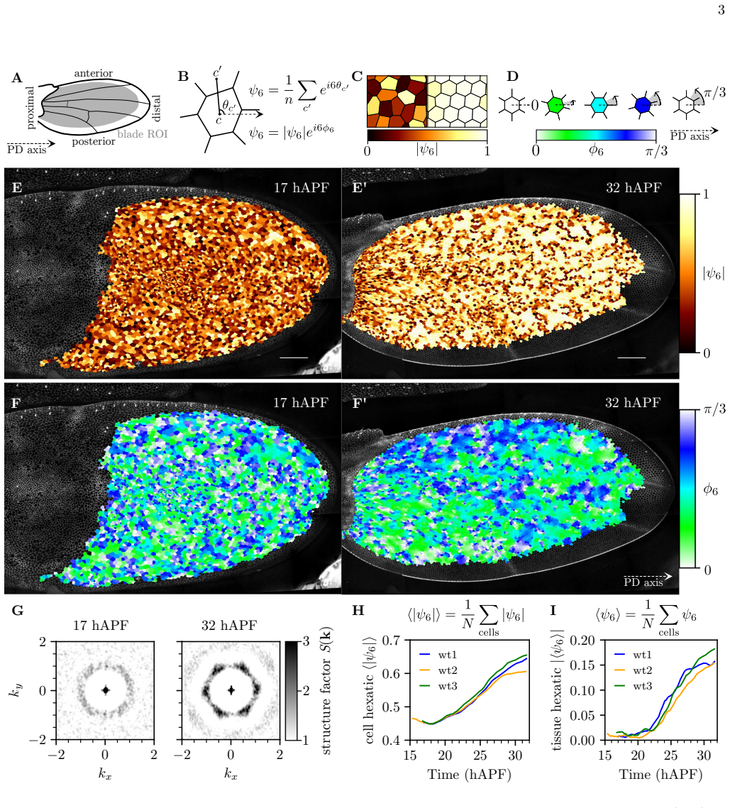
The central claim is that the disorder-to-crystal transition in the developing fly wing is controlled by cell size polydispersity, defined as the normalized standard deviation of the cell area distribution, and not by tissue shear flow. In a vertex model where cells are assigned preferred areas spread over an interval of width $\Delta$, the authors find a sharp transition near $\Delta \simeq 0.12$: below this critical value the packing is crystalline, above it the packing is disordered, with the translational and orientational order parameters peaking at slightly different polydispersities ($\Delta_t = 0.120 \pm 0.001$, $\Delta_6 = 0.125 \pm 0.001$), hinting at a hexatic phase. Experimentally, the authors measure cell area polydispersity in wild-type wings and in distal ablation, proximal ablation, $cdc2$, $dumpy$, and planar cell polarity mutants. They find that polydispersity decreases during pupal development in wild-type and distal ablation wings, that local hexatic order emerges when $\Delta_{\mathrm{ex}}$ falls below roughly $0.2$, and that proximal ablation keeps polydispersity high and blocks crystallization. Shear flow, they conclude, does not drive the transition itself but significantly accelerates the alignment of locally ordered crystallites, which is why tissue-scale hexatic order fails to develop when shear is suppressed.
Load-bearing premise
The measured polydispersity $\Delta_{\mathrm{ex}}$, computed from cell areas after a one-hour moving average and excluding cells adjacent to veins and dividing cells, is assumed to faithfully represent the model's preferred-area spread $\Delta$, and the noise magnitude $\Lambda_F = 0.35\Lambda_0$ is chosen because it reproduces the experimental decoupling of local and tissue hexatic order; if either choice is off, the apparent crossing of the model's critical polydispersity with the experimental ordering onset could be coincidental.
Editorial extensions
If this is right
- Crystallization can proceed without tissue shear: distal ablation and $dumpy$ mutant wings, where shear flow is largely suppressed, still show a strong increase in local hexatic order.
- Shear's role is kinetic, not thermodynamic: it aligns already-formed crystallites and raises tissue-scale hexatic order, but it cannot induce ordering when polydispersity is high.
- Cell divisions are not required for crystallization: the $cdc2$ mutant, with division arrested, crystallizes similarly to wild-type.
- The transition is sharp in the model and the experimental onset of ordering near $\Delta_{\mathrm{ex}} \simeq 0.2$ is consistent with a quench across a phase transition.
- Reducing polydispersity over time in the model recapitulates the experimental rise of local hexatic order and the shear dependence of tissue hexatic order.
Reading between the lines
- A direct, testable prediction of this picture is that artificially maintaining a broad cell size distribution should block crystallization even when tissue shear flows are intact; this could be probed with perturbations that disrupt cell size homeostasis without arresting divisions.
- Because polydispersity also controls relaxation times in the model, the paper implies that cell size heterogeneity could tune tissue fluidity and solidity during morphogenesis, connecting to jamming and glass-transition ideas in other epithelia.
- The proximity of the translational and orientational transition points suggests a KTHNY-like two-step melting scenario in a biological tissue, but the finite system sizes leave open whether the hexatic phase is genuine or a finite-size effect.
- The mechanism that reduces polydispersity during development is left open; the authors note it may reflect cell-cycle arrest and uniform mechanical state, or a proximal coordinating cue, and distinguishing these would sharpen the causal story.
Signed reviews
Editorial analysis
A structured set of objections, weighed in public.
Referee Report
Summary. The paper studies ordering of the Drosophila pupal wing epithelium and argues that a disorder-to-crystal transition is controlled by cell size polydispersity rather than by tissue shear flow. The authors quantify local and tissue-scale hexatic order in wild-type wings and in distal ablation, cdc2, dumpy, and PCP mutant conditions, finding that local crystalline order emerges even when shear flow is largely suppressed, whereas tissue-scale alignment of crystallites is weak without shear. They introduce a vertex model with quenched preferred-area polydispersity and bond-tension noise, and show, with finite-size scaling and defect analyses, a crystal-to-hexatic-to-liquid transition as a function of polydispersity Δ, with a critical value around Δ≈0.12. Kinetic simulations with and without imposed shear reproduce the decoupling of local and tissue hexatic order observed in distal ablation experiments. Experimentally, a smoothed measure of cell-area polydispersity Δ_ex decreases over pupal development, and a proximal ablation condition with persistently high Δ_ex fails to crystallize. The paper concludes that reduction of cell size heterogeneity controls the transition, while shear accelerates crystallite alignment but does not itself induce order.
Significance. If the central claim holds, the paper identifies cell size polydispersity as a control parameter for tissue-scale crystalline ordering, connecting the physics of polydisperse two-dimensional melting to epithelial morphogenesis. The work is careful and unusually broad in its perturbations: distal ablation, cdc2, dumpy, and several PCP mutants are analyzed, and the crystallite-size distributions and alignment strengths are quantified. The vertex-model phase diagram is supported by finite-size scaling, susceptibility-like variances, and topological-defect densities, and the kinetic simulations make a concrete, falsifiable prediction that shear accelerates alignment but does not generate order for high polydispersity. A notable strength is that the order parameters used in experiments and simulations are defined consistently, including the patch-wise removal of average cell elongation. The main weakness is the quantitative link between the experimentally measured Δ_ex and the model's preferred-area polydispersity Δ, which is not directly validated; this affects the strength of the claim that the observed decrease in Δ_ex causes the ordering rather than being a consequence of it.
major comments (4)
- [Sec. III.A and SI Sec. 5, Eq. S27] The identification of the measured cell-size polydispersity Δ_ex with the model's quenched preferred-area polydispersity Δ is not established. Δ_ex is computed from smoothed actual cell areas (SI Eq. S27), whereas Δ is an input energy-landscape parameter. In the vertex model, actual cell areas are not equal to preferred areas: they are shifted and narrowed by mechanical interactions and by the bond-tension noise that drives ordering. A decrease in Δ_ex during development could therefore be a consequence of the tissue becoming ordered rather than an independent decrease of the proposed control parameter. The proximal ablation experiment is consistent with the claim but is a single, pleiotropic perturbation. The paper should provide a calibration: compute Δ_ex from vertex-model cell areas using the same moving-average and exclusion protocol, and show that it tracks the input Δ across the phase diagram, and further test whether Δ_ex changes as order develops at fixed input Δ. Without this mapping, the apparent crossing of the experimental onset near Δ_ex≈0.2 and the model transition near Δ≈0.12–0.2 (Figs. 4G and 5G) is not decisive.
- [Sec. IV, Fig. 5] The proximal ablation experiment is presented as the critical causal test, but it does not isolate polydispersity as the manipulated variable. Ablation at the hinge–blade junction changes tissue attachment, stress distribution, and potentially signaling, and Δ_ex remains an outcome variable in that experiment. The observed failure to crystallize and the elevated Δ_ex are equally consistent with the alternative that the ablation prevents ordering through a mechanical or signaling route which in turn keeps Δ_ex high. To support the causal direction, the authors should either identify a perturbation that specifically changes cell-size heterogeneity without altering tissue mechanics or explicitly model the PA condition and show that the elevated Δ_ex alone, with the same mechanics as WT, reproduces the lack of crystallization.
- [Sec. III.C, SI Sec. 6.3] The quantitative comparison between model kinetics and experiments depends on the chosen noise magnitude Λ_F=0.35Λ0, which is selected because it reproduces the experimentally observed decoupling of local and tissue hexatic order (SI Fig. S11). Since Λ_F is not measured independently, the placement of the experimental trajectory relative to the model's critical polydispersity is partly selected by this choice. The authors should quantify how sensitive the predicted ordering threshold and the kinetics are to Λ_F, and should test whether the conclusion that polydispersity controls the transition remains robust for a range of noise levels. A direct estimate of Λ_F from cell-area or junction-length fluctuations in the imaging data would strengthen the comparison.
- [Sec. III.B and SI Sec. 4.3, 4.6] The quoted critical polydispersities Δ_t=0.120±0.001 and Δ_6=0.125±0.001 appear to come from finite-size simulations, while SI Fig. S9 shows that susceptibility peak positions shift with system size. If the quoted values are not extrapolated to the thermodynamic limit, the comparison with the experimental onset around Δ_ex≈0.2 is quantitatively compromised. Please state explicitly at which system size the main-text transition points are evaluated and provide the N→∞ extrapolation if available. In addition, the kinetics simulations reduce Δ(t) over time (SI Eq. S30), but the implementation of this reduction is not described: if preferred areas are dynamically reassigned or rescaled, the quenched disorder that defines the steady-state phase diagram is effectively annealed, which could alter the transition. Clarify the implementation and discuss its effect on the kinetic conclusions.
minor comments (5)
- [Abstract] The phrase 'even if tissue shear have been inhibited' should be 'even if tissue shear flow has been inhibited'; similar grammar corrections are needed in several places (e.g., 'the dumpy mutant wing the average cell hexatic' in SI Sec. 2.2).
- [Fig. 4 and SI Sec. 6.1] The main text uses cumulative shear strain ν=1/2, while the SI describes a simple shear implemented via Lees–Edwards boundary conditions and then states that a total pure shear strain γ=1/2 was imposed. Please make the notation and the shear protocol consistent throughout.
- [SI Sec. 5, Fig. S10H] The robustness of Δ_ex to the smoothing window T is reassuring, but the authors should also state whether the exclusion of dividing cells affects the late-time Δ_ex values in WT, since division rates decline over the same period; this would clarify whether the Δ_ex decrease could partly reflect the division-rate decrease.
- [Sec. II, Fig. 2] In the distal ablation experiment, 'largely reduces' shear flow is not the same as the zero-shear condition simulated in the vertex model. A sentence quantifying the residual shear rate in the DA wing (or referencing its measurement from the original study) would help the reader judge how directly the no-shear simulations map onto the experiment.
- [SI Sec. 4.4, Fig. S7E] The radial hexatic correlation function in the crystal phase is described as remaining constant, but the plotted data appear to decay near the box-size scale; the text should acknowledge that system-size effects limit the ability to distinguish algebraic from exponential decay, which is already mentioned later but should be stated at the first presentation of the plot.
Circularity Check
Kinetic claims are partly fitted-input predictions, but the central polydispersity phase diagram is an independent model result.
-
fitted input called prediction
[SI §6.3 (Effect of Tension Fluctuation on Crystallization); used in main-text Sec. III.C and the Abstract's shear claim]
"Second, at an intermediate value of ΛF = 0.35Λ0 we find that the cell hexatic increases equally rapidly with and without tissue shear flow, but the tissue hexatic increases significantly only in presence of tissue shear flow. This is the scenario that corresponds to the experimental observations in wild-type and distally ablated fly wings and is discussed in the main text."
The vertex-model noise magnitude is chosen specifically because it reproduces the experimental decoupling of local hexatic order (present without shear) from tissue-scale hexatic order (present only with shear). The paper then presents the same decoupling as its kinetic conclusion: 'although tissue shear does not control the transition, it significantly enhances the rate of tissue-scale ordering.' The prediction is the fit target, so this part of the claim is forced by parameter selection rather than independently predicted.
-
fitted input called prediction
[SI §6.2 (Polydispersity kinetics); used in main-text Sec. III.C and Abstract for the 'slow ordering kinetics' consistency claim]
"The relaxation rate is set as kΔ = 5/T, where T denotes the total simulation duration. With this choice we generate a similar time-evolution of polydispersity as in the fly wing, compare Fig. 4 E and G of the main text."
The time-dependent polydispersity input Δ(t) of the model is fit to the experimentally measured Δ_ex(t) curve (Fig. 4G). The resulting increase in ⟨|ψ6|⟩ and |⟨ψ6⟩| is then reported as consistency between model and experiment ('The observed dynamics of tissue crystallisation is consistent with the slow ordering kinetics we observe in the vertex model'). Since the driving function was selected to match the measured polydispersity decline, the agreement of the order-parameter time course is not an independent test of the kinetics.
full rationale
The central phase-transition claim is not circular: the vertex model is simulated with a prescribed preferred-area spread Δ (input), and a crystal-to-liquid transition is found as a function of Δ using standard order parameters and finite-size scaling, with model parameters taken from earlier published fly-wing mechanics work (Farhadifar et al. 2007). The experimental observation that measured polydispersity Δ_ex falls during development and that a proximal ablation with persistently high Δ_ex fails to crystallize provides correlational support that is outside the model's fitted values. However, two kinetic steps are partially circular. First, the noise amplitude Λ_F = 0.35Λ_0 is selected in SI §6.3 precisely because it reproduces the wild-type vs. distal-ablation decoupling of local and tissue hexatic order; the main text then presents the same decoupling as a conclusion about shear accelerating alignment without controlling crystallization. Second, the quench rate kΔ = 5/T in SI §6.2 is tuned so that the model's Δ(t) mimics the experimental Δ_ex(t), and the resulting order-parameter time course is described as 'consistent' with experiments. These are fitted inputs renamed as predictions. A further caveat, not scored as a circular step because no equation equates the two quantities, is that Δ_ex is computed from smoothed actual cell areas while the model's Δ is a quenched preferred-area input; the paper does not calibrate the mapping, so the experimental control parameter could partly reflect the consequences of ordering rather than an independent driving variable. Overall the central model prediction is independent, but the kinetic narrative is partly constructed from the data it claims to explain, warranting a partial-circularity score of 4.
Assumptions & free parameters
free parameters (4)
- Preferred-area polydispersity Delta =
0 to 0.3; critical Delta* ≈ 0.12 at Lambda_F/Lambda_0 = 0.42
- Bond tension fluctuation magnitude Lambda_F/Lambda_0 =
0.35 for kinetic simulations; 0.42 for phase diagram; scanned 0.2 to 0.6
- Polydispersity relaxation rate k_Delta =
k_Delta = 5/T, T = 10^4 simulation time units
- Smoothing window T for experimental Delta_ex =
T = 1 hour
assumptions (5)
- domain assumption The vertex model work function W (Eq. 4) with area and perimeter elasticity and bond tension is a valid description of the fly wing epithelium.
- domain assumption Ornstein-Uhlenbeck bond tension fluctuations (Eq. S7) capture the active mechanical noise in the tissue.
- domain assumption The experimental smoothed cell area polydispersity Delta_ex corresponds to the model's preferred area polydispersity Delta.
- domain assumption Simple shear via Lees-Edwards boundary conditions approximates the tissue shear flow that is inhibited in distal ablation and dumpy experiments.
- standard math KTHNY theory of two-dimensional melting is the correct interpretive framework for the observed crystal-hexatic-liquid transitions.
Cite this review
Pith. "Pith review of Cell size heterogeneity controls crystallization of the developing fruit fly wing." pith.science (2026). https://pith.science/paper/D6J2FXVF
@misc{pith2026250505437,
  author       = {Pith},
  title        = {Pith review of: Cell size heterogeneity controls crystallization of the developing fruit fly wing},
  year         = {2026},
  howpublished = {\url{https://pith.science/paper/D6J2FXVF}},
  note         = {Machine review of arXiv:2505.05437}
}
read the original abstract
A fundamental question in biology is to understand how patterns and shapes emerge from the collective interplay of large numbers of cells. Cells forming two-dimensional epithelial tissues behave as active materials that undergo remodeling and spontaneous shape changes. Focusing on the fly wing as a model system, we find that the cellular packing in the wing epithelium transitions from a disordered packing to an ordered, crystalline packing. While previous studies propose a role of tissue shear flow in establishing the ordered cell packing in the fly wing, we reveal a role of cell size heterogeneity. Indeed, we find that even if tissue shear have been inhibited, cell packings in the fruit fly wing epithelium transition from disordered to an ordered packing. We propose that the transition is controlled by the cell size heterogeneity, which is quantified by the cell size polydispersity. To explore the role of cell size polydispersity in controlling cellular packings, we implement polydispersity in a vertex model of epithelial tissues. Through numerical simulations of this model, we show that there is a critical value of cell size polydispersity above which cellular packings are disordered and below which they form a crystalline packing. By analyzing experimental data, we find that cell size polydispersity decreases during fly wing development. The observed dynamics of tissue crystallisation is consistent with the slow ordering kinetics we observe in the vertex model. Therefore, although tissue shear does not control the transition, it significantly enhances the rate of tissue-scale ordering by facilitating alignment of locally ordered crystallites. Our results identify cell size heterogeneity as a control parameter, in both the vertex model and the fruit fly wing epithelium, controlling the transition between ordered and disordered cellular packings.
Figures


Figures from the paper (2 more)


Reference graph
Works this paper leans on
-
[1]
Jeffrey M. Copeland, Ian Bosdet, J. Douglas Freeman, Ming Guo, Sharon M. Gorski, and Bruce A. Hay, “Ech- inus, required for interommatidial cell sorting and cell death in the drosophila pupal retina, encodes a protein with homology to ubiquitin-specific proteases,” BMC De- velopmental Biology 7, 82 (2007)
work page 2007
-
[2]
Daple coordinates organ-wide and cell-intrinsic polar- ity to pattern inner-ear hair bundles,
Kimberly Siletti, Basile Tarchini, and A. J. Hudspeth, “Daple coordinates organ-wide and cell-intrinsic polar- ity to pattern inner-ear hair bundles,” Proceedings of the National Academy of Sciences 114, E11170–E11179 (2017)
work page 2017
-
[3]
Julien Dubrulle, Michael J. McGrew, and Olivier Pourqui´ e, “Fgf signaling controls somite boundary po- sition and regulates segmentation clock control of spa- tiotemporal hox gene activation,” Cell 106, 219–232 (2001)
work page 2001
-
[4]
A β -catenin gra- dient links the clock and wavefront systems in mouse embryo segmentation,
Alexander Aulehla, Winfried Wiegraebe, Valerie Baubet, Matthias B. Wahl, Chuxia Deng, Makoto Taketo, Mark Lewandoski, and Olivier Pourqui´ e, “A β -catenin gra- dient links the clock and wavefront systems in mouse embryo segmentation,” Nature Cell Biology 10, 186–193 (2008)
work page 2008
-
[5]
Andrew C. Oates, Luis G. Morelli, and Sa´ ul Ares, “Pat- terning embryos with oscillations: Structure, function and dynamics of the vertebrate segmentation clock,” De- velopment (Cambridge, England) 139, 625–639 (2012)
work page 2012
-
[6]
Cell- autonomous timing drives the vertebrate segmentation clock’s wave pattern,
Laurel A Rohde, Arianne Bercowsky-Rama, Guillaume Valentin, Sundar Ram Naganathan, Ravi A Desai, Petr Strnad, Daniele Soroldoni, and Andrew C Oates, “Cell- autonomous timing drives the vertebrate segmentation clock’s wave pattern,” elife (2024), 10.7554/elife.93764.2
-
[7]
D. Gubb and A. Garc ´ ıa-Bellido, “A genetic analysis of the determination of cuticular polarity during develop- ment in drosophila melanogaster,” Development 68, 37– 57 (1982)
work page 1982
-
[8]
thesis, Techniche Universit¨ at Dresden (2005)
Anne-Kathrin Classen, Hexagonal packing of Drosophila wing epithelial cells by the Planar Cell Polarity pathway , Ph.D. thesis, Techniche Universit¨ at Dresden (2005)
work page 2005
Show all 68 references
-
[9]
Hexagonal Packing of Drosophila Wing Epithelial Cells by the Planar Cell Polarity Path- way,
Anne-Kathrin Classen, Kurt I. Anderson, Eric Marois, and Suzanne Eaton, “Hexagonal Packing of Drosophila Wing Epithelial Cells by the Planar Cell Polarity Path- way,” Developmental Cell 9, 805–817 (2005)
2005
-
[10]
Dynamics of skele- tal pattern formation in developing chick limb,
Stuart A. Newman and H. L. Frisch, “Dynamics of skele- tal pattern formation in developing chick limb,” Science 205, 662–668 (1979)
1979
-
[11]
Extracellular matrix environment influences chondrogenic pattern formation in limb bud micromass culture: Experimental verifica- tion of theoretical models,
Takashi Miura and Kohei Shiota, “Extracellular matrix environment influences chondrogenic pattern formation in limb bud micromass culture: Experimental verifica- tion of theoretical models,” The Anatomical Record 258, 100–107 (2000)
2000
-
[12]
Wnt and dkk determine hair follicle spacing through a reaction-diffusion mechanism,
Stefanie Sick, Stefan Reinker, Jens Timmer, and Thomas Schlake, “Wnt and dkk determine hair follicle spacing through a reaction-diffusion mechanism,” Science 314, 1447–1450 (2006)
2006
-
[13]
Reac- tion–diffusion models of within-feather pigmentation pat- terning,
Richard O. Prum and Scott Williamson, “Reac- tion–diffusion models of within-feather pigmentation pat- terning,” Proceedings of the Royal Society of London. Series B: Biological Sciences 269, 781–792 (2002)
2002
-
[14]
Ordering, metastabil- ity and phase transitions in two-dimensional systems,
J M Kosterlitz and D J Thouless, “Ordering, metastabil- ity and phase transitions in two-dimensional systems,” Journal of Physics C: Solid State Physics 6, 1181 (1973)
1973
-
[15]
Dislocation- mediated melting in two dimensions,
David R. Nelson and B. I. Halperin, “Dislocation- mediated melting in two dimensions,” Phys. Rev. B 19, 2457–2484 (1979)
1979
-
[16]
Melting and the vector coulomb gas in two dimensions,
A. P. Young, “Melting and the vector coulomb gas in two dimensions,” Phys. Rev. B 19, 1855–1866 (1979)
1979
-
[17]
Order in two-dimensional binary random ar- rays,
David R. Nelson, Michael Rubinstein, and Frans Spaepen, “Order in two-dimensional binary random ar- rays,” Philosophical Magazine A 46, 105–126 (1982)
1982
-
[18]
Equilibrium Phase Be- havior of Polydisperse Hard Spheres,
Moreno Fasolo and Peter Sollich, “Equilibrium Phase Be- havior of Polydisperse Hard Spheres,” Physical Review Letters 91, 068301 (2003)
2003
-
[19]
Dispersity-Driven Melting Transition in Two-Dimensional Solids,
M Reza Sadr-Lahijany, Purusattam Ray, and H Eu- gene Stanley, “Dispersity-Driven Melting Transition in Two-Dimensional Solids,” Physical Review Letters 79, 4 (1997)
1997
-
[20]
The influence of cell mechanics, cell-cell interactions, and proliferation on epithelial packing,
Reza Farhadifar, Jens-Christian R¨ oper, Benoit Aigouy, Suzanne Eaton, and Frank J¨ ulicher, “The influence of cell mechanics, cell-cell interactions, and proliferation on epithelial packing,” Current Biology 17, 2095–2104 (2007)
2007
-
[21]
Mechanics and remodelling of cell packings in epithelia,
D. B. Staple, R. Farhadifar, J. C. R¨ oper, B. Aigouy, S. Eaton, and F. J¨ ulicher, “Mechanics and remodelling of cell packings in epithelia,” The European Physical Jour- nal E 33, 117–127 (2010)
2010
-
[22]
A density-independent rigidity transition in biological tissues,
Dapeng Bi, J. H. Lopez, J. M. Schwarz, and M. Lisa Manning, “A density-independent rigidity transition in biological tissues,” Nature Physics 11, 1074–1079 (2015)
2015
-
[23]
Motility-driven glass and jamming 11 transitions in biological tissues,
Dapeng Bi, Xingbo Yang, M. Cristina Marchetti, and M. Lisa Manning, “Motility-driven glass and jamming 11 transitions in biological tissues,” Physical Review X 6 (2016), 10.1103/PhysRevX.6.021011
2016 doi
-
[24]
Hexatic phase in a model of active biological tissues,
Anshuman Pasupalak, Li Yan-Wei, Ran Ni, and Mas- simo Pica Ciamarra, “Hexatic phase in a model of active biological tissues,” Soft Matter 16, 3914–3920 (2020)
2020
-
[25]
Cell division and motility enable hexatic order in biological tissues,
Yiwen Tang, Siyuan Chen, Mark J. Bowick, and Dapeng Bi, “Cell division and motility enable hexatic order in biological tissues,” Phys. Rev. Lett. 132, 218402 (2024)
2024
-
[26]
A fluid-to-solid jam- ming transition underlies vertebrate body axis elonga- tion,
Alessandro Mongera, Payam Rowghanian, Hannah J. Gustafson, Elijah Shelton, David A. Kealhofer, Em- met K. Carn, Friedhelm Serwane, Adam A. Lucio, James Giammona, and Otger Camp` as, “A fluid-to-solid jam- ming transition underlies vertebrate body axis elonga- tion,” Nature 561,...
2018
-
[27]
Embryonic tissues as ac- tive foams,
Sangwoo Kim, Marie Pochitaloff, Georgina A. Stooke- Vaughan, and Otger Camp` as, “Embryonic tissues as ac- tive foams,” Nature Physics 17, 859–866 (2021)
2021
-
[28]
Cell cycle dynamics control fluidity of the developing mouse neu- roepithelium,
Laura Bocanegra-Moreno, Amrita Singh, Edouard Han- nezo, Marcin Zagorski, and Anna Kicheva, “Cell cycle dynamics control fluidity of the developing mouse neu- roepithelium,” Nature Physics 19, 1050–1058 (2023)
2023
-
[29]
The mechanical anisotropy in a tissue promotes ordering in hexagonal cell packing,
Kaoru Sugimura and Shuji Ishihara, “The mechanical anisotropy in a tissue promotes ordering in hexagonal cell packing,” Development 140, 4091–4101 (2013)
2013
-
[30]
Cell Flow Reorients the Axis of Pla- nar Polarity in the Wing Epithelium of Drosophila,
Beno ˆ ıt Aigouy, Reza Farhadifar, Douglas B. Staple, An- dreas Sagner, Jens-Christian R¨ oper, Frank J¨ ulicher, and Suzanne Eaton, “Cell Flow Reorients the Axis of Pla- nar Polarity in the Wing Epithelium of Drosophila,” Cell 142, 773–786 (2010)
2010
-
[31]
The balance of prickle/spiny-legs isoforms controls the amount of cou- pling between core and fat pcp systems,
Matthias Merkel, Andreas Sagner, Franz Sebastian Gru- ber, Raphael Etournay, Corinna Blasse, Eugene Myers, Suzanne Eaton, and Frank J¨ ulicher, “The balance of prickle/spiny-legs isoforms controls the amount of cou- pling between core and fat pcp systems,” Current Biology 24, ...
2014
-
[33]
Tissuem- iner: A multiscale analysis toolkit to quantify how cel- lular processes create tissue dynamics,
Rapha¨ el Etournay, Matthias Merkel, Marko Popovi´ c, Holger Brandl, Natalie A Dye, Benoit Aigouy, Guillaume Salbreux, Suzanne Eaton, and Frank J¨ ulicher, “Tissuem- iner: A multiscale analysis toolkit to quantify how cel- lular processes create tissue dynamics,” eLife 5 (2016...
2016 doi
-
[34]
Core pcp mutations affect short-time mechanical prop- erties but not tissue morphogenesis in the drosophila pu- pal wing,
Romina Piscitello-G´ omez, Franz S Gruber, Abhijeet Kr- ishna, Charlie Duclut, Carl D Modes, Marko Popovi´ c, Frank J¨ ulicher, Natalie A Dye, and Suzanne Eaton, “Core pcp mutations affect short-time mechanical prop- erties but not tissue morphogenesis in the drosophila pu- pal...
2023
-
[35]
Coarsening dynamics of phase-separating systems,
A. J. Bray, “Coarsening dynamics of phase-separating systems,” Philosophical Transactions of the Royal So- ciety of London. Series A: Mathematical, Physical and Engineering Sciences 361, 781–792 (2003)
2003
-
[36]
Functional morphology of insect wings,
Robin J. Wootton, “Functional morphology of insect wings,” Annual Review of Entomology 37, 113–140 (1992). Supplement Information: Cell size heterogeneity controls crystallizat ion of the developing fruit fly wing Kartik Chhajed, 1 Franz S. Gruber, 2 Rapha¨ el Etournay,3 Natali...
1992
-
[37]
Cell hexatic order Commonly, the hexatic orientational order in a parti- cle system is defined by connections between positions of particle centers
QUANTIFICA TION OF ORDER IN CELLULAR P ACKING 1.1. Cell hexatic order Commonly, the hexatic orientational order in a parti- cle system is defined by connections between positions of particle centers. In a packing of cells in an epithelial tissues this definition yields for a cel...
-
[38]
Crystallite size distribution In the developing fly wing, we observe formation of highly ordered regions separated by cells with low cell hexatic magnitude |È6|
CR YST ALLIZA TION UNDER REDUCED SHEAR FLOW AND INHIBITED CELL DIVISION 2.1. Crystallite size distribution In the developing fly wing, we observe formation of highly ordered regions separated by cells with low cell hexatic magnitude |È6|. To quantitatively describe these ordere...
-
[39]
If the |È6,c| < |È0|, where |È0| is a threshold value, the algorithm is finished
Cell c with the highest hexatic order parameter magnitude is selected. If the |È6,c| < |È0|, where |È0| is a threshold value, the algorithm is finished. Otherwise, cells neighboring the cell c are recur- sively included in a candidate list if they satisfy the criterion ℜ[È6,cÈ∗...
-
[40]
From the candidate list, pruning is done on cells with lowest ℜ[È6,cïÈ∗ 6,c′ ðc] that does not satisfy Eq. S6. An example of this step is shown in Fig. S2 A step II. Steps I and II are iteratively applied, starting each itera- tion with the unassigned cell that has the highest...
-
[41]
Cell Area Heterogeneity We introduce polydispersity in the vertex model by assigning preferred cell areas, A0,c, to randomly selected cells
VER TEX MODEL 3.1. Cell Area Heterogeneity We introduce polydispersity in the vertex model by assigning preferred cell areas, A0,c, to randomly selected cells. These A0,c values are uniformly distributed within the interval [(1 − √ 3∆) A0, (1 + √ 3∆) A0], where ∆ repre- 5 15 2...
-
[42]
QUANTIFICA TION OF MEL TING TRANSITION 4.1. T ranslational order parameter We introduce translational order parameter ïÈtð de- fined as ïÈtð = 1 N ∑ cells Èt, È t = 1 2 ∑ ⃗ g∈g ei⃗ g·⃗ r, (S8) where N is the total number of cells, and ⃗ rrepresents the geometric center position...
-
[43]
Inspired by our experimental observations, we mea- sured both the mean and variance of the average cell hexatic magnitude, ï|È6|ð (Fig
001 indicated by solid line. Inspired by our experimental observations, we mea- sured both the mean and variance of the average cell hexatic magnitude, ï|È6|ð (Fig. 3D and Fig. S6 K). No- tably, the variance of the average cell hexatic magnitude exhibits a peak at ∆ = 0 .123 ±...
-
[44]
However, bio- logical tissues are more complex, with spatial gradients in cell areas, and different cell types such as veins, that 11 0
QUANTIFYING POL YDISPERSITY IN DEVELOPING FRUIT FL Y WINGS Our numerical model explores the transition from or- der to disorder in a homogeneous tissue. However, bio- logical tissues are more complex, with spatial gradients in cell areas, and different cell types such as veins,...
-
[45]
In our simulations, we use 28 × 28 = 784 cells, comparable to the number of cells observed in the distal L4-L5 region after 21 hAPF
DYNAMICAL VER TEX MODEL SIMULA TIONS WITH TISSUE SHEAR FLOW Here we describe the dynamical vertex model simula- tions we use to explore the role of tissue shear flow in the crystallisation. In our simulations, we use 28 × 28 = 784 cells, comparable to the number of cells observ...
-
[46]
We inves- tigated whether perturbing PCP affects cellular packing by analyzing segmented datasets from a previous study [ 19] focusing on the core PCP system
ROBUSTNESS OF CR YST ALLIZA TION AGAINST PLANAR CELL POLARITY PER TURBA TIONS In the Drosophila wing, epithelial cells exhibit large scale order in polarity within the plane of the tissue, also known as planar cell polarity (PCP) [ 4, 19, 20]. We inves- tigated whether perturb...
-
[47]
HEXA TIC ORDER IN ADUL T FRUIT FL Y WING To assess whether hexatic order (main Fig. 1) per- sists in adult wings after expansion [ 21], we quantified hexatic order soon after eclosion and expansion in two adult wings expressing Neuroglian-GFP, using a protein trap described in ...
-
[48]
Etournay, M
R. Etournay, M. Popovi´ c, M. Merkel, A. Nandi, C. Blasse, B. Aigouy, H. Brandl, G. Myers, G. Salbreux, F. J¨ ulicher, and et al., Interplay of cell dynamics and ep- ithelial tension during morphogenesis of the drosophila pupal wing, eLife 4, 10.7554/eLife.07090 (2015)
2015 doi
-
[49]
Merkel, R
M. Merkel, R. Etournay, M. Popovi´ c, G. Salbreux, S. Eaton, and F. J¨ ulicher, Triangles bridge the scales: Quantifying cellular contributions to tissue deformation, Physical Review E 95, 10.1103/PhysRevE.95.032401 (2017)
2017 doi
-
[50]
Cohen, L
R. Cohen, L. Amir-Zilberstein, M. Hersch, S. Woland, O. Loza, S. Taiber, F. Matsuzaki, S. Bergmann, K. B. Avraham, and D. Sprinzak, Mechanical forces drive or- dered patterning of hair cells in the mammalian inner ear, Nature Communications 11, 5137 (2020)
2020
-
[51]
Merkel, From cells to tissues: Remodeling and polarity reorientation in epithelial tissues , Ph.D
M. Merkel, From cells to tissues: Remodeling and polarity reorientation in epithelial tissues , Ph.D. thesis, Techniche Universit¨ at Dresden (2014)
2014
-
[52]
Curran, C
S. Curran, C. Strandkvist, J. Bathmann, M. de Gennes, A. Kabla, G. Salbreux, and B. Baum, Myosin ii controls junction fluctuations to guide epithelial tissue ordering, Developmental Cell 43, 480 (2017)
2017
-
[53]
Farhadifar, J.-C
R. Farhadifar, J.-C. R¨ oper, B. Aigouy, S. Eaton, and F. J¨ ulicher, The influence of cell mechanics, cell-cell inter- actions, and proliferation on epithelial packing, Current Biology 17, 2095–2104 (2007)
2007
-
[54]
B. E. Warren, X-Ray Diffraction (Dover Publications, INC, New York, 1969) Chap. 3
1969
-
[55]
N. D. Mermin, Crystalline order in two dimensions, Phys- ical Review 176, 250 (1968)
1968
-
[56]
J. M. Kosterlitz and D. J. Thouless, Ordering, metasta- bility and phase transitions in two-dimensional systems, Journal of Physics C: Solid State Physics 6, 1181 (1973)
1973
-
[57]
D. R. Nelson and B. I. Halperin, Dislocation-mediated melting in two dimensions, Phys. Rev. B 19, 2457 (1979)
1979
-
[58]
A. P. Young, Melting and the vector coulomb gas in two dimensions, Phys. Rev. B 19, 1855 (1979)
1979
-
[59]
Keta and S
Y.-E. Keta and S. Henkes, Long-range order in two- dimensional systems with fluctuating active stresses, arXiv:2410.14840 [cond-mat.soft] (2024)
2024 arXiv
-
[60]
Digregorio, D
P. Digregorio, D. Levis, L. F. Cugliandolo, G. Gonnella, and I. Pagonabarraga, Unified analysis of topological de- fects in 2d systems of active and passive disks, Soft Mat- ter 18, 566 (2022)
2022
-
[61]
Y. Tang, S. Chen, M. J. Bowick, and D. Bi, Cell division and motility enable hexatic order in biological tissues, Phys. Rev. Lett. 132, 218402 (2024)
2024
-
[62]
M. J. Bowick and L. Giomi, Two-dimensional matter: order, curvature and defects, Advances in Physics 58, 449 (2009)
2009
-
[63]
Pasupalak, L
A. Pasupalak, L. Yan-Wei, R. Ni, and M. Pica Ciamarra, Hexatic phase in a model of active biological tissues, Soft Matter 16, 3914 (2020)
2020
-
[64]
Bocanegra-Moreno, A
L. Bocanegra-Moreno, A. Singh, E. Hannezo, M. Zagorski, and A. Kicheva, Cell cycle dynamics control fluidity of the developing mouse neuroepithe- lium, Nature Physics 19, 1050 (2023)
2023
-
[65]
Merkel, A
M. Merkel, A. Sagner, F. S. Gruber, R. Etournay, C. Blasse, E. Myers, S. Eaton, and F. J¨ ulicher, The bal- ance of prickle/spiny-legs isoforms controls the amount of coupling between core and fat pcp systems, Current 17 Biology 24, 2111 (2014)
2014
-
[66]
Piscitello-G´ omez, F
R. Piscitello-G´ omez, F. S. Gruber, A. Krishna, C. Duclut, C. D. Modes, M. Popovi´ c, F. J¨ ulicher, N. A. Dye, and S. Eaton, Core pcp mutations affect short-time mechanical properties but not tissue morphogenesis in the drosophila pupal wing, eLife 12, e85581 (2023)
2023
-
[67]
Aigouy, R
B. Aigouy, R. Farhadifar, D. B. Staple, A. Sagner, J.-C. R¨ oper, F. J¨ ulicher, and S. Eaton, Cell Flow Reorients the Axis of Planar Polarity in the Wing Epithelium of Drosophila, Cell 142, 773 (2010)
2010
-
[68]
Hadjaje, I
S. Hadjaje, I. Andrade-Silva, M. J. Dalbe, R. Clement, and J. Marthelot, Mechanics of drosophila wing deploy- ment, Nature Communications 15, 10577 (2024)
2024
-
[69]
F. S. Gruber, Large-Scale Analysis of the Influence of Planar Cell Polarity on Wing Morphogenesis in Drosophila melanogaster, Ph.D. thesis, Techniche Univer- sit¨ at Dresden (2016)
2016
Reviewed August 15, 2026 · model on record in the stance chip above.
Discussion (0). Continue with ORCID to comment.