REVIEW 4 major objections 5 minor 73 references
A Deep Learning Based Method for Fast Registration of Cardiac Magnetic Resonance Images
T0 review · 4 major / 5 minor · reviewed 2026-08-06 · deepseek-v4-flash
Pith's one-line read FLIR, a cascade of lightweight U-Nets, registers cardiac MR volumes with fidelity comparable to state-of-the-art methods at a fraction of their inference cost, with strain values that repeat between test and retest acquisitions.
desk verdict A clearly written thesis whose headline speed claim is contradicted by its own tables; the efficiency analysis has value but the central comparison is broken by a units error. read the letter →
The pith
A machine-rendered reading of the paper's core claim, the machinery that carries it, and where it could break.
The reading
What carries the argument
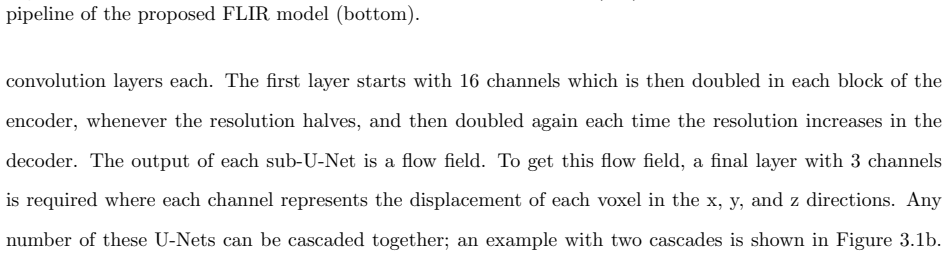
The load-bearing mechanism is a cascaded stack of compact U-Nets, each built so that convolutions run mostly at low resolution: the first layers use a 3x3x3 kernel with stride 2, and channel counts double as resolution halves, keeping full-resolution operations to a minimum. Each cascade outputs a displacement field $\phi$; the warped moving image $I_W$ is scored against the fixed image $I_F$ with the correlation-coefficient loss $L_{\text{corrcoef}} = 1 - \text{CorrCoef}[I_W, I_F]$, plus a total-variation smoothness term $L_{TV}$ that penalizes discontinuities in the field. The per-cascade similarity loss is back-propagated to every previous cascade, following the VTN training format. Two domain assumptions carry the design: same-acquisition SSFP volumes need no affine registration because patient motion would corrupt the images, and the left ventricle can be treated as a cylinder centered in the volume when converting Cartesian strain to radial, circumferential, and longitudinal strain. The companion strain calculator converts the flow field into finite strain via the deformation gradient and the right Cauchy-Green tensor, in both Lagrangian and Eulerian frames.
What would settle it
Register a batch of cardiac volume pairs to which small synthetic rigid offsets have been applied (for example 2-5 mm translations or 1-2 degree rotations between the fixed and moving volumes), then measure whether FLIR's LV dice and test-retest strain agreement fall below VoxelMorph's; a drop would confirm that the no-affine assumption, not the cascade design, is carrying part of the result.
Extended reading notes
Core claim
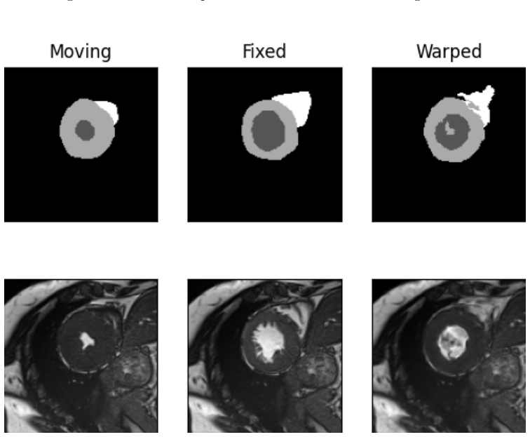
The central claim is that a single-pass, volumetric deep learning registration model can match the fidelity of state-of-the-art cardiac CMR registration without paying its inference cost, provided the architecture is engineered around a domain assumption: the two volumes come from one clinically usable SSFP acquisition, so no affine alignment is needed. FLIR is a cascade of U-Net sub-networks whose first convolutions use a stride of 2, so most operations run on downsampled volumes; each cascade predicts a small displacement field, the warped image is scored against the fixed image with the correlation-coefficient loss, and a total-variation term keeps the fields smooth. On the author's evaluation, 5xFLIR reaches a left-ventricle dice of 0.882 versus 0.835 for VoxelMorph on the same data, the largest fidelity gain of the compared methods, while 3xFLIR runs at about 1.7x VoxelMorph's CPU time compared with roughly 2.0x and 3.1x for RC-Net 2x and VR-Net relative to their own baselines. The same flow fields produce repeatable strain estimates, with average test-retest differences of 0.332 for radial, 0.344 for circumferential, and 0.032 for longitudinal strain, and the strain maps flag abnormal longitudinal strain in patients whose tissue is unhealthy, consistent with the physiology the thesis relies on. The stated goal of the thesis is that such a fast, optimized pipeline makes strain quantification from SSFP images a credible path to inferring unhealthy tissue without gadolinium contrast.
Load-bearing premise
The load-bearing assumption is that any two volumes registered by FLIR come from the same clinically usable SSFP acquisition, so that no affine alignment is needed; if patient motion, breath-hold shifts, or slice misregistration actually separate the volumes, the deformable field alone must absorb them and the reported speed-fidelity tradeoff would not transfer.
Editorial extensions
If this is right
- Fidelity scales with cascade count: one cascade runs faster than VoxelMorph but scores lower, while three and five cascades surpass VoxelMorph on left-ventricle, myocardium, and right-ventricle dice.
- The 3-cascade model reaches fidelity close to RC-Net and VR-Net at about 1.7x VoxelMorph's CPU time, versus roughly 2.0x and 3.1x for those competitors, making the accuracy-per-runtime tradeoff the model's central advantage.
- The deformations are stable enough for strain quantification: average test-retest differences are 0.332 (radial), 0.344 (circumferential), and 0.032 (longitudinal), so longitudinal strain in particular repeats tightly.
- The no-affine assumption means same-acquisition cardiac registration does not need alignment cascades, which is what lets the whole pipeline stay light.
- Fast inference on mid-level hardware makes cohort-scale processing feasible, which the thesis cites as a precondition for research and clinical adoption.
Reading between the lines
- The same architecture should transfer to other intra-acquisition cardiac time-point pairs, not only end-diastole to end-systole, because neither design decision depends on which two phases are being registered.
- The strain repeatability result is the strongest evidence for clinical utility, but the thesis does not compare its strain values against tagging-based or expert-measured ground truth; such a comparison would be the natural validation next step.
- The paper itself notes that peak strain readings are contaminated by large values in the blood pool; masking the myocardium with a segmentation before computing strain would likely tighten the radial and circumferential test-retest differences further.
- Other volumetric registration problems whose inputs come from a single acquisition — for example some fetal or fast abdominal sequences — could adopt the same 'drop the affine stage' shortcut and capture a similar speedup.
Editorial analysis
A structured set of objections, weighed in public.
Referee Report
Summary. The manuscript (an MSc thesis posted on arXiv) proposes FLIR, a cascaded U-Net for unsupervised 3D registration of cardiac cine SSFP images at end-diastole and end-systole, together with a strain calculator that converts the predicted deformation into radial, circumferential, and longitudinal strain. The method is evaluated against a VoxelMorph baseline trained on the same ACDC/UKBB data, compared with published RC-Net, VR-Net, and VoxelMorph numbers from Jia et al., and tested for strain repeatability on a 10-patient test-retest dataset. The central claim is that FLIR achieves registration fidelity comparable to state-of-the-art methods while taking a fraction of the inference time. The paper's own measurements, however, do not support this claim: the only FLIR variant faster than VoxelMorph is 1xFLIR, which has lower Dice, while all variants that match or exceed VoxelMorph Dice are slower. The cross-paper speed comparison also contains a units error and uses non-controlled conditions.
Significance. If the speed-fidelity claim were established, the work would be practically useful: a lightweight cascaded registration model with a documented strain calculator and test-retest strain repeatability could lower the computational barrier to large-scale CMR strain analysis. The paper has some genuine strengths: the VoxelMorph-Lite ablation cleanly isolates the runtime cost of full-resolution layers, the strain-calculator unit tests in Appendix A are a reproducible verification artifact, and the limitation discussion in Section 5.2 is candid. However, the central contribution—a model that is both faster than VoxelMorph and at least as accurate—is not present in the paper's own tables, and the external speed comparison relies on cross-dataset numbers with an order-of-magnitude unit error. As presented, the main claim is not established, although the underlying engineering (efficient cascaded U-Nets for cardiac registration) is a reasonable starting point for further work.
major comments (4)
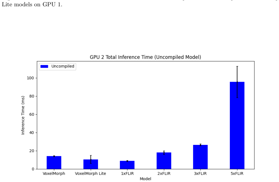
- [§4.2.2, Tables 4.1 and 4.2] The central claim that FLIR performs registration in a fraction of the time while maintaining fidelity is contradicted by the paper's own measurements. In Table 4.1, 1xFLIR has lower LV Dice than VoxelMorph (0.801 vs 0.835) and is the only FLIR variant that is faster on CPU (47.2 ms vs 76.2 ms in Table 4.2). Every configuration that matches or exceeds VoxelMorph LV Dice—2x, 3x, and 5x FLIR—is slower on CPU (98.9, 133.1, and 238.0 ms vs 76.2 ms), with the same ordering on GPU1 and GPU2. Thus the results contain no operating point that is simultaneously faster than VoxelMorph and at least as accurate; the abstract and the Section 5.1 conclusion overstate what Table 4.2 supports.
- [§4.2.2, Table 4.6 and Table 2.3] The speed comparison against RC-Net and VR-Net is invalid as reported. Table 4.6 labels the values from Jia et al. as milliseconds, but Table 2.3, the source of those exact numbers (5.97, 0.10, 11.95, 0.21, etc.), reports seconds. The table therefore misstates the external runtimes by three orders of magnitude. Additionally, the RC-Net/VR-Net Dice and timing figures in Tables 4.4–4.6 come from different datasets (UKBB, 3DCMR), different preprocessing, and different hardware than the FLIR evaluation, so the relative-slowdown comparisons in Section 4.2.3 and Figures 4.8–4.11 are not controlled comparisons. The conclusion that FLIR has smaller overhead than RC-Net/VR-Net rests on these inconsistent numbers.
- [§3.1 and §5.2] The decision to remove all affine registration stages is load-bearing for the method's speed, but the underlying assumption is untested. The thesis assumes that for any clinically usable SSFP sequence there should be no affine deformation (§3.1), yet ED and ES volumes from the same study can still be affected by breath-hold position changes, through-plane motion, and slice misregistration. Section 5.2 explicitly limits the evaluation to artifact-free data. Without an experiment or analysis showing that residual affine components are negligible for the target data, the claimed speed advantage may not transfer to realistic acquisitions, because the deformable network would have to compensate for global transformations itself.
- [§4.3.2, Tables 4.7–4.11] The test-retest strain analysis contains an unexplained duplication and a metrics problem. In Table 4.7, Patient 10 has identical test and retest values for all three strain components (0.763, 0.932, 0.818), giving zero differences in Table 4.8; for a genuine retest acquisition this is implausible and is not commented on. More broadly, the average test-retest differences reported in Table 4.9 for radial (0.332±0.236) and circumferential (0.344±0.356) strain are large relative to the myocardial strain values the method is intended to measure, and the authors show in Section 4.3.2 that the nominally peak strains are dominated by blood-pool artifacts unless the most negative values are selected (Tables 4.10–4.11). The claim that strain values are very consistent is therefore only supported for longitudinal strain and only after a post-hoc redefinition of the peak metric.
minor comments (5)
- [Table 4.3] The compiled GPU1 total timings (e.g., 0.0269 ms for VoxelMorph) are about two orders of magnitude smaller than the uncompiled values in Table 4.2 (2.193 ms for the same configuration); the measurement protocol, including whether data transfer and warping are included, should be stated.
- [Table 4.10] In Table 4.10, the Patient 3 retest value 0.441 is positive under a column labeled 'Peak Negative Radial Strain'; if this is a sign typo, it should be corrected.
- [Tables 4.7–4.11] The strain tables do not state the units of the reported strain values or define whether 'peak' means maximum absolute value, maximum positive value, or most negative value; this makes the values in Table 4.7 difficult to interpret.
- [Throughout] Several typographical errors remain, including 'Convolutoinal' in the list of abbreviations, 'start-of-the-art' in Section 2.7, and 'inevitability loss' in Section 3.1 (presumably 'invertibility loss').
- [§2.5.1] The abbreviation LDDMM is used in the discussion of Quicksilver without definition or citation; a brief definition and reference would help readers outside the registration subfield.
Circularity Check
No load-bearing circular reasoning; the registration and strain claims are empirically evaluated against external dice and test-retest consistency targets.
full rationale
The FLIR thesis contains no self-citations and no fitted parameter that is renamed as a prediction. The registration model is trained with an unsupervised correlation-coefficient plus total-variation loss, and its fidelity is then judged by dice overlap of warped segmentations; dice is not part of the training objective, so high dice is not guaranteed by construction. The loss weights and cascade count are ordinary model-selection choices, not quantities defined in terms of the reported outcomes. The strain repeatability evaluation compares strain values from paired test and retest acquisitions, which is an external consistency check rather than a definitional identity. The only substantive criticism suggested by the paper's own tables is that the speed-versus-fidelity claim lacks a single FLIR operating point that is both faster than VoxelMorph and at least as accurate; that is a correctness and experimental-design concern, not circularity. Under the specified criteria, the derivation chain is self-contained and no circular step can be exhibited with quotes.
Assumptions & free parameters
free parameters (2)
- Loss weights ω_corrcoef and ω_TV =
not reported
- Number of cascades (1, 2, 3, 5) =
5 used for strain experiments
assumptions (4)
- domain assumption Images from the same SSFP acquisition are already aligned, so affine registration is unnecessary.
- domain assumption The center of the left ventricle is at the center of the volume for cylindrical coordinate conversion.
- domain assumption Test-retest acquisitions of the same patient should yield nearly identical strain values, so the difference between them is a valid measure of accuracy.
- standard math Pseudo-inverse F^{-1} = (F^T F)^{-1} F^T is used to compute the Eulerian strain tensor.
Cite this review
Pith. "Pith review of A Deep Learning Based Method for Fast Registration of Cardiac Magnetic Resonance Images." pith.science (2026). https://pith.science/paper/ERMDZR3P
@misc{pith2026250619167,
  author       = {Pith},
  title        = {Pith review of: A Deep Learning Based Method for Fast Registration of Cardiac Magnetic Resonance Images},
  year         = {2026},
  howpublished = {\url{https://pith.science/paper/ERMDZR3P}},
  note         = {Machine review of arXiv:2506.19167}
}
read the original abstract
Image registration is used in many medical image analysis applications, such as tracking the motion of tissue in cardiac images, where cardiac kinematics can be an indicator of tissue health. Registration is a challenging problem for deep learning algorithms because ground truth transformations are not feasible to create, and because there are potentially multiple transformations that can produce images that appear correlated with the goal. Unsupervised methods have been proposed to learn to predict effective transformations, but these methods take significantly longer to predict than established baseline methods. For a deep learning method to see adoption in wider research and clinical settings, it should be designed to run in a reasonable time on common, mid-level hardware. Fast methods have been proposed for the task of image registration but often use patch-based methods which can affect registration accuracy for a highly dynamic organ such as the heart. In this thesis, a fast, volumetric registration model is proposed for the use of quantifying cardiac strain. The proposed Deep Learning Neural Network (DLNN) is designed to utilize an architecture that can compute convolutions incredibly efficiently, allowing the model to achieve registration fidelity similar to other state-of-the-art models while taking a fraction of the time to perform inference. The proposed fast and lightweight registration (FLIR) model is used to predict tissue motion which is then used to quantify the non-uniform strain experienced by the tissue. For acquisitions taken from the same patient at approximately the same time, it would be expected that strain values measured between the acquisitions would have very small differences. Using this metric, strain values computed using the FLIR method are shown to be very consistent.
Figures


Figures from the paper (43 more)








Reference graph
Works this paper leans on
-
[1]
Deep learning using rectied linear units (relu).arXiv preprint arXiv:1803.08375, 2018
Abien Fred Agarap. Deep learning using rectied linear units (relu).arXiv preprint arXiv:1803.08375, 2018
arXiv 2018
-
[2]
University of Salford (United Kingdom), 2017
Ali Majeed Hasan Al-Waeli.An automated system for the classication and segmentation of brain tumours in MRI images based on the modied grey level co-occurrence matrix. University of Salford (United Kingdom), 2017
work page 2017
-
[3]
Daniel C Alexander and James C Gee. Elastic matching of diusion tensor images.Computer Vision and Image Understanding, 77(2):233–250, 2000
work page 2000
-
[4]
John mccarthy: father of ai.IEEE Intelligent Systems, 17(5):84–85, 2002
Scott L Andresen. John mccarthy: father of ai.IEEE Intelligent Systems, 17(5):84–85, 2002
work page 2002
-
[5]
Cardiovascular magnetic resonance in the evaluation of heart failure.Heart, 93(8):985–992, 2007
Ravi G Assomull, Dudley J Pennell, and Sanjay K Prasad. Cardiovascular magnetic resonance in the evaluation of heart failure.Heart, 93(8):985–992, 2007
work page 2007
-
[6]
Types of machine learning algorithms.New advances in machine learning, 3:19–48, 2010
Taiwo Oladipupo Ayodele. Types of machine learning algorithms.New advances in machine learning, 3:19–48, 2010
work page 2010
-
[7]
Guha Balakrishnan, Amy Zhao, Mert R Sabuncu, John Guttag, and Adrian V Dalca. Voxelmorph: a learning framework for deformable medical image registration.IEEE transactions on medical imaging, 38(8):1788–1800, 2019
work page 2019
-
[8]
An empirical study on generalizations of the relu activation function
Chaity Banerjee, Tathagata Mukherjee, and Eduardo Pasiliao Jr. An empirical study on generalizations of the relu activation function. InProceedings of the 2019 ACM Southeast Conference, pages 164–167, 2019
work page 2019
Show all 73 references
-
[9]
How do i do it? speckle-tracking echocardiography.Indian heart journal, 65(1):117, 2013
Manish Bansal and Ravi R Kasliwal. How do i do it? speckle-tracking echocardiography.Indian heart journal, 65(1):117, 2013
2013
-
[10]
Some recent applications of reinforcement learning
Andrew G Barto, Philip S Thomas, and Richard S Sutton. Some recent applications of reinforcement learning. InProceedings of the Eighteenth Yale Workshop on Adaptive and Learning Systems, 2017. 73
2017
-
[11]
Olivier Bernard, Alain Lalande, Clement Zotti, Frederick Cervenansky, Xin Yang, Pheng-Ann Heng, Irem Cetin, Karim Lekadir, Oscar Camara, Miguel Angel Gonzalez Ballester, et al. Deep learning tech- niques for automatic mri cardiac multi-structures segmentation and diagnosis: is...
2018
-
[12]
Optimizing the dice score and jaccard index for medical image segmentation: Theory and practice
Jeroen Bertels, Tom Eelbode, Maxim Berman, Dirk Vandermeulen, Frederik Maes, Raf Bisschops, and Matthew B Blaschko. Optimizing the dice score and jaccard index for medical image segmentation: Theory and practice. InMedical Image Computing and Computer Assisted Intervention–MIC...
2019
-
[13]
From compressed-sensing to articial intelligence-based cardiac mri reconstruction.Frontiers in cardiovascular medicine, 7:17, 2020
Aur´ elien Bustin, Niccolo Fuin, Ren´ e M Botnar, and Claudia Prieto. From compressed-sensing to articial intelligence-based cardiac mri reconstruction.Frontiers in cardiovascular medicine, 7:17, 2020
2020
-
[14]
Detection of myocardial scar using t2* in a clinical setting.Journal of Cardiovascular Magnetic Reso- nance, 17:1–2, 2015
Britta Butzbach, Bernhard Schnackenburg, Christoph Jacoby, Malte Kelm, and Mirja Neizel-Wittke. Detection of myocardial scar using t2* in a clinical setting.Journal of Cardiovascular Magnetic Reso- nance, 17:1–2, 2015
2015
-
[15]
Steady-state free precession in nuclear magnetic resonance.Physical Review, 112(5):1693, 1958
HY Carr. Steady-state free precession in nuclear magnetic resonance.Physical Review, 112(5):1693, 1958
1958
-
[16]
Applying self- supervised learning to medicine: review of the state of the art and medical implementations
Alexander Chowdhury, Jacob Rosenthal, Jonathan Waring, and Renato Umeton. Applying self- supervised learning to medicine: review of the state of the art and medical implementations. In Informatics, volume 8, page 59. MDPI, 2021
2021
-
[17]
Patch-based discrete registration of clinical brain images
Adrian V Dalca, Andreea Bobu, Natalia S Rost, and Polina Golland. Patch-based discrete registration of clinical brain images. InPatch-Based Techniques in Medical Imaging: Second International Workshop, Patch-MI 2016, Held in Conjunction with MICCAI 2016, Athens, Greece, Octobe...
2016
-
[18]
Strain and strain rate imaging by echocardiography-basic concepts and clinical applicability.Current cardiology reviews, 5(2):133–148, 2009
Michael Dandel, Hans Lehmkuhl, Christoph Knosalla, Nino Suramelashvili, and Roland Hetzer. Strain and strain rate imaging by echocardiography-basic concepts and clinical applicability.Current cardiology reviews, 5(2):133–148, 2009
2009
-
[19]
The history of echocardiography.Ultrasound in medicine & biology, 30(12):1565–1644, 2004
Inge Edler and Kjell Lindstr¨ om. The history of echocardiography.Ultrasound in medicine & biology, 30(12):1565–1644, 2004
2004
-
[20]
Strain imaging echocardiography: what imag- ing cardiologists should know.Current cardiology reviews, 13(2):118–129, 2017
Dagmar F Hernandez-Suarez and Angel L´ opez-Candales. Strain imaging echocardiography: what imag- ing cardiologists should know.Current cardiology reviews, 13(2):118–129, 2017. 74
2017
-
[21]
Fda drug safety communication: Fda warns that gadolinium-based contrast agents (gbcas) are retained in the body; requires new class warning
US FDA. Fda drug safety communication: Fda warns that gadolinium-based contrast agents (gbcas) are retained in the body; requires new class warning. 2018, 2018
2018
-
[22]
Registration of 3-d intraoperative mr images of the brain using anite-element biomechanical model
Matthieu Ferrant, Arya Nabavi, Benoit Macq, Ferenc A Jolesz, Ron Kikinis, and Simon K Wareld. Registration of 3-d intraoperative mr images of the brain using anite-element biomechanical model. IEEE transactions on medical imaging, 20(12):1384–1397, 2001
2001
-
[23]
Recent advances in convolutional neural networks
Jiuxiang Gu, Zhenhua Wang, Jason Kuen, Lianyang Ma, Amir Shahroudy, Bing Shuai, Ting Liu, Xingxing Wang, Gang Wang, Jianfei Cai, et al. Recent advances in convolutional neural networks. Pattern recognition, 77:354–377, 2018
2018
-
[24]
Digital selection and analogue amplication coexist in a cortex-inspired silicon circuit.nature, 405(6789):947–951, 2000
Richard HR Hahnloser, Rahul Sarpeshkar, Misha A Mahowald, Rodney J Douglas, and H Sebastian Seung. Digital selection and analogue amplication coexist in a cortex-inspired silicon circuit.nature, 405(6789):947–951, 2000
2000
-
[25]
Machine learning and articial intelligence: denitions, applications, and future directions.Current reviews in musculoskeletal medicine, 13:69–76, 2020
J Matthew Helm, Andrew M Swiergosz, Heather S Haeberle, Jaret M Karnuta, Jonathan L Schaer, Viktor E Krebs, Andrew I Spitzer, and Prem N Ramkumar. Machine learning and articial intelligence: denitions, applications, and future directions.Current reviews in musculoskeletal m...
2020
-
[26]
A clinicians guide to tissue doppler imaging.Circulation, 113(10):e396–e398, 2006
Carolyn Y Ho and Scott D Solomon. A clinicians guide to tissue doppler imaging.Circulation, 113(10):e396–e398, 2006
2006
-
[27]
Mechanics of materials: Strainmechanics of slender structures: Boston university, 2013
Douglas Holmes. Mechanics of materials: Strainmechanics of slender structures: Boston university, 2013
2013
-
[28]
Unet 3+: A full-scale connected unet for medical image segmentation
Huimin Huang, Lanfen Lin, Ruofeng Tong, Hongjie Hu, Qiaowei Zhang, Yutaro Iwamoto, Xianhua Han, Yen-Wei Chen, and Jian Wu. Unet 3+: A full-scale connected unet for medical image segmentation. In ICASSP 2020-2020 IEEE international conference on acoustics, speech and signal pro...
2020
-
[29]
Myocardial tagging with mr imaging: overview of normal and pathologicndings.Radiographics, 32(5):1381–1398, 2012
Mi-Young Jeung, Philippe Germain, Pierre Croisille, Soraya El ghannudi, Catherine Roy, and Afshin Gangi. Myocardial tagging with mr imaging: overview of normal and pathologicndings.Radiographics, 32(5):1381–1398, 2012
2012
-
[30]
Learning a model-driven variational network for deformable image registration.IEEE Transactions on Medical Imaging, 41(1):199–212, 2021
Xi Jia, Alexander Thorley, Wei Chen, Huaqi Qiu, Linlin Shen, Iain B Styles, Hyung Jin Chang, Ales Leonardis, Antonio De Marvao, Declan P ORegan, et al. Learning a model-driven variational network for deformable image registration.IEEE Transactions on Medical Imaging, 41(1):19...
2021
-
[31]
Real-time volumetric echocar- diography: the technology and the possibilities.Echocardiography, 17(8):773–779, 2000
Joseph Kisslo, Bohdan Firek, Takahiro Ota, Duk Hyun Kang, Craig E Fleishman, George Stetten, Jennifer Li, Chikai J Ohazama, David Adams, Carolyn Landolfo, et al. Real-time volumetric echocar- diography: the technology and the possibilities.Echocardiography, 17(8):773–779, 2000
2000
-
[32]
Richard A Lerski, K Straughan, LR Schad, D Boyce, S Bl¨ uml, and I Zuna. Viii. mr image texture analysis—an approach to tissue characterization.Magnetic resonance imaging, 11(6):873–887, 1993
1993
-
[33]
A survey of convolutional neural networks: analysis, applications, and prospects.IEEE transactions on neural networks and learning systems, 2021
Zewen Li, Fan Liu, Wenjie Yang, Shouheng Peng, and Jun Zhou. A survey of convolutional neural networks: analysis, applications, and prospects.IEEE transactions on neural networks and learning systems, 2021
2021
-
[34]
Rectier nonlinearities improve neural network acoustic models
Andrew L Maas, Awni Y Hannun, Andrew Y Ng, et al. Rectier nonlinearities improve neural network acoustic models. InProc. icml, volume 30, page 3. Atlanta, GA, 2013
2013
-
[35]
Articial intelligence and texture analysis in cardiac imaging.Current cardiology reports, 22:1–9, 2020
Manoj Mannil, Matthias Eberhard, Jochen von Spiczak, Walter Heindel, Hatem Alkadhi, and Bettina Baessler. Articial intelligence and texture analysis in cardiac imaging.Current cardiology reports, 22:1–9, 2020
2020
-
[36]
Sophie Mavrogeni, Juerg Schwitter, Albert van Rossum, Robin Nijveldt, Antony Aletras, Genovefa Kolovou, Gerald Pohost, and Joao Lima. Cardiac magnetic resonance imaging in myocardial inamma- tion in autoimmune rheumatic diseases: an appraisal of the diagnostic strengths and l...
2018
-
[37]
Acute myocardial infarction
Oren J Mechanic, Michael Gavin, and Shamai A Grossman. Acute myocardial infarction. 2017
2017
-
[38]
Writing Group Members, Thomas Thom, Nancy Haase, Wayne Rosamond, Virginia J Howard, John Rumsfeld, Teri Manolio, Zhi-Jie Zheng, Katherine Flegal, Christopher ODonnell, et al. Heart disease and stroke statistics—2006 update: a report from the american heart association statist...
2006
-
[39]
Introduction to articial intelligence in medicine.Minimally Invasive Therapy & Allied Technologies, 28(2):73–81, 2019
Yoav Mintz and Ronit Brodie. Introduction to articial intelligence in medicine.Minimally Invasive Therapy & Allied Technologies, 28(2):73–81, 2019
2019
-
[40]
Denisa Muraru, Alice Niero, Hugo Rodriguez-Zanella, Diana Cherata, and Luigi Badano. Three- dimensional speckle-tracking echocardiography: benets and limitations of integrating myocardial me- chanics with three-dimensional imaging.Cardiovascular diagnosis and therapy, 8(1):101, 2018
2018
-
[41]
A primer for un- derstanding radiology articles about machine learning and deep learning.Diagnostic and Interventional Imaging, 101(12):765–770, 2020
Takeshi Nakaura, Toru Higaki, Kazuo Awai, Osamu Ikeda, and Yasuyuki Yamashita. A primer for un- derstanding radiology articles about machine learning and deep learning.Diagnostic and Interventional Imaging, 101(12):765–770, 2020. 76
2020
-
[42]
Ulf Neisius, Hossam El-Rewaidy, Selcuk Kucukseymen, Connie W Tsao, Jennifer Mancio, Shiro Nakamori, Warren J Manning, and Reza Nezafat. Texture signatures of native myocardial t1 as novel imaging markers for identication of hypertrophic cardiomyopathy patients without scar.Jo...
2020
-
[43]
Theory, simulation, and compensation of physiological motion artifacts in functional mri
Douglas C Noll and Walter Schneider. Theory, simulation, and compensation of physiological motion artifacts in functional mri. InProceedings of 1st International Conference on Image Processing, volume 3, pages 40–44. IEEE, 1994
1994
-
[44]
Medical image registration: A review.Computer methods in biomechanics and biomedical engineering, 17:73–93, 01 2014
Francisco Oliveira and Joao Tavares. Medical image registration: A review.Computer methods in biomechanics and biomedical engineering, 17:73–93, 01 2014
2014
-
[45]
Fisp—a new fast mri sequence.Electromedica, 54(1):15–18, 1986
A Oppelt, R Graumann, H Barfuss, H Fischer, W Hartl, W Schajor, et al. Fisp—a new fast mri sequence.Electromedica, 54(1):15–18, 1986
1986
-
[46]
A review of deep learning in image recognition
Myeongsuk Pak and Sanghoon Kim. A review of deep learning in image recognition. In2017 4th in- ternational conference on computer applications and information processing technology (CAIPT), pages 1–3. IEEE, 2017
2017
-
[47]
Unsupervised learning of goal spaces for intrinsically motivated goal exploration.arXiv preprint arXiv:1803.00781, 2018
Alexandre P´ er´ e, S´ ebastien Forestier, Olivier Sigaud, and Pierre-Yves Oudeyer. Unsupervised learning of goal spaces for intrinsically motivated goal exploration.arXiv preprint arXiv:1803.00781, 2018
2018 arXiv
-
[48]
Strain and strain rate echocardiography
Cristina Pislaru, Theodore P Abraham, and Marek Belohlavek. Strain and strain rate echocardiography. Current opinion in cardiology, 17(5):443–454, 2002
2002
-
[49]
Physics of mri: a primer.Journal of magnetic resonance imaging, 35(5):1038–1054, 2012
Donald B Plewes and Walter Kucharczyk. Physics of mri: a primer.Journal of magnetic resonance imaging, 35(5):1038–1054, 2012
2012
-
[50]
Cardiac magnetic resonance imaging: aone-stop-shop evaluation of myocardial dysfunction.Current opinion in cardiology, 17(6):663–670, 2002
Michael Poon, Valentin Fuster, and Zahi Fayad. Cardiac magnetic resonance imaging: aone-stop-shop evaluation of myocardial dysfunction.Current opinion in cardiology, 17(6):663–670, 2002
2002
-
[51]
End-to-end deep learning nonrigid motion-corrected reconstruction for highly accelerated free-breathing coronary mra.Magnetic Resonance in Medicine, 86(4):1983–1996, 2021
Haikun Qi, Reza Hajhosseiny, Gastao Cruz, Thomas Kuestner, Karl Kunze, Radhouene Neji, Ren´ e Botnar, and Claudia Prieto. End-to-end deep learning nonrigid motion-corrected reconstruction for highly accelerated free-breathing coronary mra.Magnetic Resonance in Medicine, 86(4):...
1983
-
[52]
3-d facial expression represen- tation using b-spline statistical shape model
Wei Quan, Bogdan Matuszewski, Lik Shark, and Djamel Ait-Boudaoud. 3-d facial expression represen- tation using b-spline statistical shape model. 2007. 77
2007
-
[53]
Evaluating the potential of chelation therapy to prevent and treat gadolinium deposition from mri contrast agents.Scientic reports, 8(1):4419, 2018
Julian A Rees, Gauthier J-P Deblonde, Dahlia D An, Camille Ansoborlo, Stacey S Gauny, and Rebecca J Abergel. Evaluating the potential of chelation therapy to prevent and treat gadolinium deposition from mri contrast agents.Scientic reports, 8(1):4419, 2018
2018
-
[54]
U-net: Convolutional networks for biomedical image segmentation
Olaf Ronneberger, Philipp Fischer, and Thomas Brox. U-net: Convolutional networks for biomedical image segmentation. InMedical Image Computing and Computer-Assisted Intervention–MICCAI 2015: 18th International Conference, Munich, Germany, October 5-9, 2015, Proceedings, Part I...
2015
-
[55]
Diagnostic and prognostic utility of cardiovascular magnetic resonance imaging in light-chain cardiac amyloidosis.The American journal of cardiology, 103(4):544– 549, 2009
Frederick L Ruberg, Evan Appelbaum, Ravin Davido, Al Ozono, Kraig V Kissinger, Caitlin Harrigan, Martha Skinner, and Warren J Manning. Diagnostic and prognostic utility of cardiovascular magnetic resonance imaging in light-chain cardiac amyloidosis.The American journal of ca...
2009
-
[56]
Magnetization evo- lution in balanced steady-state free precession with continuously moving table.Physics in Medicine & Biology, 52(8):N173, 2007
Randall B Staord, Mohammad Sabati, Houman Mahallati, and Richard Frayne. Magnetization evo- lution in balanced steady-state free precession with continuously moving table.Physics in Medicine & Biology, 52(8):N173, 2007
2007
-
[57]
Uk biobank: an open access resource for identifying the causes of a wide range of complex diseases of middle and old age.PLoS medicine, 12(3):e1001779, 2015
Cathie Sudlow, John Gallacher, Naomi Allen, Valerie Beral, Paul Burton, John Danesh, Paul Downey, Paul Elliott, Jane Green, Martin Landray, et al. Uk biobank: an open access resource for identifying the causes of a wide range of complex diseases of middle and old age.PLoS medi...
2015
-
[58]
Introduction: The challenge of reinforcement learning
Richard S Sutton. Introduction: The challenge of reinforcement learning. InReinforcement learning, pages 1–3. Springer, 1992
1992
-
[59]
Unsupervised machine learning for networking: Techniques, applications and research challenges.IEEE access, 7:65579–65615, 2019
Muhammad Usama, Junaid Qadir, Aunn Raza, Hunain Arif, Kok-Lim Alvin Yau, Yehia Elkhatib, Amir Hussain, and Ala Al-Fuqaha. Unsupervised machine learning for networking: Techniques, applications and research challenges.IEEE access, 7:65579–65615, 2019
2019
-
[60]
A survey on semi-supervised learning.Machine learning, 109(2):373–440, 2020
Jesper E Van Engelen and Holger H Hoos. A survey on semi-supervised learning.Machine learning, 109(2):373–440, 2020
2020
-
[61]
Principles of magnetic resonance imaging.Clinics in developmental medicine, pages 1–1, 1995
Theodore Villafana. Principles of magnetic resonance imaging.Clinics in developmental medicine, pages 1–1, 1995
1995
-
[62]
Signicance of late gadolinium en- hancement in cardiovascular magnetic resonance imaging (cmr).HERZ-MUNICH-, 32(2):129, 2007
M V ˆAhringer, Heiko Mahrholdt, Ali Yilmaz, and Udo Sechtem. Signicance of late gadolinium en- hancement in cardiovascular magnetic resonance imaging (cmr).HERZ-MUNICH-, 32(2):129, 2007. 78
2007
-
[63]
Volumetric magnetic resonance imaging: clinical applications and contributions to the understanding of temporal lobe epilepsy.Archives of neurology, 54(12):1521–1531, 1997
Craig Watson, Cliord R Jack, and Fernando Cendes. Volumetric magnetic resonance imaging: clinical applications and contributions to the understanding of temporal lobe epilepsy.Archives of neurology, 54(12):1521–1531, 1997
1997
-
[64]
Prog- nostic value of lge-cmr in hcm: a meta-analysis.JACC: Cardiovascular Imaging, 9(12):1392–1402, 2016
Zhen Weng, Jialu Yao, Raymond H Chan, Jun He, Xiangjun Yang, Yafeng Zhou, and Yang He. Prog- nostic value of lge-cmr in hcm: a meta-analysis.JACC: Cardiovascular Imaging, 9(12):1392–1402, 2016
2016
-
[65]
Hinrich B Winther, Christian Hundt, Bertil Schmidt, Christoph Czerner, Johann Bauersachs, Frank Wacker, and Jens Vogel-Claussen.ν-net: deep learning for generalized biventricular mass and function parameters using multicenter cardiac mri data.JACC: Cardiovascular Imaging, 11(7...
2018
-
[66]
Reluplex made more practical: Leaky relu
Jin Xu, Zishan Li, Bowen Du, Miaomiao Zhang, and Jing Liu. Reluplex made more practical: Leaky relu. In2020 IEEE Symposium on Computers and communications (ISCC), pages 1–7. IEEE, 2020
2020
-
[67]
Convolutional neural networks: an overview and application in radiology.Insights into imaging, 9:611–629, 2018
Rikiya Yamashita, Mizuho Nishio, Richard Kinh Gian Do, and Kaori Togashi. Convolutional neural networks: an overview and application in radiology.Insights into imaging, 9:611–629, 2018
2018
-
[68]
Quicksilver: Fast predictive image registration–a deep learning approach.NeuroImage, 158:378–396, 2017
Xiao Yang, Roland Kwitt, Martin Styner, and Marc Niethammer. Quicksilver: Fast predictive image registration–a deep learning approach.NeuroImage, 158:378–396, 2017
2017
-
[69]
Articial intelligence-based myocardial texture analysis in etiological dierentiation of left ventricular hypertrophy.Annals of Translational Medicine, 9(2), 2021
Fei Yu, Haibo Huang, Qihui Yu, Yuqing Ma, Qi Zhang, and Bo Zhang. Articial intelligence-based myocardial texture analysis in etiological dierentiation of left ventricular hypertrophy.Annals of Translational Medicine, 9(2), 2021
2021
-
[70]
Motion artifacts in mri: A complex problem with many partial solutions.Journal of Magnetic Resonance Imaging, 42(4):887–901, 2015
Maxim Zaitsev, Julian Maclaren, and Michael Herbst. Motion artifacts in mri: A complex problem with many partial solutions.Journal of Magnetic Resonance Imaging, 42(4):887–901, 2015
2015
-
[71]
Fast and robust iterative closest point.IEEE Transactions on Pattern Analysis and Machine Intelligence, 44(7):3450–3466, 2021
Juyong Zhang, Yuxin Yao, and Bailin Deng. Fast and robust iterative closest point.IEEE Transactions on Pattern Analysis and Machine Intelligence, 44(7):3450–3466, 2021
2021
-
[72]
Recursive cascaded networks for unsupervised medical image registration
Shengyu Zhao, Yue Dong, Eric I Chang, Yan Xu, et al. Recursive cascaded networks for unsupervised medical image registration. InProceedings of the IEEE/CVF international conference on computer vision, pages 10600–10610, 2019
2019
-
[73]
Unsupervised 3d end-to- end medical image registration with volume tweening network.IEEE journal of biomedical and health informatics, 24(5):1394–1404, 2019
Shengyu Zhao, Tingfung Lau, Ji Luo, I Eric, Chao Chang, and Yan Xu. Unsupervised 3d end-to- end medical image registration with volume tweening network.IEEE journal of biomedical and health informatics, 24(5):1394–1404, 2019. 79 Appendix A 3D Strain Calculator Unit Tests 80 Fi...
2019
Reviewed August 6, 2026 · model on record in the stance chip above.
Discussion (0). Continue with ORCID to comment.