REVIEW 1 major objections 5 minor 18 references
Proportional Sensitivity in Generative Adversarial Network (GAN)-Augmented Brain Tumor Classification Using Convolutional Neural Network
T0 review · 1 major / 5 minor · reviewed 2026-08-06 · deepseek-v4-flash
Pith's one-line read Adding 10% synthetic MRI images yields the best tumor-classification accuracy in this study's experiments.
desk verdict A useful GAN-ratio sweep undermined by inconsistent data-split arithmetic; the trend is plausible, but the headline numbers are not currently reproducible. read the letter →
The pith
A machine-rendered reading of the paper's core claim, the machinery that carries it, and where it could break.
The reading
What carries the argument
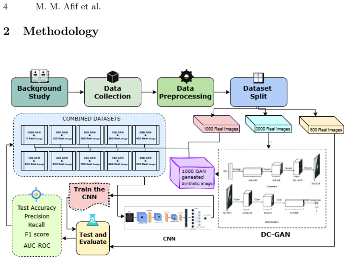
The machinery is a DCGAN (a deep convolutional generative adversarial network) trained over 1,000 epochs to synthesize 1,000 brain MRI images, which are then blended with real images into 11 training sets covering real-to-GAN ratios from 100:0 to 0:100. Each training set contains exactly 1,000 images, and a fixed custom CNN with three convolutional layers, ReLU activations, max-pooling, dropout, and a sigmoid output is trained on each and evaluated on a real test set. The ratio grid is the load-bearing device: it makes the performance decline visible as a function of synthetic share, and the evaluation metrics (accuracy, precision, recall, F1-score, AUC) tie that decline to the model's sensitivity to domain shift.
What would settle it
Compare the actual file names in the BR35H 'yes' and 'no' folders with the images assigned to GAN training, CNN training, and testing. If any real test image was also used to train or validate the GAN or the CNN, the reported accuracy numbers cannot be interpreted as generalization to unseen real scans.
Extended reading notes
Core claim
The paper's central claim is that CNN performance on real brain tumor MRI is sensitive to the proportion of GAN-generated images mixed into the training set. With 900 real and 100 synthetic images (10% GAN), the custom CNN reaches 95.20% accuracy, 94.31% precision, 96.20% recall, 95.25% F1-score, and an AUC of 0.98, slightly better than the 94.70% accuracy of fully real training. Every further increase in synthetic share lowers accuracy, down to 65.40% at 100% GAN, with AUC falling to 0.71. The authors interpret this as small-scale GAN augmentation acting as a regularizer, while heavy reliance on synthetic data introduces a domain shift that hurts generalization to real scans.
Load-bearing premise
The load-bearing premise is that the real images used for GAN training, CNN training, and testing are disjoint; the paper's stated counts (1,000 for CNN training, 3,000 for GAN training, and 500 or 1,000 test images) exceed the 3,000 images in the dataset, so overlap is possible and would make the test set not independent.
Editorial extensions
If this is right
- A small dose of GAN-generated images, around 10% of the training set, can slightly improve accuracy over all-real training, suggesting a mild regularizing effect.
- Increasing the synthetic share beyond roughly 50% leads to a pronounced drop in accuracy, recall, and F1-score, so synthetic data should not dominate training in this setting.
- Even at 100% synthetic training, the model retains some discriminative ability (65.4% accuracy, AUC 0.71), meaning GAN images carry real signal but not enough to replace real scans.
- The monotone decline across all metrics gives practitioners a concrete warning that domain shift from synthetic to real data grows with synthetic proportion.
- Future generators, such as diffusion models or domain-adapted GANs, would need to close the remaining realism gap before higher synthetic ratios become safe.
Reading between the lines
- Editorial inference: because the reported partition sizes (1,000 for CNN training, 3,000 for GAN training, and 500 or 1,000 for testing) do not sum to the 3,000 available images, the performance differences could partly reflect data leakage rather than the synthetic ratio itself.
- Editorial inference: the 0.5 percentage-point gap between the 10%-GAN and 0%-GAN conditions is small, so the claimed regularizing benefit may not survive repeated runs with different random seeds.
- Editorial inference: a direct testable extension would repeat the ratio grid on a fully disjoint split, with held-out real images never shown to the GAN or the CNN, to confirm whether the monotone decline in accuracy is robust.
- Editorial inference: the optimal synthetic share likely depends on GAN fidelity and dataset size; with higher-quality generators, the performance peak could shift toward larger synthetic proportions.
Signed reviews
Editorial analysis
A structured set of objections, weighed in public.
Referee Report
Summary. The paper trains a DCGAN on the BR35H brain MRI dataset, generates 1,000 synthetic images, and constructs 11 training sets with real-to-GAN ratios from 100:0 to 0:100. A custom CNN with three convolutional layers is trained on each mixture and evaluated on a real test set. The reported best performance is at 10% GAN (95.20% accuracy), with performance generally declining as the synthetic fraction increases, down to 65.40% at 100% GAN. The authors conclude that small amounts of GAN data can regularize training, while heavy reliance on synthetic data hurts generalization.
Significance. If the central trend holds, the paper addresses a practically important question for medical imaging: how much synthetic data should be mixed with real data when training classifiers. The systematic sweep over 11 ratios, the presentation of full metric tables, and the reproducible architecture description are strengths. However, the core quantitative claims currently rest on an internally inconsistent data-split description and on single-run results without uncertainty estimates. The finding that 10% GAN is better than 0% GAN is a 0.5 percentage-point difference that may be within run-to-run noise, so the paper's main recommendation is not yet established at the level of rigor expected for a journal publication.
major comments (1)
- [§2.2, §3.1] The quality of the synthetic images is not quantitatively assessed. The paper relies only on visual inspection and loss curves, but no Frechet Inception Distance, Inception Score, or similar measure is reported. Given that the central argument is about how synthetic data quality affects downstream CNN performance, a quantitative quality measure would substantially strengthen the interpretation of the ratio-dependent results. If such measures are unavailable, the authors should at least acknowledge this limitation explicitly in the Discussion.
minor comments (5)
- [§2.1] The sentence 'The dataset was partitioned into 1,000 images for further training, 3,000 for GAN training, and 500 reserved as an independent test set' is also internally unclear because 'further training' is not defined; please rewrite to give exact counts for each role and clarify that all subsets are disjoint.
- [Abstract and §2.2] The notation alternates between 'DCGAN' and 'DC-GAN'; please use one consistent abbreviation throughout.
- [Table 2] The column header 'Data Distribution' would be clearer as 'Real:GAN Ratio' or 'Percentage of GAN Data'.
- [Algorithm 1] Line 11 says 'Optionally evaluate on validation set and store metrics,' but the paper does not describe how validation is used (e.g., for early stopping or model selection). Please specify whether validation influenced the final model or was only used for reporting.
- [References] Several references are missing spaces or have typographical issues (e.g., 'Synthetic Data· Brain Tumor Classification' in the keywords line and the reference formatting in Ref. [8]). Please run a final copyediting pass.
Circularity Check
No significant circularity: the paper's central results are direct empirical measurements on a held-out test set, not predictions derived from fitted inputs or self-citations.
full rationale
The paper reports a controlled experiment: it trains a CNN on 11 real/GAN image ratios and evaluates each on the same real test set. The main claims—that accuracy declines with increasing GAN proportion and that the 10% GAN ratio gives the best observed accuracy—are read directly from Table 2's measured metrics. There is no fitted parameter that is later renamed as a prediction, no equation that defines the output in terms of the input, and no uniqueness theorem or load-bearing result imported from prior work. The Section 3.3 explanation that a small GAN fraction may act as a regularizer is post-hoc interpretation, not a derivation, so it does not create circularity. The paper cites two works involving overlapping authors (references [2] and [6]) but only as methodological references for evaluation metrics and related deep learning approaches; no central argument depends on them. The internal inconsistency in the reported data split (1,000 + 3,000 + 500 = 4,500 from 3,000 images, and a later statement of a 1,000-image test set) is a serious correctness and reproducibility concern, but it is an empirical validity issue, not circular reasoning. Accordingly, the circularity score is 0.
Assumptions & free parameters
free parameters (1)
- Optimal real-to-GAN ratio =
10% GAN (900 real, 100 GAN)
assumptions (4)
- domain assumption BR35H dataset labels (tumor vs. non-tumor) are correct.
- domain assumption GAN-generated images are assigned the class they were generated for.
- domain assumption The reported train, validation, GAN-training, and test partitions are disjoint.
- domain assumption The CNN and DCGAN hyperparameters are sufficient for the observed performance.
Cite this review
Pith. "Pith review of Proportional Sensitivity in Generative Adversarial Network (GAN)-Augmented Brain Tumor Classification Using Convolutional Neural Network." pith.science (2026). https://pith.science/paper/G6FAZGHY
@misc{pith2026250617165,
  author       = {Pith},
  title        = {Pith review of: Proportional Sensitivity in Generative Adversarial Network (GAN)-Augmented Brain Tumor Classification Using Convolutional Neural Network},
  year         = {2026},
  howpublished = {\url{https://pith.science/paper/G6FAZGHY}},
  note         = {Machine review of arXiv:2506.17165}
}
read the original abstract
Generative Adversarial Networks (GAN) have shown potential in expanding limited medical imaging datasets. This study explores how different ratios of GAN-generated and real brain tumor MRI images impact the performance of a CNN in classifying healthy vs. tumorous scans. A DCGAN was used to create synthetic images which were mixed with real ones at various ratios to train a custom CNN. The CNN was then evaluated on a separate real-world test set. Our results indicate that the model maintains high sensitivity and precision in tumor classification, even when trained predominantly on synthetic data. When only a small portion of GAN data was added, such as 900 real images and 100 GAN images, the model achieved excellent performance, with test accuracy reaching 95.2%, and precision, recall, and F1-score all exceeding 95%. However, as the proportion of GAN images increased further, performance gradually declined. This study suggests that while GANs are useful for augmenting limited datasets especially when real data is scarce, too much synthetic data can introduce artifacts that affect the model's ability to generalize to real world cases.
Figures


Figures from the paper (3 more)


Reference graph
Works this paper leans on
-
[1]
Diagnostics14(16), 1714 (2024)
Aamir, M., Namoun, A., Munir, S., Aljohani, N., Alanazi, M.H., Alsahafi, Y., Alotibi, F.: Brain tumor detection and classification using an optimized convolu- tional neural network. Diagnostics14(16), 1714 (2024)
work page 2024
-
[2]
Ahmmed, M.M., Noman, A.A., Afif, M.M., Kabir, K.M.T., Rahman, M.M., Mah- mud, M.: A model-mediated stacked ensemble approach for depression prediction among professionals (2025),https://arxiv.org/abs/2506.14459 12 M. M. Afif et al
work page Pith review arXiv 2025
-
[3]
Scientific Data 11, 259 (2024)
Akbar, M.U., Larsson, M., Blystad, I., Eklund, A.: Brain tumor segmentation using synthetic mr images - a comparison of gans and diffusion models. Scientific Data 11, 259 (2024)
work page 2024
-
[4]
Springer (2006)
Bishop, C.M.: Pattern Recognition and Machine Learning. Springer (2006)
2006
-
[5]
Computers in Biology and Medicine111, 103345 (2019)
Deepak, S., Ameer, P.: Brain tumor classification using deep cnn features via trans- fer learning. Computers in Biology and Medicine111, 103345 (2019)
work page 2019
-
[6]
Eity, S.N., Afif, M.M., Fairooz, T., Ahmmed, M.M., Miah, M.S.U.: Dgg-xnet: A hybrid deep learning framework for multi-class brain disease classification with explainable ai (2025),https://arxiv.org/abs/2506.14367
work page Pith review arXiv 2025
-
[7]
Advances in neural infor- mation processing systems27 (2014)
Goodfellow, I., Pouget-Abadie, J., Mirza, M., Xu, B., Warde-Farley, D., Ozair, S., Courville, A., Bengio, Y.: Generative adversarial nets. Advances in neural infor- mation processing systems27 (2014)
2014
-
[8]
HASHIRA: Br35h :: Brain tumor detection 2020 dataset (jan 2025), https://universe.roboflow.com/hashira-fhxpj/br35h-:: -brain-tumor-detection-2020, visited on 2025-06-16
work page 2020
Show all 18 references
-
[9]
International Agency for Research on Cancer (IARC): Globocan 2020: Estimated cancer incidence, mortality and prevalence worldwide in 2020 (2020), global Cancer Observatory
2020
-
[10]
Neural Computing and Applications37, 8731–8758 (2025)
Karpakam, S., Kumareshan, N.: Enhanced brain tumor detection and classification using a deep image recognition generative adversarial network (dir-gan). Neural Computing and Applications37, 8731–8758 (2025)
2025
-
[11]
Proceedings of the IEEE86(11), 2278–2324 (1998)
LeCun, Y., Bottou, L., Bengio, Y., Haffner, P.: Gradient-based learning applied to document recognition. Proceedings of the IEEE86(11), 2278–2324 (1998)
1998
-
[12]
Multimedia Tools and Appli- cations 84(5), 2261–2292 (2025)
Mijwil, M.M.: Smart architectures: computerized classification of brain tumors from mri images utilizing deep learning approaches. Multimedia Tools and Appli- cations 84(5), 2261–2292 (2025)
2025
-
[13]
Scientific Reports 12(1), 9141 (2022)
Mukherkjee, D., Saha, P., Kaplun, D., Sinitca, A., Sarkar, R.: Brain tumor im- age generation using an aggregation of gan models with style transfer. Scientific Reports 12(1), 9141 (2022)
2022
-
[14]
Heliyon10(20), e38997 (2024)
Nazir, M.I., Akter, A., Wadud, M.A.H., Uddin, M.A.: Utilizing customized cnn for brain tumor prediction with explainable ai. Heliyon10(20), e38997 (2024)
2024
-
[15]
Biomedical Sig- nal Processing and Control74, 103537 (2022)
Neelima, G., Chigurukota, D.R., Maram, B., Girirajan, B.: Optimal deepmrseg based tumor segmentation with gan for brain tumor classification. Biomedical Sig- nal Processing and Control74, 103537 (2022)
2022
-
[16]
arXiv preprint arXiv:1511.06434 (2015)
Radford,A.,Metz,L.,Chintala,S.:Unsupervisedrepresentationlearningwithdeep convolutional generative adversarial networks. arXiv preprint arXiv:1511.06434 (2015)
2015 arXiv
-
[17]
BMC Medical Informatics and Decision Making23(1), 16 (2023)
Saeedi, S., Rezayi, S., Keshavarz, H., Niakan Kalhori, S.R.: Mri-based brain tumor detection using convolutional deep learning methods and chosen machine learning techniques. BMC Medical Informatics and Decision Making23(1), 16 (2023)
2023
-
[18]
World Health Organization: Cancer (2023), fact sheet, Updated January 2023
2023
Reviewed August 6, 2026 · model on record in the stance chip above.
Discussion (0). Continue with ORCID to comment.