REVIEW 2 major objections 1 minor 36 references
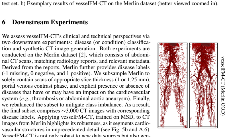
vesselFM-CT is the first model to segment all blood vessels in 3D CT images from major arteries down to tiny mesenteric vessels.
Reviewed by Pith at T0; open to challenge. T0 means a machine referee read the full paper against a public rubric. the ladder, T0–T4 →
T0 review · grok-4.3
2026-06-27 17:17 UTC pith:K3RBUG7S
load-bearing objection The paper reframes vessel segmentation as a full-system task but the abstract supplies no metrics or ablations to show the iterative training and TubeLoss actually deliver robustness across vessel scales. the 2 major comments →
vesselFM-CT: Segmenting All Blood Vessels in CT Images for System-Level Cardiovascular Analysis
The pith
A machine-rendered reading of the paper's core claim, the machinery that carries it, and where it could break.
Core claim
We introduce vesselFM-CT, the first model capable of robustly segmenting all blood vessels in 3D CT images, ranging from the largest components of the cardiovascular system to even minuscule mesenteric vessels. VesselFM-CT is trained via an iterative, multi-step process and optimizes our proposed TubeLoss loss function, effectively addressing the inherent heterogeneity of the cardiovascular system.
What carries the argument
vesselFM-CT model trained iteratively with TubeLoss to segment vessels across wide ranges of size, topology, and local anatomy in a single pass.
Load-bearing premise
The iterative training process and TubeLoss together cover every vessel size, branching pattern, and background variation without vessel-type-specific models or manual exclusions.
What would settle it
A held-out CT volume containing an extreme mix of large arteries and sub-millimeter mesenteric vessels where the model misses more than a small fraction of the total vessel length.
If this is right
- The model outperforms all tested baselines on full-vessel segmentation.
- It produces automated, precise extraction of the entire cardiovascular system from CT images.
- The output supports downstream automated disease classification.
- The segmented vessels enable generation of synthetic CT images.
Where Pith is reading between the lines
- Full-vessel maps could support population-scale studies tracking how vascular topology changes with age or disease across the body.
- The same training strategy might transfer to MR angiography or ultrasound volumes without new loss functions.
- Extracted vessel graphs could feed directly into fluid-dynamics simulations of systemic blood flow.
Editorial analysis
A structured set of objections, weighed in public.
Referee Report
Summary. The paper introduces vesselFM-CT as the first model for robustly segmenting all blood vessels in 3D CT images, from aorta-scale to minuscule mesenteric vessels. It uses an iterative multi-step training process and a proposed TubeLoss function to handle vessel heterogeneity in radius, topology, and anatomical backgrounds, claiming outperformance over baselines and enabling system-level cardiovascular analysis, automated disease classification, and synthetic CT generation.
Significance. If the robustness and outperformance claims hold with supporting evidence, the work would enable previously unavailable system-level vascular analysis in CT, with potential clinical impact on diagnostics and research into cardiovascular physiology. The shift from narrow-scope vessel segmentation to comprehensive coverage addresses a recognized limitation in the field.
major comments (2)
- [Abstract] Abstract: The claim that vesselFM-CT 'outperforms all baselines' and 'robustly' segments vessels across all scales lacks any supporting quantitative results, ablation studies, radius-stratified metrics, or dataset descriptions. No evidence is provided to verify that the iterative training and TubeLoss close the generalization gap noted in prior literature.
- [Abstract] Abstract: The description of the multi-step iterative process and TubeLoss does not specify mechanisms for handling extreme class imbalance, false-positive rates on tiny vessels, or variations in background without vessel-type-specific adaptations or post-hoc exclusions, leaving the central 'all vessels' robustness claim unverified.
minor comments (1)
- [Abstract] The abstract states the training approach but supplies no details on implementation, loss formulation, or evaluation protocol, which are required to assess the contribution.
Simulated Author's Rebuttal
We thank the referee for the constructive comments on the abstract. We agree that the abstract should better support its claims with evidence and will revise it accordingly while preserving its concise nature.
read point-by-point responses
-
Referee: [Abstract] Abstract: The claim that vesselFM-CT 'outperforms all baselines' and 'robustly' segments vessels across all scales lacks any supporting quantitative results, ablation studies, radius-stratified metrics, or dataset descriptions. No evidence is provided to verify that the iterative training and TubeLoss close the generalization gap noted in prior literature.
Authors: We acknowledge that the abstract, as currently written, does not include quantitative results or explicit references to supporting analyses. The manuscript contains these elements in its experimental evaluation, including baseline comparisons, ablations on the iterative training and TubeLoss, and dataset details. We will revise the abstract to incorporate key quantitative findings (such as overall performance metrics) and brief mentions of the datasets and ablation outcomes to substantiate the outperformance and robustness claims. revision: yes
-
Referee: [Abstract] Abstract: The description of the multi-step iterative process and TubeLoss does not specify mechanisms for handling extreme class imbalance, false-positive rates on tiny vessels, or variations in background without vessel-type-specific adaptations or post-hoc exclusions, leaving the central 'all vessels' robustness claim unverified.
Authors: The abstract provides only a high-level summary of the approach. The full manuscript details how TubeLoss addresses class imbalance and tubular structures while the iterative process mitigates background variations. We agree that the abstract could more explicitly reference these mechanisms and will revise it to include a concise statement on how these components contribute to handling small vessels and background heterogeneity without type-specific adaptations. revision: yes
Circularity Check
No derivation chain; empirical model proposal with no self-referential reductions
full rationale
The manuscript proposes vesselFM-CT as an empirical segmentation model trained via an iterative multi-step process and TubeLoss. No equations, first-principles derivations, or predictions appear in the abstract or described claims. The central assertion (robust segmentation of all vessels) is presented as an empirical outcome of the training procedure rather than a derived result that reduces to its own inputs by construction. No self-citations, fitted parameters renamed as predictions, or ansatzes are load-bearing in a mathematical sense. This is a standard non-finding for an applied ML paper without a formal derivation chain.
Axiom & Free-Parameter Ledger
read the original abstract
The vascular network in the human body is characterized by blood vessels exhibiting drastic structural variations in radius, length, topological properties, and branching patterns. This heterogeneity, together with location-specific anatomical background variations, poses a significant challenge for robust, large-scale analysis of the entire cardiovascular system. As a result, most research has focused on narrow, isolated segments of the vascular network. While such targeted studies provide valuable insights, they inherently limit the ability to assess the systemic health and functional integrity of the vascular network as a whole. In this work, we aim to bridge this gap to advance both clinical diagnostics and our fundamental understanding of vascular physiology. We propose the task of segmenting all vessels in CT images, ranging from the largest components of the cardiovascular system to even minuscule mesenteric vessels. To this end, we introduce vesselFM-CT, the first model capable of robustly segmenting all blood vessels in 3D CT images. VesselFM-CT is trained via an iterative, multi-step process and optimizes our proposed TubeLoss loss function, effectively addressing the inherent heterogeneity of the cardiovascular system. We demonstrate that vesselFM-CT outperforms all baselines and enables automated, precise extraction of the cardiovascular system from CT images, thereby unlocking a wide range of clinical and technical perspectives, including automated disease classification and synthetic CT image generation.
Figures




Reference graph
Works this paper leans on
-
[1]
The medical segmentation decathlon.Nature communications, 13(1):4128, 2022
Michela Antonelli, Annika Reinke, Spyridon Bakas, Keyvan Farahani, Annette Kopp-Schneider, Bennett A Landman, Geert Litjens, Bjoern Menze, Olaf Ronneberger, Ronald M Summers, et al. The medical segmentation decathlon.Nature communications, 13(1):4128, 2022
2022
-
[2]
Merlin: a computed tomography vision–language foundation model and dataset.Nature, pages 1–11, 2026
Louis Blankemeier, Ashwin Kumar, Joseph Paul Cohen, Jiaming Liu, Longchao Liu, Dave Van Veen, Syed Jamal Safdar Gardezi, Hongkun Yu, Magdalini Paschali, Zhihong Chen, et al. Merlin: a computed tomography vision–language foundation model and dataset.Nature, pages 1–11, 2026
2026
-
[3]
MONAI: An open-source framework for deep learning in healthcare
M Jorge Cardoso, Wenqi Li, Richard Brown, Nic Ma, Eric Kerfoot, Yiheng Wang, Benjamin Murrey, Andriy Myronenko, Can Zhao, Dong Yang, et al. Monai: An open-source framework for deep learning in healthcare.arXiv preprint arXiv:2211.02701, 2022
work page internal anchor Pith review Pith/arXiv arXiv 2022
-
[4]
Deep learning-driven pulmonary artery and vein seg- mentation reveals demography-associated vasculature anatomical differences.Nature Communications, 16(1):2262, 2025
Yuetan Chu, Gongning Luo, Longxi Zhou, Shaodong Cao, Guolin Ma, Xianglin Meng, Juexiao Zhou, Changchun Yang, Dexuan Xie, Dan Mu, et al. Deep learning-driven pulmonary artery and vein seg- mentation reveals demography-associated vasculature anatomical differences.Nature Communications, 16(1):2262, 2025
2025
-
[5]
Maisi: Medical ai for synthetic imaging
Pengfei Guo, Can Zhao, Dong Yang, Ziyue Xu, Vishwesh Nath, Yucheng Tang, Benjamin Simon, Mason Belue, Stephanie Harmon, Baris Turkbey, et al. Maisi: Medical ai for synthetic imaging. In2025 IEEE/CVF Winter Conference on Applications of Computer Vision (WACV), pages 4430–4441. IEEE, 2025
2025
-
[6]
Generalist foundation models from a multimodal dataset for 3d computed tomography.Nature Biomedical Engineering, pages 1–19, 2026
Ibrahim Ethem Hamamci, Sezgin Er, Chenyu Wang, Furkan Almas, Ayse Gulnihan Simsek, Sevval Nil Esirgun, Irem Dogan, Omer Faruk Durugol, Benjamin Hou, Suprosanna Shit, et al. Generalist foundation models from a multimodal dataset for 3d computed tomography.Nature Biomedical Engineering, pages 1–19, 2026
2026
-
[7]
Mri-derived quantification of hepatic vessel-to-volume ratios in chronic liver disease using a deep learning approach.European Radiology Experimental, 9(1):75, 2025
Alexander Herold, Daniel Sobotka, Lucian Beer, Nina Bastati, Sarah Poetter-Lang, Michael Weber, Thomas Reiberger, Mattias Mandorfer, Georg Semmler, Benedikt Simbrunner, et al. Mri-derived quantification of hepatic vessel-to-volume ratios in chronic liver disease using a deep learning approach.European Radiology Experimental, 9(1):75, 2025
2025
-
[8]
Automatic segmentation of cardiovascular structures on chest ct data sets: an update of the totalsegmentator.European journal of radiology, 185:112006, 2025
Daniel Hinck, Martin Segeroth, Jules Miazza, Denis Berdajs, Jens Bremerich, Jakob Wasserthal, and Maurice Pradella. Automatic segmentation of cardiovascular structures on chest ct data sets: an update of the totalsegmentator.European journal of radiology, 185:112006, 2025
2025
-
[9]
nnu-net revisited: A call for rigorous validation in 3d medical image segmentation
Fabian Isensee, Tassilo Wald, Constantin Ulrich, Michael Baumgartner, Saikat Roy, Klaus Maier-Hein, and Paul F Jaeger. nnu-net revisited: A call for rigorous validation in 3d medical image segmentation. InInternational Conference on Medical Image Computing and Computer-Assisted Intervention, pages 488–498. Springer, 2024
2024
-
[10]
Adam: A Method for Stochastic Optimization
Diederik P Kingma and Jimmy Ba. Adam: A method for stochastic optimization.arXiv preprint arXiv:1412.6980, 2014
work page internal anchor Pith review Pith/arXiv arXiv 2014
-
[11]
Skeleton recall loss for connec- tivity conserving and resource efficient segmentation of thin tubular structures
Yannick Kirchhoff, Maximilian R Rokuss, Saikat Roy, Balint Kovacs, Constantin Ulrich, Tassilo Wald, Maximilian Zenk, Philipp V ollmuth, Jens Kleesiek, Fabian Isensee, et al. Skeleton recall loss for connec- tivity conserving and resource efficient segmentation of thin tubular structures. InEuropean Conference on Computer Vision, pages 218–234. Springer, 2024
2024
-
[12]
Automatic coronary artery segmentation and diagnosis of stenosis by deep learning based on computed tomographic coronary angiography.European Radiology, 32(9):6037–6045, 2022
Yiming Li, Yu Wu, Jingjing He, Weili Jiang, Jianyong Wang, Yong Peng, Yuheng Jia, Tianyuan Xiong, Kaiyu Jia, Zhang Yi, et al. Automatic coronary artery segmentation and diagnosis of stenosis by deep learning based on computed tomographic coronary angiography.European Radiology, 32(9):6037–6045, 2022
2022
-
[13]
Emanuele Muscogiuri, Marly van Assen, Giovanni Tessarin, Alexander C Razavi, Max Schoebinger, Michael Wels, Mehmet Akif Gulsun, Puneet Sharma, George SK Fung, and Carlo N De Cecco. Clinical validation of a deep learning algorithm for automated coronary artery disease detection and classification using a heterogeneous multivendor coronary computed tomograp...
2025
-
[14]
Global Burden of Cardiovascular Diseases and Risks 2023 Collaborators. Global, regional, and national burden of cardiovascular diseases and risk factors in 204 countries and territories, 1990-2023.Journal of the American College of Cardiology, 86(22):2167–2243, 2025
2023
-
[15]
Automated lung vessel segmentation reveals blood vessel volume redistribution in viral pneumonia.European journal of radiology, 150:110259, 2022
Julien Poletti, Michael Bach, Shan Yang, Raphael Sexauer, Bram Stieltjes, David C Rotzinger, Jens Bremerich, Alexander Walter Sauter, and Thomas Weikert. Automated lung vessel segmentation reveals blood vessel volume redistribution in viral pneumonia.European journal of radiology, 150:110259, 2022
2022
-
[16]
Sparse Representation Learning for Vessels
Chinmay Prabhakar, Bastian Wittmann, Paul Büschl, Hongwei Bran Li, Bjoern Menze, and Suprosanna Shit. Sparse representation learning for vessels.arXiv preprint arXiv:2605.01382, 2026
work page internal anchor Pith review Pith/arXiv arXiv 2026
-
[17]
V oxtell: Free-text promptable universal 3d medical image segmentation, 2025
Maximilian Rokuss, Moritz Langenberg, Yannick Kirchhoff, Fabian Isensee, Benjamin Hamm, Constantin Ulrich, Sebastian Regnery, Lukas Bauer, Efthimios Katsigiannopulos, Tobias Norajitra, and Klaus Maier- Hein. V oxtell: Free-text promptable universal 3d medical image segmentation, 2025
2025
-
[18]
Vlahavas
Konstantinos Sechidis, Grigorios Tsoumakas, and Ioannis P. Vlahavas. On the stratification of multi-label data. InECML/PKDD, 2011
2011
-
[19]
Rapid model- guided design of organ-scale synthetic vasculature for biomanufacturing.Science, 388(6752):1198–1204, 2025
Zachary A Sexton, Dominic Rütsche, Jessica E Herrmann, Andrew R Hudson, Soham Sinha, Jianyi Du, Daniel J Shiwarski, Anastasiia Masaltseva, Fredrik Samdal Solberg, Jonathan Pham, et al. Rapid model- guided design of organ-scale synthetic vasculature for biomanufacturing.Science, 388(6752):1198–1204, 2025
2025
-
[20]
Sexton, Dominic Rütsche, Jessica E
Zachary A. Sexton, Dominic Rütsche, Jessica E. Herrmann, Andrew R. Hudson, Soham Sinha, Jianyi Du, Daniel J. Shiwarski, Anastasiia Masaltseva, Fredrik Samdal Solberg, Jonathan Pham, Jason M. Szafron, Sean M. Wu, Adam W. Feinberg, Mark A. Kylar-Scott, and Alison L. Marsden. svVascularize: Initial Release of SVV Package, April 2025. Zenodo.https://doi.org/1...
-
[21]
Centerline boundary dice loss for vascular segmentation
Pengcheng Shi, Jiesi Hu, Yanwu Yang, Zilve Gao, Wei Liu, and Ting Ma. Centerline boundary dice loss for vascular segmentation. InInternational Conference on Medical Image Computing and Computer-Assisted Intervention, pages 46–56. Springer, 2024
2024
-
[22]
cldice-a novel topology-preserving loss function for tubular structure segmentation
Suprosanna Shit, Johannes C Paetzold, Anjany Sekuboyina, Ivan Ezhov, Alexander Unger, Andrey Zhylka, Josien PW Pluim, Ulrich Bauer, and Bjoern H Menze. cldice-a novel topology-preserving loss function for tubular structure segmentation. InProceedings of the IEEE/CVF Conference on Computer Vision and Pattern Recognition, pages 16560–16569, 2021
2021
-
[23]
Automated segmentation and quantification of the healthy and diseased aorta in ct angiographies using a dedicated deep learning approach.European Radiology, 32(1):690–701, 2022
Malte Maria Sieren, Cornelia Widmann, Nick Weiss, Jan Hendrik Moltz, Florian Link, Franz Wegner, Erik Stahlberg, Marco Horn, Thekla Helene Oecherting, Jan Peter Goltz, et al. Automated segmentation and quantification of the healthy and diseased aorta in ct angiographies using a dedicated deep learning approach.European Radiology, 32(1):690–701, 2022
2022
-
[24]
Amber L Simpson, Michela Antonelli, Spyridon Bakas, Michel Bilello, Keyvan Farahani, Bram Van Gin- neken, Annette Kopp-Schneider, Bennett A Landman, Geert Litjens, Bjoern Menze, et al. A large annotated medical image dataset for the development and evaluation of segmentation algorithms.arXiv preprint arXiv:1902.09063, 2019
work page internal anchor Pith review Pith/arXiv arXiv 1902
-
[25]
Segmentation of 71 anatomical structures necessary for the evaluation of guideline-conforming clinical target volumes in head and neck cancers
Alexandra Walter, Philipp Hoegen-Saßmannshausen, Goran Stanic, Joao Pedro Rodrigues, Sebastian Adeberg, Oliver Jäkel, Martin Frank, and Kristina Giske. Segmentation of 71 anatomical structures necessary for the evaluation of guideline-conforming clinical target volumes in head and neck cancers. Cancers, 16(2), 2024
2024
-
[26]
Totalsegmentator: robust segmentation of 104 anatomic structures in ct images.Radiology: Artificial Intelligence, 5(5):e230024, 2023
Jakob Wasserthal, Hanns-Christian Breit, Manfred T Meyer, Maurice Pradella, Daniel Hinck, Alexander W Sauter, Tobias Heye, Daniel T Boll, Joshy Cyriac, Shan Yang, et al. Totalsegmentator: robust segmentation of 104 anatomic structures in ct images.Radiology: Artificial Intelligence, 5(5):e230024, 2023
2023
-
[27]
Knowledge-augmented deep learning for segmenting and detecting cerebral aneurysms with ct angiography: a multicenter study.Radiology, 312(2):e233197, 2024
Jianyong Wei, Xinyu Song, Xiaoer Wei, Zhiwen Yang, Lisong Dai, Mengfei Wang, Zheng Sun, Yidong Jin, Chune Ma, Chunhong Hu, et al. Knowledge-augmented deep learning for segmenting and detecting cerebral aneurysms with ct angiography: a multicenter study.Radiology, 312(2):e233197, 2024
2024
-
[28]
Simulation-based segmentation of blood vessels in cerebral 3d octa images
Bastian Wittmann, Lukas Glandorf, Johannes C Paetzold, Tamaz Amiranashvili, Thomas Wälchli, Daniel Razansky, and Bjoern Menze. Simulation-based segmentation of blood vessels in cerebral 3d octa images. InInternational Conference on Medical Image Computing and Computer-Assisted Intervention, pages 645–655. Springer, 2024
2024
-
[29]
vesselFM: A Foundation Model for Universal 3D Blood Vessel Segmentation
Bastian Wittmann, Yannick Wattenberg, Tamaz Amiranashvili, Suprosanna Shit, and Bjoern Menze. vesselFM: A Foundation Model for Universal 3D Blood Vessel Segmentation. InProceedings of the IEEE/CVF Conference on Computer Vision and Pattern Recognition, pages 20874–20884, 2025. 11
2025
-
[30]
Cardiovascular Diseases (CVDs) Fact Sheet
World Health Organization (WHO). Cardiovascular Diseases (CVDs) Fact Sheet. https://www.who. int/en/news-room/fact-sheets/detail/cardiovascular-diseases-(cvds) , 2025. Accessed: 24.02.2026
2025
-
[31]
Cads: A comprehensive anatomical dataset and segmentation for whole-body anatomy in computed tomography, 2025
Murong Xu, Tamaz Amiranashvili, Fernando Navarro, et al. Cads: A comprehensive anatomical dataset and segmentation for whole-body anatomy in computed tomography, 2025
2025
-
[32]
Benchmarking the CoW with the TopCoW Challenge: Topology-Aware Anatomical Segmentation of the Circle of Willis for CTA and MRA, 2025
Kaiyuan Yang, Fabio Musio, Yihui Ma, et al. Benchmarking the CoW with the TopCoW Challenge: Topology-Aware Anatomical Segmentation of the Circle of Willis for CTA and MRA, 2025
2025
-
[33]
Yushkevich, Joseph Piven, Heather Cody Hazlett, Rachel Gimpel Smith, Sean Ho, James C
Paul A. Yushkevich, Joseph Piven, Heather Cody Hazlett, Rachel Gimpel Smith, Sean Ho, James C. Gee, and Guido Gerig. User-guided 3D active contour segmentation of anatomical structures: Significantly improved efficiency and reliability.Neuroimage, 31(3):1116–1128, 2006.www.itksnap.org
2006
-
[34]
Adding conditional control to text-to-image diffusion models
Lvmin Zhang, Anyi Rao, and Maneesh Agrawala. Adding conditional control to text-to-image diffusion models. InProceedings of the IEEE/CVF international conference on computer vision, pages 3836–3847, 2023
2023
-
[35]
A foundation model for joint segmentation, detection and recognition of biomedical objects across nine modalities.Nature methods, 22(1):166–176, 2025
Theodore Zhao, Yu Gu, Jianwei Yang, Naoto Usuyama, Ho Hin Lee, Sid Kiblawi, Tristan Naumann, Jianfeng Gao, Angela Crabtree, Jacob Abel, et al. A foundation model for joint segmentation, detection and recognition of biomedical objects across nine modalities.Nature methods, 22(1):166–176, 2025
2025
-
[36]
Complete cardiovascular system in CT
Theodore Zhao, Sid Kiblawi, Naoto Usuyama, Ho Hin Lee, Sam Preston, Hoifung Poon, and Mu Wei. Boltzmann attention sampling for image analysis with small objects. InProceedings of the Computer Vision and Pattern Recognition Conference, pages 25950–25959, 2025. 12 A Technical Appendices and Supplementary Material A.1 TubeLoss Implementation Details TubeLoss...
discussion (0)
Sign in with ORCID, Apple, or X to comment. Anyone can read and Pith papers without signing in.