REVIEW 4 major objections 4 minor 47 references
Confinement effects on protein stability in a freezing water environment
T0 review · 4 major / 4 minor · reviewed 2026-08-02 · deepseek-v4-flash
Pith's one-line read Freezing water confines a protein's shape instead of unfolding it, and densifies its hydration layer.
desk verdict A plausible, novel MD observation that ice Ih confinement restricts Yfh1's conformational ensemble and densifies its hydration shell, but the equilibrium interpretation is undercut by single liquid controls, a post hoc analysis window, and possible kinetic trapping. read the letter →
The pith
A machine-rendered reading of the paper's core claim, the machinery that carries it, and where it could break.
The reading
What carries the argument
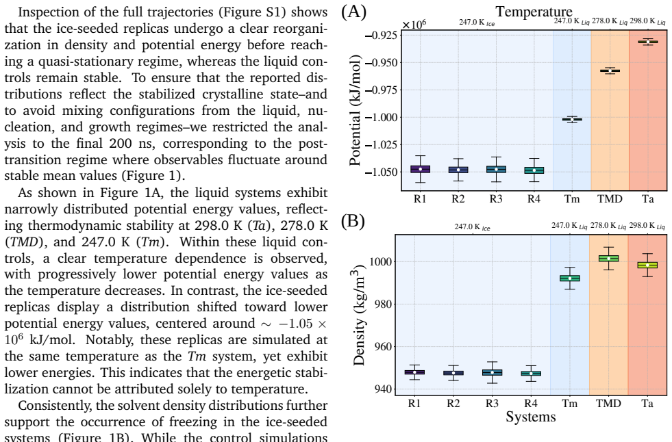
The central mechanism is the ice lattice itself as a geometric constraint. The simulations use a fixed hexagonal ice seed surrounded by liquid water; below the model's melting point the seed grows into an extended ice Ih lattice around the protein, while leaving a liquid-like interfacial layer. This bulk crystallization is validated by solvent density, potential energy, and local bond-order parameters. The load-bearing analyses are principal component analysis (PCA) and dihedral PCA (dPCA) of backbone motion, which map the free-energy landscape, together with surface hydration density—water molecules normalized by solvent-accessible surface area—and hydrogen-bond counts.
What would settle it
If a frozen Yfh1 sample showed the same broad conformational ensemble as supercooled liquid at the same temperature, the confinement claim would fail; in simulation, extending the frozen trajectories well beyond 200 ns and observing the protein cross between the discrete minima would indicate kinetic trapping, not thermodynamic confinement.
Extended reading notes
Core claim
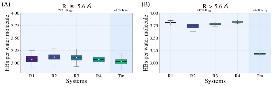
The paper reports that as the aqueous environment of yeast frataxin crystallizes into hexagonal ice, the protein's conformational ensemble contracts. Principal component analysis and dihedral principal component analysis of 1-microsecond trajectories show a shift from a continuous, connected free-energy landscape in liquid water to a discretized landscape with well-separated minima in the frozen replicas. The radius of gyration and secondary-structure content indicate partial expansion of the protein without global unfolding. Interfacial water within about 5.6 Å of the protein retains liquid-like hydrogen bonding, but its surface density (water molecules per unit solvent-accessible surface a
Load-bearing premise
The claim rests on treating the final 200 ns of each trajectory as a quasi-stationary ensemble for the protein, even though the protein undergoes significant conformational transitions during the first ~500 ns of ice growth; if those states are still relaxing, the observed contraction of conformational space reflects slow non-equilibrium trapping rather than a thermodynamic consequence of ice.
Editorial extensions
If this is right
- Low-temperature protein behavior should be described by solvent phase, not just temperature: the same protein at the same temperature shows a broad liquid ensemble or a confined crystalline ensemble depending on whether water is liquid or ice.
- Cryopreservation by freezing may protect proteins by reducing conformational sampling and trapping substates, rather than by immobilizing the native fold; the hydration shell remains dense and liquid-like at the interface.
- Cold-denaturation mechanisms derived for supercooled liquid water do not transfer directly to frozen systems, because ice adds a mechanical/geometric constraint that can suppress the unfolding that supercooling alone would promote.
- Simulations of proteins at low temperature should explicitly model ice growth; treating the solvent as a supercooled liquid misses the dominant effect of solvent structure.
Reading between the lines
- If the confinement is genuinely geometric, the accessible substates should depend on the shape and orientation of the ice cavity around the protein; varying the seed geometry or protein orientation relative to the crystal planes could test this directly.
- The densification and persistence of interfacial water under ice may be a general feature of ice-confined solutes, not specific to Yfh1; the same mechanism could apply to other proteins, nucleic acids, or nanoparticles in frozen environments.
- The restriction of the analysis to the final 200 ns means the discrete minima could be kinetic traps rather than equilibrium states; longer simulations or enhanced-sampling methods would distinguish true confinement from slow relaxation after ice growth.
Signed reviews
Editorial analysis
A structured set of objections, weighed in public.
Referee Report
Summary. This paper uses ~1 µs atomistic MD simulations of yeast frataxin (Yfh1) in TIP4P/2005 water to compare the protein's behavior when the solvent crystallizes into ice Ih (four ice-seeded replicas, R1–R4) with single liquid-water controls at 298 K (Ta), 278 K (TMD), and near the model melting point (Tm). Ice formation is validated by solvent density, potential energy, and local bond-order parameters W4/W6. The authors then use PCA, dPCA, and free-energy landscapes built from the 1 µs trajectories, together with SASA, radius of gyration, hydrogen-bonding, and hydration-density analyses restricted mostly to the final 200 ns (or final 100 ns for hydration persistence), to conclude that ice formation does not drive global unfolding but instead restricts conformational sampling, discretizes the conformational landscape, and preserves/densifies the interfacial hydration layer. The central claim is that solvent structure, not temperature alone, governs protein behavior under freezing conditions.
Significance. If established, the result would address an experimentally difficult regime—protein behavior in a growing ice lattice—and would have implications for cryopreservation and for understanding cold denaturation. The paper has several strengths: the explicit ice-growth setup with four ice replicas, the multi-observable validation of the liquid–solid transition (density, energy, W4/W6), and the direct liquid controls at three temperatures. However, the central equilibrium interpretation is currently not secured: the analysis window is justified by solvent stationarity rather than protein stationarity, the free-energy landscapes are computed from non-stationary full trajectories, and each liquid condition rests on a single trajectory with no run-to-run error bars. These issues affect the main conclusion and require re-analysis rather than mere editing.
major comments (4)
- [§3.1, §3.2, Fig. 5] The paper justifies the final-200 ns analysis window using plateaus in solvent density and potential energy (§3.1), but the protein's conformational observables are not shown to be stationary in that window. §3.2 explicitly says that the first ~500 ns contain 'significant conformational transitions' and are 'followed by progressive confinement within specific regions of the essential subspace.' The dPCA projections in Fig. 5 are shown over the full 1 µs and continue to gate into discrete minima toward the end. A 'progressive confinement' that continues past 500 ns means the 800–1000 ns window may still be part of a slow relaxation after ice growth, not a quasi-stationary ensemble. The observed contraction of conformational space could then be kinetic trapping rather than an ice-induced equilibrium restriction. Please quantify protein stationarity inside the final 200 ns (block analysis o
- [§2.4, Eq. (1)] Eq. (1) applies the Boltzmann relation to the joint distribution of PC1/PC2 from the full 1 µs trajectory. Because the trajectory includes the liquid-to-ice transition and the slow protein drift discussed in §3.2, the resulting histogram is not a Boltzmann equilibrium population; barrier heights and basin depths in the 'free energy landscapes' of Fig. 5 are not thermodynamically interpretable. Please compute the landscapes only from quasi-stationary portions of the trajectories (or use an equilibrium sampling scheme), and consider reporting them as conformational density plots rather than free energies.
- [Table 1, §2.2, Figs. 6–7] Table 1 lists only one liquid-control trajectory for each of Ta, TMD, and Tm, and each liquid system is analyzed as a single trajectory. Figures 6 and 7 therefore compare temporal fluctuations within one trajectory (ice: four replicas) with temporal fluctuations in one liquid trajectory; no run-to-run or bootstrapped error bars are provided. The four ice replicas all start from the same PDB structure 2GA5, so they demonstrate reproducibility of one relaxation pathway rather than independent equilibrium samples. At a minimum, add multiple independent liquid simulations or block-bootstrap uncertainties, and state explicitly whether the R1–R4 replicas differ in protein initial conditions or only in solvent/seed arrangement.
- [§3.3, Conclusions] The title and Conclusions use the word 'stability,' but no stability free energy is computed; the paper measures conformational sampling, SASA, Rg, and hydrogen bonding. The last paragraph of §3.3 itself notes that current force fields are parameterized to maintain the native fold over accessible timescales, so the preservation of secondary structure does not establish thermodynamic stability. I recommend either computing a direct free-energy/stability metric (e.g., from equilibrium sampling or reweighting) or restricting the conclusions to 'restriction of conformational sampling' rather than 'stability.'
minor comments (4)
- [§2.2] Typo: 'The Computations were carried out...' should read 'Computations were carried out...'.
- [§2.3 / Figs. S3–S4] The text says residues 1–19 were 'excluded from all structural analyses,' but Fig. S3 and parts of the SASA/Rg analysis appear to report values for the full protein. Please clarify which analyses use the globular domain only and which use all atoms.
- [Fig. 3] The inset panel is described as showing W6 vs W4, but the axis labels are 'W4 (A.U.)' and 'W6 (A.U.)'. Please make the axis labeling and legend consistent, and state whether the W4/W6 values in the main panel come from the final 200 ns window.
- [§3.3 / Fig. 6E] Minor notation issue: the text reports Tm Rg ≈ 1.414 nm while the figure y-axis is labeled generically; a uniform significant-figures convention would improve readability.
Circularity Check
No significant circularity: the ice-vs-liquid comparison is an independent simulation design; the main weakness is non-equilibrium sampling, not circular reasoning.
full rationale
The paper does not fit a parameter to a target observable and then rename it as a prediction. Its central comparison is between four ice-seeded replicas and liquid controls under the same force field; the ice-liquid distinction is imposed by the seed, not derived from the protein observables. PCA/dPCA/FEL are descriptive reductions of the trajectories, so a landscape computed from a trajectory and then used to describe that same trajectory is not a circular derivation—it is a visualization/summary. No fitted quantities enter Eq. (1); ΔG is a Boltzmann reweighting of the histogram. The only self-citation ([41], Espinosa et al.) is used to justify excluding the disordered N-terminal residues from dPCA, a technical preprocessing choice, not a load-bearing uniqueness claim or ansatz. The paper itself flags the force-field limitation in Section 3.3 ('the preservation of the overall secondary-structure content across all conditions is consistent with the known behavior of current biomolecular force fields...'), which is a stated limitation, not circularity. The legitimate concern—that the final 200 ns window is justified by solvent plateaus while the protein may still be relaxing ('progressive confinement within specific regions of the essential subspace' continues after 500 ns)—is a sampling/stationarity risk that affects interpretation, but it does not reduce any equation or claim to its own input. Accordingly, circularity score is at most 1, reflecting only the minor self-citation, with the central claim retaining independent empirical content.
Assumptions & free parameters
free parameters (4)
- Analysis window = final 200 ns per trajectory =
200 ns (of 1 µs)
- Persistence threshold for hydration waters =
50% of last 100 ns
- Hydration shell cutoffs =
2.8 Å and 5.6 Å
- N-terminal truncation for dPCA =
residues 1-19 excluded
assumptions (5)
- domain assumption TIP4P/2005 water model faithfully represents ice Ih growth, water density anomaly, and protein-water interactions at low temperature.
- domain assumption Amber03w force field preserves the native fold over microsecond timescales, so secondary-structure preservation is not a force-field artifact.
- domain assumption One 1 µs trajectory per liquid condition is sufficient to estimate equilibrium conformational distributions and free-energy landscapes.
- ad hoc to paper Stationary solvent observables in the final 200 ns imply a quasi-stationary protein conformational ensemble.
- domain assumption Fixed prismatic ice seed with periodic boundary conditions reproduces bulk freezing around a protein without artifacts from the fixed lattice or cell geometry.
Cite this review
Pith. "Pith review of Confinement effects on protein stability in a freezing water environment." pith.science (2026). https://pith.science/paper/K5SGKOOB
@misc{pith2026260713966,
  author       = {Pith},
  title        = {Pith review of: Confinement effects on protein stability in a freezing water environment},
  year         = {2026},
  howpublished = {\url{https://pith.science/paper/K5SGKOOB}},
  note         = {Machine review of arXiv:2607.13966}
}
read the original abstract
Understanding how proteins behave at low temperatures remains a central challenge in biophysics, with direct implications for cold denaturation and cryopreservation. While cold denaturation of proteins in the supercooled liquid regime has been studied extensively, the behavior of a protein embedded in a growing ice lattice remains largely inaccessible to experiments. Here we use molecular dynamics simulations that explicitly capture ice Ih formation to characterize the conformational dynamics of yeast frataxin (Yfh1) as its aqueous environment crystallizes. Using four independent ice-seeded replicas and liquid-water controls at three temperatures, we first validate the liquid-solid transition through convergent changes in solvent density, potential energy, and local bond-order parameters (W4, W6). Principal component analysis (PCA), dihedral PCA (dPCA), and free-energy landscapes then reveal that crystallization of the solvent markedly reshapes the accessible conformational space, shifting it from a continuous, highly connected regime in liquid water toward a discretized landscape dominated by confined states. Complementary analyses of solvent-accessible surface area (SASA), radius of gyration, and hydrogen bonding indicate a solvent-driven reorganization of protein-water interactions: although first-shell water remains liquid-like, its surface density increases under freezing, while conformational sampling contracts. Together, these results indicate that protein behavior at low temperatures is governed not by temperature alone but by the structural organization of the surrounding water. By imposing geometrical constraints on the solvent, ice formation restricts conformational sampling while preserving -- and even densifying -- the interfacial hydration layer, highlighting the role of water structure as a determinant of protein stability under freezing conditions.
Figures


Figures from the paper (4 more)


Reference graph
Works this paper leans on
-
[1]
R. B. Simpson and W. Kauzmann,Journal of the American Chemical Society, 1953,75, 5139– 5152
1953
-
[2]
P. L. Privalov,Critical reviews in biochemistry and molecular biology, 1990,25, 281–306
1990
-
[3]
C.-J. Tsai, J. V. Maizel and R. Nussinov,Criti- cal reviews in biochemistry and molecular biology, 2002,37, 55–69
2002
-
[4]
T. R. Sosnick and M. C. Baxa,Annual Review of Biophysics, 2025,54, 17–34
2025
-
[5]
P. A. Temussi, S. R. Martin and A. Pastore,Quar- terly Reviews of Biophysics, 2025,58, e2
2025
-
[6]
Adrover, V
M. Adrover, V. Esposito, G. Martorell, A. Pastore and P. A. Temussi,Journal of the American Chem- ical Society, 2010,132, 16240–16246
2010
-
[7]
Adrover, G
M. Adrover, G. Martorell, S. R. Martin, D. Urosev, P. V. Konarev, D. I. Svergun, X. Daura, P. Temussi and A. Pastore,Journal of molecular biology, 2012,417, 413–424
2012
-
[8]
Sanfelice and P
D. Sanfelice and P. A. Temussi,Biophysical chem- istry, 2016,208, 4–8
2016
Show all 47 references
-
[9]
Puglisi, G
R. Puglisi, G. Karunanithy , D. F. Hansen, A. Pa- store and P. A. Temussi,Communications chem- istry, 2021,4, 127
2021
-
[10]
Sanfelice, E
D. Sanfelice, E. Morandi, A. Pastore, N. Nicco- lai and P. A. Temussi,ChemPhysChem, 2015,16, 3599–3602
2015
-
[11]
Puglisi, P
R. Puglisi, P. Cioni, E. Gabellieri, G. Presciuttini, A. Pastore and P. A. Temussi,Biophysical Journal, 2022,121, 1502–1511
2022
-
[12]
Alfano, D
C. Alfano, D. Sanfelice, S. R. Martin, A. Pastore and P. A. Temussi,Nature communications, 2017, 8, 15428
2017
-
[13]
Campanile and G
M. Campanile and G. Graziano,Biophysica, 2024, 4, 507–516
2024
-
[14]
Hishida, R
M. Hishida, R. Anjum, T. Anada, D. Murakami and M. Tanaka,The Journal of Physical Chemistry B, 2022,126, 2466–2475
2022
-
[15]
Muller,Accounts of Chemical Research, 1990, 23, 23–28
N. Muller,Accounts of Chemical Research, 1990, 23, 23–28
1990
-
[16]
Graziano and B
G. Graziano and B. Lee,The Journal of Physical Chemistry B, 2005,109, 8103–8107
2005
-
[17]
Graziano,Physical Chemistry Chemical Physics, 2014,16, 21755–21767
G. Graziano,Physical Chemistry Chemical Physics, 2014,16, 21755–21767
2014
-
[18]
Riccio and G
A. Riccio and G. Graziano,Proteins: Structure, Function, and Bioinformatics, 2011,79, 1739– 1746
2011
-
[19]
C. L. Dias, T. Ala-Nissila, J. Wong-Ekkabut, I. Vat- tulainen, M. Grant and M. Karttunen,Cryobiol- ogy, 2010,60, 91–99
2010
-
[20]
C. L. Dias,Physical review letters, 2012,109, 048104
2012
-
[21]
M. J. Kuiper, C. J. Morton, S. E. Abraham and A. Gray-Weale,Elife, 2015,4, e05142
2015
-
[22]
W. L. Jorgensen, J. Chandrasekhar, J. D. Madura, R. W. Impey and M. L. Klein,The Journal of chem- ical physics, 1983,79, 926–935
1983
-
[23]
Y. He, S. L. Alam, S. V. Proteasa, Y. Zhang, E. Lesuisse, A. Dancis and T. L. Stemmler,Bio- chemistry, 2004,43, 16254–16262
2004
-
[24]
J. L. Abascal and C. Vega,The Journal of chemical physics, 2005,123, 234505
2005
-
[25]
Conde, M
M. Conde, M. Rovere and P. Gallo,The Journal of chemical physics, 2017,147, 244506
2017
-
[26]
Blazquez and C
S. Blazquez and C. Vega,The Journal of Chemical Physics, 2022,156, 216101
2022
-
[27]
Bussi, D
G. Bussi, D. Donadio and M. Parrinello,The Jour- nal of chemical physics, 2007,126, 014101
2007
-
[28]
H. J. Berendsen, J. v. Postma, W. F. van Gun- steren, A. DiNola and J. Haak,The Journal of chemical physics, 1984,81, 3684–3690
1984
-
[29]
R. G. Pereyra, I. Szleifer and M. A. Carignano,The Journal of chemical physics, 2011,135, 034508
2011
-
[30]
D. J. Evans and B. L. Holian,The Journal of chem- ical physics, 1985,83, 4069–4074
1985
-
[31]
Parrinello and A
M. Parrinello and A. Rahman,Journal of Applied physics, 1981,52, 7182–7190
1981
-
[32]
Essmann, L
U. Essmann, L. Perera, M. L. Berkowitz, T. Dar- den, H. Lee and L. G. Pedersen,The Journal of chemical physics, 1995,103, 8577–8593
1995
-
[33]
Harvey and G
M. Harvey and G. De Fabritiis,Journal of chemical theory and computation, 2009,5, 2371–2377
2009
-
[34]
B. Hess, H. Bekker, H. J. Berendsen and J. G. Fraaije,Journal of computational chemistry, 1997,18, 1463–1472
1997
-
[35]
R. B. Best and J. Mittal,The journal of physical chemistry B, 2010,114, 14916–14923
2010
-
[36]
M. J. Abraham, T. Murtola, R. Schulz, S. Páll, J. C. Smith, B. Hess and E. Lindahl,SoftwareX, 2015,1, 19–25. 10
2015
-
[37]
Y. Mu, P. H. Nguyen and G. Stock,Proteins: Struc- ture, Function, and Bioinformatics, 2005,58, 45– 52
2005
-
[38]
Altis, P
A. Altis, P. H. Nguyen, R. Hegger and G. Stock, The Journal of chemical physics, 2007,126, 244111
2007
-
[39]
P. J. Steinhardt, D. R. Nelson and M. Ronchetti, Physical Review B, 1983,28, 784–805
1983
-
[40]
Y. Wang, S. Teitel and C. Dellago,The Journal of Chemical Physics, 2005,122, 214722
2005
-
[41]
Y. R. Espinosa, J. R. Grigera and E. R. Caffarena, Proteins: Structure, Function, and Bioinformatics, 2017,85, 125–136
2017
-
[42]
Arsiccio and R
A. Arsiccio and R. Pisano,Journal of Pharmaceu- tical Sciences, 2020,109, 2116–2130
2020
-
[43]
J. Li, X. Lin and Z. Zhen,European Journal of Pharmaceutics and Biopharmaceutics, 2025,214, 114764
2025
-
[44]
R. Fang, R. H. Bogner, S. L. Nail and M. J. Pikal, Journal of Pharmaceutical Sciences, 2020,109, 1896–1904
2020
-
[45]
Jin and W
T. Jin and W. Zhuang,JACS Au, 2025,5, 6254– 6264
2025
-
[46]
Reátegui and A
E. Reátegui and A. Aksan,Physical Chemistry Chemical Physics, 2010,12, 10161–10172
2010
-
[47]
S. Wang, M. Wang, L. Han, Y. Sun, W. Cai and X. Shao,Spectrochimica Acta Part A: Molec- ular and Biomolecular Spectroscopy, 2022,267, 120581. 11 Supplementary Material Confinement effects on protein stability in a freezing water environment Yanis R. Espinosaa,b H. Ariel Alvare...
2022
Reviewed August 2, 2026 · model on record in the stance chip above.
Discussion (0). Continue with ORCID to comment.