REVIEW 3 major objections 5 minor 1 cited by
AnyECG: Foundational Models for Multitask Cardiac Analysis in Real-World Settings
T0 review · 3 major / 5 minor · reviewed 2026-08-12 · deepseek-v4-flash
Pith's one-line read AnyECG is a pretrained ECG foundation model that turns any real-world recording into discrete rhythm codes and, after fine-tuning, outperforms task-specific state-of-the-art models on four cardiac tasks.
desk verdict Plausible architecture, but the evaluation as written does not support the 'any real-world ECG' claim because the test set appears to be inside the pretraining corpus. read the letter →
The pith
A machine-rendered reading of the paper's core claim, the machinery that carries it, and where it could break.
The reading
What carries the argument
The load-bearing object is the Rhythm Codebook, a vector-quantized lookup table of normalized codewords that maps each fixed-duration ECG patch to its nearest code, turning continuous, noisy waveforms into discrete tokens. It is trained with a multi-view synergistic decoder: a morphology decoder reconstructs the raw time-domain signal, a frequency decoder reconstructs discrete wavelet transform coefficients, and a demography decoder predicts patient attributes, with codebook and commitment losses stabilizing quantization. The second stage runs masked modeling over these code indices, and the Cardio-Sparse Attention mask (each patch attends only to patches from the same lead or the same temporal position, within a tolerance window) is the mechanism that keeps ultra-long sequences computationally feasible.
What would settle it
Compare AnyECG's anomaly detection on a database of subtle ST-segment and T-wave abnormalities recorded at a native sampling rate above 300 Hz, running one version on signals resampled to 300 Hz and another on native-resolution signals. If the downsampled version loses materially on these cases, the paper's any-real-world-ECG-data promise is falsified.
Extended reading notes
Core claim
The paper argues that ECG analysis does not need a separate specialized model per task or per device. AnyECG first trains a tokenizer whose vector-quantized codebook indexes each one-second ECG patch to a rhythm code; three decoding objectives—waveform reconstruction, wavelet coefficient reconstruction, and demographic attribute prediction—force those codes to carry morphology, frequency, and patient-context information while discarding noise. The full model is then pretrained by masked modeling: random patches are replaced by a mask token and the model must predict the original rhythm-code index from the surrounding unmasked patches, which teaches it rhythm-event associations. Throughout, Cardio-Sparse Attention restricts each patch to attend only to same-lead and same-time-position patches with a small tolerance, cutting computation on uninformative segments. Across four downstream tasks, the AnyECG-B, AnyECG-L, and AnyECG-XL variants beat the non-pretrained baselines and the prior open ECG foundation model, with the average gain reported as 6%.
Load-bearing premise
Resampling every ECG to 300 Hz and cutting it into fixed one-second patches preserves all diagnostically relevant waveform detail, including subtle ST-T changes and low-amplitude pathological features.
Editorial extensions
If this is right
- One fine-tuned AnyECG backbone can replace separate specialized models for anomaly detection, arrhythmia classification, corrupted lead generation, and ultra-long ECG analysis.
- Because the rhythm codebook is trained on heterogeneous, noisy data, downstream fine-tuning on a new device or population should need less task-specific data than training from scratch.
- Cardio-Sparse Attention makes transformer-based processing of ultra-long recordings practical, so the architecture can scale toward 24-hour Holter-style inputs.
- The reported average gain of 6% is the quantitative claim that independent runs on the same four tasks would either confirm or refute.
Reading between the lines
- The strongest untested boundary of the paper is preprocessing: a direct ablation of native sampling rates versus 300 Hz would reveal whether the claim of universal coverage depends on that choice.
- The rhythm codebook could be reused as a discrete token vocabulary for tasks the paper does not explore, such as generating natural-language descriptions of an ECG or retrieving similar clinical cases.
- A more demanding test than the four reported tasks would be a truly unseen wearable device with a low native sampling rate and heavy motion artifact, since pretraining still includes mostly hospital-style high-rate recordings.
Signed reviews
Editorial analysis
A structured set of objections, weighed in public.
Referee Report
Summary. The paper introduces AnyECG, a family of ECG foundation models (254M–1.7B parameters) that use a two-stage self-supervised pretraining: a Rhythm Quantizer that maps fixed-duration ECG patches to discrete codebook tokens via morphology, wavelet-frequency, and demographic proxy tasks, followed by masked modeling of token sequences. The authors claim that jointly pretraining on seven heterogeneous ECG datasets yields a model that, after fine-tuning, outperforms specialized baselines on anomaly detection, arrhythmia classification, corrupted lead generation, and ultra-long ECG analysis, with an average improvement of 6%.
Significance. AnyECG addresses a timely and important problem: constructing a single ECG encoder that works across devices, sampling rates, lead counts, and noise levels. The proposed components (Cardio-Sparse Attention, multi-view quantizer) are technically interesting, and the paper reports ablations supporting the contribution of each loss term. If the empirical results were trustworthy, this would be a valuable contribution to the ECG foundation model literature. However, the evaluation protocol has a load-bearing data-leakage problem, and several tables are internally inconsistent, so the significance cannot be assessed from the current manuscript.
major comments (3)
- [Section IV-A, Table I, Section IV-C] The central generalization claim is not supported because the downstream evaluation appears to use the same data as pretraining. Section IV-A states that the evaluation used 'a comprehensive set of ECG datasets that include all available unlabeled data during pre-training' and that 'for various downstream tasks, we mixed all datasets together.' Section IV-C states that pretraining was conducted on seven different sources, and Table I lists exactly seven datasets, including the 'Undisclosed Database,' which is described in the text as a 'geographically distinct test set.' No patient-level or record-level split between pretraining and downstream test sets is described anywhere. Therefore, the reported results may be inflated by memorization of test subjects, and the claimed generalization to real-world data is not demonstrated. The authors must re-run the evaluation on a held-out set that was never used in pretraining, or explicitly state and justify the split if one exists.
- [Tables III and IV] Tables III and IV contain numerically identical rows for multiple baselines on two different tasks. For example, DENS-ECG, ContraWR, CNN-Transformer, FFCL, and ST-Transformer have the same four numbers in both tables, yet the column labels differ: Table III reports AUROC and Weighted F1 Score as the third and fourth columns, while Table IV reports Weighted F1 Score and Precision. Either the experiments were accidentally run on the same task and mislabeled, or the tables were copied incorrectly. This makes it impossible to verify the task-specific baseline comparisons and undermines the credibility of the experimental results.
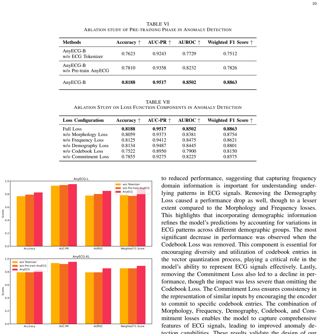
- [Abstract and Section IV-D] The abstract claims 'an average performance improvement of 6% across four critical tasks,' but the manuscript never defines how this average is computed or which numbers are averaged. More importantly, many of the claimed gains are within the reported standard deviations (e.g., Table III, AnyECG-XL vs. RNN1D for AUROC; Table IV, AnyECG-XL vs. DENS-ECG for accuracy). No statistical significance tests are reported, and the arrhythmia detection accuracies are very low for all methods (≤0.35), which raises questions about the task setup. The authors should provide a clear aggregation rule, error bars on the aggregate, and significance tests, and should discuss the practical relevance of the absolute performance.
minor comments (5)
- [Section III-A] The statement that '300 Hz is considered sufficient for diagnosing most cardiac conditions based on the Nyquist-Shannon sampling theorem' is an overgeneralization; the Nyquist theorem applies to bandlimited signals, and the filter cutoff is 75 Hz, so resampling to 300 Hz may discard high-frequency content relevant for some diagnoses. A sensitivity analysis or a citation for the sufficiency claim would strengthen the paper.
- [Section III-A and Section IV-C] The notation is inconsistent: P is used both for the number of patches (Section III-A) and for the patch size (Section IV-C: 'we set the patch size P = 300'). This can confuse readers and should be corrected.
- [Section IV-C and Appendix] Hyperparameters such as codebook size K, masking ratio r, CSA mask width, and DWT levels Lw are mentioned but never specified; a hyperparameter table would aid reproducibility.
- [Figure 3] Figure 3 would benefit from axis labels, time scales, and voltage units; a quantitative comparison with the original signal traces would be more informative than the current visual display.
- [Throughout] Minor typos include 'AnyECGs' in the introduction (Section I) and 'Therefore, We proposed' in Section IV-D(d).
Circularity Check
Central evaluation is circular: the 'Undisclosed Database' test set is listed in Table I as one of the seven pre-training sources, and Section IV-A says downstream evaluation used all pre-training data mixed together, so the reported SOTA gains are in-domain rather than true generalization.
-
fitted input called prediction
[Section IV-A 'ECG Datasets' and Table I]
"To evaluate the performance of AnyECG and baseline models, we utilized a comprehensive set of ECG datasets that include all available unlabeled data during pre-training. ... For various downstream tasks, we mixed all datasets together to minimize biases introduced by individual datasets and to better validate the model's generalizability. ... Undisclosed Database: This dataset contributes an additional 10,000 ECG recordings, providing a geographically distinct test set to further validate the model's performance without data leakage."
The paper's own Table I lists the 'Undisclosed Dataset' (10,000 records, 'Geographically distinct test set') as one of the seven pre-training data sources, and Section IV-A states that the downstream evaluation used 'all available unlabeled data during pre-training' and that 'for various downstream tasks, we mixed all datasets together.' No subject-level or record-level exclusion of the test set from pre-training is stated. The model is therefore fine-tuned and evaluated on recordings whose raw signals were already seen during pre-training; downstream performance on these records can exploit memorized or transductive representations rather than generalization to unseen real-world ECG data.
full rationale
The main circularity is in the evaluation protocol, not in the model equations. Section IV-A explicitly says the downstream evaluation used the same datasets as pre-training ('include all available unlabeled data during pre-training'), and Table I enumerates the seven sources, including the 10,000-record 'Undisclosed Dataset' whose stated role is to be a 'geographically distinct test set.' Because the paper never describes a record-level split that removes downstream test records from pre-training, any benchmark result on these data is partly a measure of how well the model fits its own training distribution. That makes the abstract's claim of 'any real-world ECG data' generalization and the 6% average improvement over SOTA unverifiable as reported and circular in the sense that the test input was an input to the model. I do not count the identical baseline rows in Tables III and IV as circularity; they are a reporting/consistency concern. There is no load-bearing self-citation chain: the Rhythm Quantizer, Cardio-Sparse Attention, and loss components are evaluated through ablations, although those ablations inherit the same leaked-evaluation problem. If the authors can supply an explicit record-level split showing the 10,000 'Undisclosed' records were never used in pre-training, the circularity would be resolved; as written, the paper's own table and prose exhibit the overlap.
Assumptions & free parameters
free parameters (8)
- Resampling rate =
300 Hz
- Patch duration s =
300 samples (1 second)
- Bandpass and notch filter frequencies =
0.1-75 Hz bandpass; 50 Hz notch
- Codebook size K =
not reported
- Masking ratio r =
not reported
- Commitment loss weight beta =
not reported
- CSA mask width (positional tolerance) =
not reported
- DWT decomposition levels Lw =
not reported
assumptions (5)
- domain assumption Nyquist-Shannon sampling theorem applies to ECG such that 300 Hz preserves diagnostically relevant information
- domain assumption db6 wavelet denoising and the chosen bandpass/notch filters remove noise while preserving pathological waveforms
- domain assumption Patient demographic attributes (age, sex, weight) are predictable from individual 1-second patches and help the representation
- domain assumption Restricting attention to same-lead and same-position patches captures all clinically relevant rhythm-event associations
- domain assumption The mixed-dataset 80/20 split is a valid proxy for real-world generalization across devices and populations
Cite this review
Pith. "Pith review of AnyECG: Foundational Models for Multitask Cardiac Analysis in Real-World Settings." pith.science (2026). https://pith.science/paper/KI64W57Q
@misc{pith2026241117711,
  author       = {Pith},
  title        = {Pith review of: AnyECG: Foundational Models for Multitask Cardiac Analysis in Real-World Settings},
  year         = {2026},
  howpublished = {\url{https://pith.science/paper/KI64W57Q}},
  note         = {Machine review of arXiv:2411.17711}
}
read the original abstract
Electrocardiogram (ECG), a non-invasive and affordable tool for cardiac monitoring, is highly sensitive in detecting acute heart attacks. However, due to the lengthy nature of ECG recordings, numerous machine learning methods have been developed for automated heart disease detection to reduce human workload. Despite these efforts, performance remains suboptimal. A key obstacle is the inherent complexity of ECG data, which includes heterogeneity (e.g., varying sampling rates), high levels of noise, demographic-related pattern shifts, and intricate rhythm-event associations. To overcome these challenges, this paper introduces AnyECG, a foundational model designed to extract robust representations from any real-world ECG data. Specifically, a tailored ECG Tokenizer encodes each fixed-duration ECG fragment into a token and, guided by proxy tasks, converts noisy, continuous ECG features into discrete, compact, and clinically meaningful local rhythm codes. These codes encapsulate basic morphological, frequency, and demographic information (e.g., sex), effectively mitigating signal noise. We further pre-train the AnyECG to learn rhythmic pattern associations across ECG tokens, enabling the capture of cardiac event semantics. By being jointly pre-trained on diverse ECG data sources, AnyECG is capable of generalizing across a wide range of downstream tasks where ECG signals are recorded from various devices and scenarios. The experimental results show that AnyECG achieves an average performance improvement of 6% across four critical tasks-anomaly detection, arrhythmia classification, corrupted lead generation, and ultra-long ECG recognition. AnyECG learns common ECG rhythm from data and significantly outperforms state-of-the-art methods in each of these tasks.
Figures


Figures from the paper (1 more)
Forward citations
Cited by 1 Pith paper
-
FoundationalECGNet: A Lightweight Foundational Model for ECG-based Multitask Cardiac Analysis
A multi-architecture ECG classifier reports near-perfect scores on a small test set, but the evaluation is compromised by pre-split oversampling and inconsistent metric reporting.
Reference graph
Works this paper leans on
-
[1]
Large-scale training of foundation models for wearable biosignals,
S. Abbaspourazad, O. Elachqar, A. C. Miller, S. Emrani, U. Nallasamy, and I. Shapiro, “Large-scale training of foundation models for wearable biosignals,” arXiv preprint arXiv:2312.05409 , 2023
arXiv 2023
-
[2]
Y . Ansari, O. Mourad, K. Qaraqe, and E. Serpedin, “Deep learning for ecg arrhythmia detection and classification: an overview of progress for period 2017–2023,” Frontiers in Physiology, vol. 14, p. 1246746, 2023
work page 2017
-
[3]
Validation of an automated artificial intelligence system for 12-lead ecg interpretation,
R. Herman, A. Demolder, B. Vavrik, M. Martonak, V . Boza, V . Kres- nakova, A. Iring, T. Palus, J. Bahyl, O. Nelis et al. , “Validation of an automated artificial intelligence system for 12-lead ecg interpretation,” Journal of Electrocardiology, vol. 82, pp. 147–154, 2024
work page 2024
-
[4]
P. Singh and A. Sharma, “Attention-based convolutional denoising autoencoder for two-lead ecg denoising and arrhythmia classification,” IEEE Transactions on Instrumentation and Measurement , vol. 71, pp. 1–10, 2022
work page 2022
-
[5]
J. Chen, S. Huang, Y . Zhang, Q. Chang, Y . Zhang, D. Li, J. Qiu, L. Hu, X. Peng, Y . Du et al., “Congenital heart disease detection by pediatric electrocardiogram based deep learning integrated with human concepts,” Nature Communications, vol. 15, no. 1, p. 976, 2024
work page 2024
-
[6]
A. Jain, H. Tandri, D. Dalal, H. Chahal, E. Z. Soliman, R. J. Prineas, A. R. Folsom, J. A. Lima, and D. A. Bluemke, “Diagnostic and prognostic utility of ecg for left ventricular hypertrophy defined by mri in relationship to ethnicity: The multi-ethnic study of atherosclerosis (mesa),” American heart journal , vol. 159, no. 4, p. 652, 2010
work page 2010
-
[7]
Combining 1d cnn and lstm for automated myocardial infarction detection from ecg signals,
S. Prathipati and S. H. V . Malyavantham, “Combining 1d cnn and lstm for automated myocardial infarction detection from ecg signals,” in International Conference on Data Science and Big Data Analysis . Springer, 2023, pp. 561–571
work page 2023
-
[8]
S. Kucukseymen, S. Uslu, and S. Ozdemir, “Left atrial overload detec- tion in ecg using frequency domain features with convolutional neural network,” European Heart Journal , vol. 43, no. Supplement 2, pp. ehac544–427, 2022
work page 2022
Show all 50 references
-
[9]
An investigation of the contextual distribution of false pos- itives in a deep learning-based atrial fibrillation detection algorithm,
D. Kumar, S. Puthusserypady, H. Dominguez, K. Sharma, and J. E. Bardram, “An investigation of the contextual distribution of false pos- itives in a deep learning-based atrial fibrillation detection algorithm,” Expert Systems with Applications , vol. 211, p. 118540, 2023
2023
-
[10]
Ecg-based cardiac arrhythmias detection through ensemble learning and fusion of deep spatial–temporal and long-range dependency features,
S. Din, M. Qaraqe, O. Mourad, K. Qaraqe, and E. Serpedin, “Ecg-based cardiac arrhythmias detection through ensemble learning and fusion of deep spatial–temporal and long-range dependency features,” Artificial Intelligence in Medicine , vol. 150, p. 102818, 2024
2024
-
[11]
Ecg-transcovnet: A hybrid transformer model for accurate arrhythmia detection using electrocardiogram signals,
H. A. Shah, F. Saeed, M. Diyan, N. A. Almujally, and J.-M. Kang, “Ecg-transcovnet: A hybrid transformer model for accurate arrhythmia detection using electrocardiogram signals,” CAAI Transactions on Intel- ligence Technology, 2024
2024
-
[12]
Msgformer: A multi-scale grid transformer network for 12-lead ecg arrhythmia detec- tion,
C. Ji, L. Wang, J. Qin, L. Liu, Y . Han, and Z. Wang, “Msgformer: A multi-scale grid transformer network for 12-lead ecg arrhythmia detec- tion,” Biomedical Signal Processing and Control , vol. 87, p. 105499, 2024
2024
-
[13]
Apneanet: A hybrid 1dcnn-lstm architecture for detection of obstructive sleep apnea using digitized ecg signals,
G. Srivastava, A. Chauhan, N. Kargeti, N. Pradhan, and V . S. Dhaka, “Apneanet: A hybrid 1dcnn-lstm architecture for detection of obstructive sleep apnea using digitized ecg signals,” Biomedical Signal Processing and Control, vol. 84, p. 104754, 2023
2023
-
[14]
1d cnn framework on ecg signals,
B. D. Jasvitha, K. Kanagaraj, K. Murali, T. Singh, and P. Duraisamy, “1d cnn framework on ecg signals,” in 2024 3rd International Conference for Innovation in Technology (INOCON) . IEEE, 2024, pp. 1–6
2024
-
[15]
Automatic diagnosis of the 12-lead ecg using a deep neural network,
A. H. Ribeiro, M. H. Ribeiro, G. M. Paix ˜ao, D. M. Oliveira, P. R. Gomes, J. A. Canazart, M. P. Ferreira, C. R. Andersson, P. W. Macfarlane, W. Meira Jr et al. , “Automatic diagnosis of the 12-lead ecg using a deep neural network,” Nature communications, vol. 11, no. 1, p. 1760, 2020
2020
-
[16]
An end-to-end atrial fibrillation detec- tion by a novel residual-based temporal attention convolutional neural network with exponential nonlinearity loss,
Y . Gao, H. Wang, and Z. Liu, “An end-to-end atrial fibrillation detec- tion by a novel residual-based temporal attention convolutional neural network with exponential nonlinearity loss,” Knowledge-Based Systems, vol. 212, p. 106589, 2021
2021
-
[17]
Automatic segmentation of atrial fibrillation and flutter in single-lead electrocardiograms by self-supervised learning and transformer architecture,
D. Yun, H.-L. Yang, S. Kwon, S.-R. Lee, K. Kim, K. Kim, H.-C. Lee, C.-W. Jung, Y . S. Kim, and S. S. Han, “Automatic segmentation of atrial fibrillation and flutter in single-lead electrocardiograms by self-supervised learning and transformer architecture,” Journal of the Amer...
2024
-
[18]
Improving ballistocardiogram-based continuous heart rate variability monitoring: A self-supervised learning approach,
C. Wu, J. Qiu, and G. Shen, “Improving ballistocardiogram-based continuous heart rate variability monitoring: A self-supervised learning approach,” Biomedical Signal Processing and Control , vol. 89, p. 105774, 2024
2024
-
[19]
Learning with incomplete labels of multisource datasets for ecg classification,
Q. Li, Y . Liu, Z. Zhang, J. Liu, Y . Yuan, K. Wang, and R. He, “Learning with incomplete labels of multisource datasets for ecg classification,” Pattern Recognition, vol. 150, p. 110321, 2024
2024
-
[20]
Clocs: Contrastive learning of cardiac signals across space, time, and patients,
D. Kiyasseh, T. Zhu, and D. A. Clifton, “Clocs: Contrastive learning of cardiac signals across space, time, and patients,” in International Conference on Machine Learning . PMLR, 2021, pp. 5606–5615
2021
-
[21]
Adversarial spatiotemporal contrastive learning for electrocardiogram signals,
N. Wang, P. Feng, Z. Ge, Y . Zhou, B. Zhou, and Z. Wang, “Adversarial spatiotemporal contrastive learning for electrocardiogram signals,” IEEE Transactions on Neural Networks and Learning Systems , 2023
2023
-
[22]
Self-supervised representation learning from 12-lead ecg data,
T. Mehari and N. Strodthoff, “Self-supervised representation learning from 12-lead ecg data,” Computers in biology and medicine , vol. 141, p. 105114, 2022
2022
-
[23]
Maefe: Masked autoencoders family of electrocardiogram for self- supervised pretraining and transfer learning,
H. Zhang, W. Liu, J. Shi, S. Chang, H. Wang, J. He, and Q. Huang, “Maefe: Masked autoencoders family of electrocardiogram for self- supervised pretraining and transfer learning,” IEEE Transactions on Instrumentation and Measurement , vol. 72, pp. 1–15, 2022
2022
-
[24]
Guiding masked representation learning to capture spatio-temporal relationship of electrocardiogram,
Y . Na, M. Park, Y . Tae, and S. Joo, “Guiding masked representation learning to capture spatio-temporal relationship of electrocardiogram,” arXiv preprint arXiv:2402.09450 , 2024
2024 arXiv
-
[25]
An effective data enhancement method for classification of ecg arrhythmia,
S. Ma, J. Cui, C.-L. Chen, X. Chen, and Y . Ma, “An effective data enhancement method for classification of ecg arrhythmia,”Measurement, vol. 203, p. 111978, 2022. 12
2022
-
[26]
Electrocardio panorama: Synthesizing new ecg views with self-supervision,
J. Chen, X. Zheng, H. Yu, D. Z. Chen, and J. Wu, “Electrocardio panorama: Synthesizing new ecg views with self-supervision,” in In- ternational Joint Conference on Artificial Intelligence , 2021
2021
-
[27]
The discrete wavelet transform: wedding the a trous and mallat algorithms,
M. J. Shensa et al. , “The discrete wavelet transform: wedding the a trous and mallat algorithms,” IEEE Transactions on signal processing , vol. 40, no. 10, pp. 2464–2482, 1992
1992
-
[28]
Bert: Pre-training of deep bidirectional transformers for language understanding,
J. D. M.-W. C. Kenton and L. K. Toutanova, “Bert: Pre-training of deep bidirectional transformers for language understanding,” in Proceedings of naacL-HLT, vol. 1. Minneapolis, Minnesota, 2019, p. 2
2019
-
[29]
Beit: Bert pre-training of image transformers,
H. Bao, L. Dong, S. Piao, and F. Wei, “Beit: Bert pre-training of image transformers,” arXiv preprint arXiv:2106.08254 , 2021
2021 arXiv
-
[30]
Masked au- toencoders are scalable vision learners,
K. He, X. Chen, S. Xie, Y . Li, P. Doll ´ar, and R. Girshick, “Masked au- toencoders are scalable vision learners,” inProceedings of the IEEE/CVF conference on computer vision and pattern recognition , 2022, pp. 16 000–16 009
2022
-
[31]
An open access database for evaluating the algorithms of electrocardiogram rhythm and morphology abnormality detection,
F. Liu, C. Liu, L. Zhao, X. Zhang, X. Wu, X. Xu, Y . Liu, C. Ma, S. Wei, Z. He et al. , “An open access database for evaluating the algorithms of electrocardiogram rhythm and morphology abnormality detection,” Journal of Medical Imaging and Health Informatics , vol. 8, no. 7, ...
2018
-
[32]
St petersburg incart 12-lead arrhythmia database,
V . Tihonenko, A. Khaustov, S. Ivanov, A. Rivin, and E. Yakushenko, “St petersburg incart 12-lead arrhythmia database,” PhysioBank Phys- ioToolkit and PhysioNet, 2008
2008
-
[33]
Nutzung der ekg- signaldatenbank cardiodat der ptb ¨uber das internet,
R. Bousseljot, D. Kreiseler, and A. Schnabel, “Nutzung der ekg- signaldatenbank cardiodat der ptb ¨uber das internet,” 1995
1995
-
[34]
Ptb-xl, a large publicly available electro- cardiography dataset,
P. Wagner, N. Strodthoff, R.-D. Bousseljot, D. Kreiseler, F. I. Lunze, W. Samek, and T. Schaeffter, “Ptb-xl, a large publicly available electro- cardiography dataset,” Scientific data, vol. 7, no. 1, pp. 1–15, 2020
2020
-
[35]
Dens-ecg: A deep learning approach for ecg signal delineation,
A. Peimankar and S. Puthusserypady, “Dens-ecg: A deep learning approach for ecg signal delineation,” Expert systems with applications , vol. 165, p. 113911, 2021
2021
-
[36]
Self-supervised eeg representation learning for automatic sleep staging,
C. Yang, D. Xiao, M. B. Westover, and J. Sun, “Self-supervised eeg representation learning for automatic sleep staging,” arXiv preprint arXiv:2110.15278, 2021
2021 arXiv
-
[37]
Ecg-fm: An open electrocardiogram foundation model,
K. McKeen, L. Oliva, S. Masood, A. Toma, B. Rubin, and B. Wang, “Ecg-fm: An open electrocardiogram foundation model,” arXiv preprint arXiv:2408.05178, 2024
2024 arXiv
-
[38]
Bag of tricks for image classification with convolutional neural networks,
T. He, Z. Zhang, H. Zhang, Z. Zhang, J. Xie, and M. Li, “Bag of tricks for image classification with convolutional neural networks,” in Proceedings of the IEEE/CVF conference on computer vision and pattern recognition, 2019, pp. 558–567
2019
-
[39]
Transformer convolutional neural networks for automated artifact detection in scalp eeg,
W. Y . Peh, Y . Yao, and J. Dauwels, “Transformer convolutional neural networks for automated artifact detection in scalp eeg,” in 2022 44th Annual International Conference of the IEEE Engineering in Medicine & Biology Society (EMBC) . IEEE, 2022, pp. 3599–3602
2022
-
[40]
Ecg-based biometrics using recurrent neural networks,
R. Salloum and C.-C. J. Kuo, “Ecg-based biometrics using recurrent neural networks,” in 2017 IEEE International Conference on Acoustics, Speech and Signal Processing (ICASSP) . IEEE, 2017, pp. 2062–2066
2017
-
[41]
Motor imagery eeg classification algorithm based on cnn-lstm feature fusion network,
H. Li, M. Ding, R. Zhang, and C. Xiu, “Motor imagery eeg classification algorithm based on cnn-lstm feature fusion network,” Biomedical signal processing and control, vol. 72, p. 103342, 2022
2022
-
[42]
Deep learning for ecg analysis: Benchmarks and insights from ptb-xl,
N. Strodthoff, P. Wagner, T. Schaeffter, and W. Samek, “Deep learning for ecg analysis: Benchmarks and insights from ptb-xl,” IEEE journal of biomedical and health informatics , vol. 25, no. 5, pp. 1519–1528, 2020
2020
-
[43]
Transformer-based spatial-temporal feature learning for eeg decoding,
Y . Song, X. Jia, L. Yang, and L. Xie, “Transformer-based spatial-temporal feature learning for eeg decoding,” arXiv preprint arXiv:2106.11170, 2021
2021 arXiv
-
[44]
Conditional generative adversarial nets,
M. Mirza, “Conditional generative adversarial nets,” arXiv preprint arXiv:1411.1784, 2014
2014 arXiv
-
[45]
Banach wasserstein gan,
J. Adler and S. Lunz, “Banach wasserstein gan,” Advances in neural information processing systems , vol. 31, 2018
2018
-
[46]
Automated and interpretable patient ecg profiles for disease detection, tracking, and discovery,
G. H. Tison, J. Zhang, F. N. Delling, and R. C. Deo, “Automated and interpretable patient ecg profiles for disease detection, tracking, and discovery,” Circulation: Cardiovascular Quality and Outcomes , vol. 12, no. 9, p. e005289, 2019
2019
-
[47]
Biot: Biosignal transformer for cross-data learning in the wild,
C. Yang, M. Westover, and J. Sun, “Biot: Biosignal transformer for cross-data learning in the wild,” Advances in Neural Information Pro- cessing Systems, vol. 36, 2024
2024
-
[48]
Large brain model for learning generic representations with tremendous eeg data in bci,
W.-B. Jiang, L.-M. Zhao, and B.-L. Lu, “Large brain model for learning generic representations with tremendous eeg data in bci,” arXiv preprint arXiv:2405.18765, 2024
2024 arXiv
-
[49]
Foundation models for electrocardiograms,
J. Song, J.-H. Jang, B. T. Lee, D. Hong, J.-m. Kwon, and Y .- Y . Jo, “Foundation models for electrocardiograms,” arXiv preprint arXiv:2407.07110, 2024
2024 arXiv
-
[50]
Cardiogpt: An ecg interpretation generation model,
G. Fu, J. Zheng, I. Abudayyeh, C. Ani, C. Rakovski, L. Ehwerhemuepha, H. Lu, Y . Guo, S. Liu, H. Chu et al., “Cardiogpt: An ecg interpretation generation model,” IEEE Access, 2024. 13 VII. A PPENDIX A. Notations Data and Indices X ∈ RL×T Multi-channel ECG signals L Number of E...
2024
Reviewed August 12, 2026 · model on record in the stance chip above.
Discussion (0). Continue with ORCID to comment.