REVIEW 3 major objections 4 minor 65 references
Loops versus lines and the compression stiffening of cells
T0 review · 3 major / 4 minor · reviewed 2026-08-14 · deepseek-v4-flash
Pith's one-line read Mouse embryonic fibroblasts compression stiffen under uniaxial compression, and a single semiflexible polymer loop model captures the measured stress-strain curve up to about 35% strain.
desk verdict New AFM compression result and two new model mechanisms, but the stress conversion is unvalidated, so the central claim needs a control experiment. read the letter →
The pith
A machine-rendered reading of the paper's core claim, the machinery that carries it, and where it could break.
The reading
What carries the argument
The central object is the semiflexible polymer loop Hamiltonian $$H_{v+c}=\frac12 K_{cf}\sum_{\langle ij\rangle}(l_{ij}-l_0)^2+\frac12 K_{sf}\sum_{\langle ijk\rangle}(\theta_{ijk}-\theta_0)^2+\$\lambda$(A-A_0),$$ a closed chain of stretchable springs and bending springs whose enclosed area is held fixed by a Lagrange multiplier. The area constraint models an incompressible viscous cytoplasm and supplies an effective outward pressure that prevents the loop from buckling in plane, which is why the loop compression stiffens instead of softening. The dimensionless ratio $\tilde{\kappa}=K_{cf}l_0^2/K_{sf}$ controls whether stretching or bending dominates: as $\tilde{\kappa}\to 0$ the stress grows as $\gamma^3$ with no linear regime, while finite bending produces the linear small-strain response and delayed stiffening seen in the mEF data at $\tilde{\kappa}=0.768$. Transplanting the same loop concept into a two-dimensional fiber network, as area-conserving inclusions and as angular constraints at the mesh scale, generates the bending-driven stiffening mechanisms that apply when a bulk cytoskeletal network or an in vitro fibrin network is present.
What would settle it
Compress mouse embryonic fibroblasts after selectively disrupting the actomyosin cortex, for instance with an actin-depolymerizing drug, leaving the interior intact: the single-loop model predicts that compression stiffening should be abolished or shifted to substantially larger strains, whereas survival of stiffening would show that a bulk fiber network is load-bearing.
Extended reading notes
Core claim
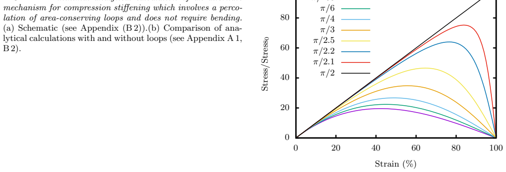
Mouse embryonic fibroblasts compression stiffen under uniaxial compression imposed by an atomic force microscope, with stiffening setting in at $\gamma_c \approx 20\%$ compressive strain, in contrast to the compression softening expected from semiflexible biopolymer networks. The paper's central quantitative claim is that, up to about 35% compressive strain, the measured stress-strain curve is well described by a single semiflexible polymer loop model of the actomyosin cortex enclosing an incompressible fluid, with only one free parameter, $\tilde{\kappa} = 0.768$, the ratio of stretching to bending stiffness of the loop. In this model, the incompressible interior acts as an outward pressure that suppresses in-plane buckling, so the loop stiffens rather than collapses; with finite bending stiffness the stress-strain curve has a linear small-strain regime followed by nonlinear stiffening, as observed. The paper additionally establishes two bulk-network mechanisms for compression stiffening: area-conserving loops, modeling fluid organelles and vesicles, interspersed in a fiber network force fibers into non-affine bending and convert the network's compression softening into stiffening even at small packing fractions, and angularly constrained crosslinks distort under compression to produce stiffening without organelles. For fibrin networks embedded with adherent beads, the area-conserving-loop model captures the stiffening and stress magnitude, with a later onset of stiffening in the model than in the experiment.
Load-bearing premise
The quantitative fit assumes that a squeezed cell behaves like independent flat slices of an incompressible fluid wrapped in a cortex, with no load-bearing fiber network spanning the cell's interior.
Editorial extensions
If this is right
- Single cells can stiffen under compression on short timescales, without waiting for cytoskeletal reorganization, giving them a fast mechanical defense against large homeostatic pressures.
- For cortex-dominated cells, uniaxial compression curves collapse onto a one-parameter family; measuring $\tilde{\kappa}$ from a compression test gives a readout of the balance between cortex stretching and bending.
- Only a small number of fluid-like organelles or vesicles, with packing fraction around 0.04, can flip a fiber network from compression softening to compression stiffening by forcing fibers to bend.
- Because the stress-strain curve depends on the packing fraction and size distribution of area-conserving loops, compression rheology can distinguish cell types or states by their internal organelle geometry.
- Cellular compression stiffening is a plausible contributor to tissue compression stiffening, consistent with the observation that decellularized tissue loses much of its stiffening response.
Reading between the lines
- Editorial inference: if the single-loop description is the operative mechanism, the onset strain $\gamma_c$ and fitted $\tilde{\kappa}$ should shift systematically when cortex composition is altered, so compression tests on cells with more or less actin-crosslinking protein would provide a direct check.
- Editorial inference: the mechanical-fingerprint result suggests compression tests could serve as a label-free assay for organelle size and vesicle content; changing organelle size osmotically should move $\gamma_c$ and the stress magnitude in a way the area-conserving-loop model can predict.
- Editorial inference: the paper's two-dimensional-to-three-dimensional argument implies a direct three-dimensional loop-shell model should produce quantitatively different stress at large strain; comparing enucleated cells with whole cells would isolate whether the deviation above 35% strain comes from the nucleus or from the percolating bulk network.
Signed reviews
Editorial analysis
A structured set of objections, weighed in public.
Referee Report
Summary. The manuscript reports AFM uniaxial compression experiments on rounded mouse embryonic fibroblasts, finding a compressive stress–strain curve that bends upward with stiffening onset near γc ≈ 20%. To explain this, the authors study three mechanical models: (i) a two-dimensional semiflexible polymer loop enclosing an incompressible area, representing the actomyosin cortex with viscous interior; (ii) a diluted triangular fiber network with embedded area-conserving loops, representing organelles and vesicles; and (iii) a fiber network with angle-constraining crosslinks. They report that the single-loop model matches the mEF experiment up to about 35% strain with one dimensionless parameter κ̃, and that the fiber-network-with-loops model captures compression stiffening observed in fibrin networks with adherent beads. Analytical calculations in the appendices support the numerical energy minimizations.
Significance. If the mEF result is a genuine material response, it establishes single-cell compression stiffening on fast timescales, in contrast to expectations from semiflexible polymer networks, and it would be directly relevant to tissue-scale compression stiffening. The study's strengths include explicit numerical simulations with error bars and system-size checks, analytical treatments of the loop model in Appendix A, a separate fibrin-bead experiment that provides a falsifiable test of the area-conserving-loop mechanism, and a PAA-gel control in Appendix B3 showing no stiffening when fiber bending is negligible. However, the load-bearing experimental stress conversion and the number of effective fitting parameters in the central model–experiment comparison must be addressed before the quantitative claims are secure.
major comments (3)
- [Section II (Whole cell compression)] The reported stress is computed as σ = F/(2πrh), with r = 12.5 µm and h the indentation depth, while the strain is ε = h/H0. This makes the reported stress proportional to F(ε)/ε. For a linear-elastic half-space indented by a sphere, Hertz contact gives F ∝ ε^{3/2} and hence the reported stress would scale as ε^{1/2}, which has decreasing slope. The observed upward curvature could therefore arise from the assumed contact-area model, finite-thickness or confinement effects, or genuine material stiffening. The PAA-gel control in Appendix B3 uses rheometer plate compression, not the AFM spherical-indenter geometry, so it does not validate the stress conversion used for the cells. A control with the identical AFM sphere protocol and the same σ = F/(2πrh) analysis applied to a passive linear-elastic material of comparable size is needed to support the paper's central experimental claim.
- [Section V, Fig. 6] The text states that the loop-model comparison has 'only one free parameter, κ̃,' but the experimental curve is transformed by subtracting the pre-stress and normalizing the stress by that same value. The subtracted offset and the stress scale are determined from the data being explained, and the model stress is separately normalized to the experimental scale. Therefore the comparison effectively uses more than one fitted quantity, and the agreement in Fig. 6 is a weaker test than claimed. The authors should either report the fit with a pre-specified offset and scale, or explicitly count the offset and scale as fitted parameters when assessing the agreement.
- [Section V (Comparison with Experiments)] The selection of the single-loop cortex model as the explanation up to 35% strain rests on the stated assumption: 'Since we do not know directly whether or not there is a bulk, rigid cytoskeletal network, let us assume there is not.' This assumption is load-bearing: if a spanning fiber network contributes appreciably to the mEF compression response, the fitted κ̃ and the loop-model agreement could be coincidental. The manuscript does not present any experimental test of this assumption, such as comparing wild-type cells with vimentin-null or actin-disrupted cells under the same compression protocol. Given that the authors themselves identify this as an unknown, the conclusion that the cortex is the dominant contributor up to 35% strain should be either explicitly tested or substantially weakened.
minor comments (4)
- [Section IV] In the paragraph following the definition of Ho+fn, the text says 'See Fig. 3b for the stress-strain curves' when discussing the fiber network without loops; the relevant panel appears to be Fig. 4b, not Fig. 3b.
- [Section II] The sentence 'The data was obtained from 10 cells and averaged over with the error bar denoting the standard deviation' should be rephrased as 'Data were averaged over 10 cells, with error bars denoting the standard deviation.'
- [Section VI] There are two typographical issues: 'mouse embryonic fibroblastas' should be 'mouse embryonic fibroblasts,' and 'experimental experimental techniques' contains a duplicated word.
- [Appendix B1 and Fig. 4b] The caption of Fig. 4b states that the analytical fits 'are scaled here to fit with the numerical data,' so the comparison between the numerical network compression and the analytical model is not parameter-free; the scaling factor should be reported.
Circularity Check
No significant circularity: the loop-model stiffening is derived from its own Hamiltonian, and the experimental compression-stiffening claim is independent of the model.
full rationale
The central experimental claim rests on AFM force-distance data converted via sigma = F/(2*pi*r*h) and epsilon = h/H0; whatever the merits of that spherical-cap area normalization, it is a data-analysis convention and not an input to the loop model. The single-loop model's stress-strain curves are obtained by numerical minimization of Eq. (1), with stiffening following from the area-conservation constraint and bending energy (Eqs. A3-A4); no experimental curve is used to define the Hamiltonian. The comparison in Fig. 6 does fit the dimensionless parameter kappa-tilde = 0.768 and normalizes by the experimental pre-stress, so the 'agreement' is a one-parameter fit rather than a parameter-free prediction; but the paper labels kappa-tilde as a free parameter and does not claim the prediction is unconditional. The self-citations ([3] and [20]) supply background or standard percolation values and are not load-bearing. The paper explicitly declines to rule out alternative mechanisms ('we cannot necessarily rule out any of the compression stiffening mechanisms') and admits fluid flow cannot yet be excluded, which further indicates the derivation is not being forced. Concerns about the spherical-cap stress normalization are correctness risks, not circularity.
Assumptions & free parameters
free parameters (4)
- κ̃ (Kcf l0^2 / Ksf) =
0.768
- Experimental pre-stress offset and stress scale =
Not tabulated; taken from the mEF curve in Fig. 1
- Area-spring stiffness ratio KA l0^2 / Kcf =
10^3
- Rigid-loop stiffness amplification =
100 times Kcf for central-force springs surrounding loops
assumptions (6)
- domain assumption A semiflexible loop enclosing an incompressible fluid does not buckle in plane because area conservation pushes the perimeter outward.
- domain assumption The mEF can be represented as independent two-dimensional cross-sections with minimal fluid flow between them during AFM compression.
- ad hoc to paper There is no bulk, rigid cytoskeletal network dominating mEF mechanics.
- domain assumption Two-dimensional stress can be compared to three-dimensional stress up to a length-factor rescaling.
- domain assumption Fibrin fibers are in the regime where bending energy is comparable to stretching energy.
- domain assumption A randomly diluted triangular lattice with open boundaries and no prestress captures the generic mechanics of the cell cytoskeleton and fibrin networks.
Cite this review
Pith. "Pith review of Loops versus lines and the compression stiffening of cells." pith.science (2026). https://pith.science/paper/L2AHXWOU
@misc{pith2026190803725,
  author       = {Pith},
  title        = {Pith review of: Loops versus lines and the compression stiffening of cells},
  year         = {2026},
  howpublished = {\url{https://pith.science/paper/L2AHXWOU}},
  note         = {Machine review of arXiv:1908.03725}
}
read the original abstract
Both animal and plant tissue exhibit a nonlinear rheological phenomenon known as compression stiffening, or an increase in moduli with increasing uniaxial compressive strain. Does such a phenomenon exist in single cells, which are the building blocks of tissues? One expects an individual cell to compression soften since the semiflexible biopolymer-based cytoskeletal network maintains the mechanical integrity of the cell and in vitro semiflexible biopolymer networks typically compression soften. To the contrary, we find that mouse embryonic fibroblasts (mEFs) compression stiffen under uniaxial compression via atomic force microscopy (AFM) studies. To understand this finding, we uncover several potential mechanisms for compression stiffening. First, we study a single semiflexible polymer loop modeling the actomyosin cortex enclosing a viscous medium modeled as an incompressible fluid. Second, we study a two-dimensional semiflexible polymer/fiber network interspersed with area-conserving loops, which are a proxy for vesicles and fluid-based organelles. Third, we study two-dimensional fiber networks with angular-constraining crosslinks, i.e. semiflexible loops on the mesh scale. In the latter two cases, the loops act as geometric constraints on the fiber network to help stiffen it via increased angular interactions. We find that the single semiflexible polymer loop model agrees well with our AFM experiments until approximately 35% compressive strain. We also find for the fiber network with area-conserving loops model that the stress-strain curves are sensitive to the packing fraction and size distribution of the area-conserving loops, thereby creating a mechanical fingerprint across different cell types. Finally, we make comparisons between this model and experiments on fibrin networks interlaced with beads as well as discuss the tissue-scale implications of cellular compression stiffening.
Figures


Figures from the paper (14 more)




Reference graph
Works this paper leans on
-
[1]
Compression softening has earlier been observed in a tensegrity model of a cell [37]. What can we expect when we include organelles as area-conserving loops into the fiber network? While working with our initial cell as a viscous medium sur- rounded by an actomyosin cortex, we saw that despite a loop of central-force springs being floppy according to Maxwel...
-
[2]
is shown for systems with occupation proba- bilityp = 0.58. When the ratio is small, Hf n+axlinks reduces to Ho+f n (with KA = 0) and compression softening is ob- served as expected. The onset of nonlinearity is not tunable by this ratio. Bottom: With Kxlink/(Kcfl2
-
[3]
For both figures, the curves are averaged over 1000 runs on an 8x8 lattice with Kcfl2 0/Ksf = 1
= 0 .1, com- pression stiffening for different occupation probabilities p is shown. For both figures, the curves are averaged over 1000 runs on an 8x8 lattice with Kcfl2 0/Ksf = 1. area-conserving loops, this network compression stiff- ens as can be inferred from Fig. 5. At small strain, the angles between fibers change within each triangu- lar loop with both ...
-
[4]
closer to unity. Note that we can explore a larger range of occu- pation probabilities than with freely-rotating crosslinks because thep above which the network is rigid is the con- nectivity percolation threshold for the triangular lattice, i.e. pc = 2 sin(π
- [5]
- [6]
-
[7]
M. Perepelyuk, L. Chin, X. Cao, A. van Oosten, V. B. Shenoy, P. A. Janmey, and R. G. Wells, PloS ONE 11, e0146588 (2016)
work page 2016
-
[8]
T. A. Engstrom, K. Pogoda, K. Cruz, P. A. Janmey, and J. M. Schwarz, Phys. Rev. E 99, 052413 (2019)
work page 2019
Show all 65 references
-
[9]
Alberts, D
B. Alberts, D. Bray, J. Lewis, M. Raff, K. Roberts, and J. Watson, Molecular Biology of the Cell (Garland, New York, 1994) pp. 907–982
1994
-
[10]
M. Guo, A. J. Ehrlicher, S. Mahammad, H. Fabich, M. H. Jensen, J. R. Moore, J. J. Fredberg, R. D. Goldman, and D. A. Weitz, Biophy. J. 105, 1562 (2013)
2013
-
[11]
M¨ ucke, L
N. M¨ ucke, L. Kreplak, R. Kirmse, T. Wedig, H. Her- rmann, U. Aebi, and J. Langowski, J. Mol. Biol. 335, 1241 (2004)
2004
-
[12]
Gittes, B
F. Gittes, B. Mickey, J. Nettleton, and J. Howard, J. Cell Biol. 120, 923 (1993)
1993
-
[13]
A. Ott, M. Magnasco, A. Simon, and A. Libchaber, Phys. Rev. E 48, R1642 (1993)
1993
-
[14]
S. H. Shabbir, M. M. Cleland, R. D. Goldman, and M. Mrksich, Biomaterials 35, 1359 (2014)
2014
-
[15]
A. S. Van Oosten, M. Vahabi, A. J. Licup, A. Sharma, P. A. Galie, F. C. MacKintosh, and P. A. Janmey, Sci. Reps. 6, 19270 (2016)
2016
-
[16]
M. G. Mendez, D. Restle, and P. A. Janmey, Biophys. J. 107, 314 (2014)
2014
-
[17]
A. E. Patteson, A. Vahabikashi, K. Pogoda, S. A. Adam, K. Mandal, M. Kittisopikul, S. Sivagurunathan, A. Gold- man, R. D. Goldman, and P. A. Janmey, Journal of Cell Biology 218, 4079 (2019)
2019
-
[18]
For a range of p <0.7, the network response is similar
= 0.347 [20]. For a range of p <0.7, the network response is similar. However, for p> 0.7, we observe a plateau in the stress-strain response occuring at intermediate strains. This plateau corresponds to a global distortion of the lattice to weaken it. This phe- nomenon may be...
2017
-
[19]
P. A. Janmey, M. E. McCormick, S. Rammensee, J. L. Leight, P. C. Georges, and F. C. MacKintosh, Nat. Mat. 6, 48 (2006)
2006
-
[20]
Storm, J
C. Storm, J. J. Pastore, F. C. MacKintosh, T. C. Luben- sky, and P. A. Janmey, Nature 435, 191 (2005)
2005
-
[21]
MacKintosh, J
F. MacKintosh, J. K¨ as, and P. Janmey, Phys. Rev. Lett. 75, 4425 (1995)
1995
-
[22]
C. P. Broedersz and F. C. MacKintosh, Rev. Mod. Phys. 86, 995 (2014)
2014
-
[23]
L. D. Landau and E. Lifshitz, Course of Theoretical Physics 3, 109 (1986)
1986
-
[24]
Pilyugina, B
E. Pilyugina, B. Krajina, A. J. Spakowitz, and J. D. Scheiber, Polymers 9, 99 (2017)
2017
-
[25]
M. Das, D. Quint, and J. Schwarz, PloS ONE 7, e35939 (2012)
2012
-
[26]
Lin and L
S. Lin and L. Gu, Materials (Basel) 8, 551 (2015)
2015
-
[27]
Hatami-Marbini, Phys
H. Hatami-Marbini, Phys. Rev. E 97, 022504 (2018)
2018
-
[28]
Picariello and et al., PNAS , 201902847 (2019)
H. Picariello and et al., PNAS , 201902847 (2019)
2019
-
[29]
A. S. van Oosten, X. Chen, L. Chin, K. Cruz, A. E. Patteson, K. Pogoda, V. B. Shenoy, and P. A. Janmey, Nature 573, 96 (2019)
2019
-
[30]
G. Y. Onoda and E. G. Liniger, Phys. Rev. Lett. 64, 2727 (1990)
1990
-
[31]
Bernal and J
J. Bernal and J. Mason, Nature 188, 910 (1960)
1960
-
[32]
J. C. Maxwell, Philos. Mag. Ser. 5 27, 294 (1864)
-
[33]
Lulevich, T
V. Lulevich, T. Zink, H.-Y. Chen, F.-T. Liu, and G.-Y. Liu, Langmuir 22, 8151 (2006)
2006
-
[34]
Arreaga, R
G. Arreaga, R. Capovilla, C. Chryssomalakos, and J. Guven, Phys. Rev. E 65, 031801 (2002)
2002
-
[35]
Hiramoto, Exp
Y. Hiramoto, Exp. Cell Res. 32, 59 (1963)
1963
-
[36]
J. Feng, H. Levine, X. Mao, and L. M. Sander, Soft matter 12, 1419 (2016)
2016
-
[37]
D. A. Fletcher and R. D. Mullins, Nature 463, 485 (2010)
2010
-
[38]
Vahabi, A
M. Vahabi, A. Sharma, A. J. Licup, A. S. G. van Oosten, P. A. Galie, P. A. Janmey, and F. C. MacKintosh, Soft Matt. 12, 5050 (2016)
2016
-
[39]
E. E. Charrier and P. A. Janmey, Meth. Enzymol. 568, 35 (2016)
2016
-
[40]
D. E. Discher, D. H. Boal, and S. K. Boey, Phys. Rev. E 55, 4762 (1997)
1997
-
[41]
Wintz, R
W. Wintz, R. Everaers, and U. Seifert, Journal de Physique I 7, 1097 (1997)
1997
-
[42]
Beysens and G
D. Beysens and G. Forgacs, Dynamical Networks in Physics and Biology: At the Frontier of Physics and Biol- ogy Les Houches Workshop, March 17–21, 1997 , Vol. 10 (Springer Science & Business Media, 2013)
1997
-
[43]
D. A. Head, A. J. Levine, and F. MacKintosh, Phys. Rev. Lett 91, 108102 (2003)
2003
-
[44]
C. P. Broedersz, X. Mao, T. C. Lubensky, and F. C. MacKintosh, Nat. Phys. 7, 983 (2011)
2011
-
[45]
Sharma, A
A. Sharma, A. J. Licup, K. A. Jansen, R. Rens, M. Shein- man, G. H. Koenderink, and F. C. MacKintosh, Nat. Phys. 12, 584 (2016)
2016
-
[46]
K. N. Dahl, A. J. S. Ribeiro, and J. Lammerding, Circ. Res. 102, 1307 (2008)
2008
-
[47]
Wu and et al., Nat
P.-H. Wu and et al., Nat. Meth. 15, 491 (2018)
2018
-
[48]
Nakamura, T
F. Nakamura, T. Osborn, C. Hartemink, J. Hartwig, and T. Stossel, J. Cell Biol. 179, 1011 (2007)
2007
-
[49]
W. Liu, L. M. Jawerth, E. A. Sparks, M. R. Falvo, R. R. Hantgan, S. T. Superfine, R. Lord, and G. M., Science 313, 634 (2006)
2006
-
[50]
O. V. Kim, R. I. Litvinov, J. W. Weisel, and M. S. Alber, Biomaterials 35, 6739 (2014)
2014
-
[51]
Fernandez and A
P. Fernandez and A. Ott, Phys. Rev. Lett. 100, 238102 (2008)
2008
-
[52]
Koster, D
S. Koster, D. A. Weitz, R. D. Goldman, U. Aebi, and H. Herrmann, Curr. Opin. Cell Biol. 32, 82 (2013)
2013
-
[53]
A. X. Cartagena-Rivera, J. S. Logue, C. M. Waterman, and R. S. Chadwick, Biophys. J. 110, 2528 (2016)
2016
-
[54]
Feric and C
M. Feric and C. P. Brangwynne, Nat. Cell Biol. 15, 1253 (2013)
2013
-
[55]
A. E. Patteson, K. Pogoda, F. J. Byfield, K. Mandal, Z. Ostrowska-Podhorodecka, E. E. Charrier, P. A. Galie, P. Deptu la, R. Bucki, C. A. McCulloch, et al., Small 15, 14 1903180 (2019)
2019
-
[56]
A. J. Licup and et al., PNAS 112, 9573 (2015)
2015
-
[57]
Ramanujan, Quart
S. Ramanujan, Quart. J. Math 45, 350 (1914)
1914
-
[58]
Ferone, B
V. Ferone, B. Kawohl, and C. Nitsch, Mathematische Annalen 365, 987 (2016). Appendix A: Cell as a viscous medium surrounded by a cortex
2016
-
[59]
We choose the arms of the loop to be oblique to the vertical compression rather than to have the arms perfectly parallel to the direction of compres- sion
Compressing a 4-gon We assume the simplest loop symmetric about the x−axis and y−axis, a 4-gon that will be vertically and uniaxially compressed. We choose the arms of the loop to be oblique to the vertical compression rather than to have the arms perfectly parallel to the dir...
-
[60]
The continuum limit loop is then assumed to be a circle at zero strain and an ellipse at finite strain
An ellipse in the continuum limit When the number of vertices in the loop is large and ˜κ < 1, then numerical minimization yields elliptical shapes with compressive strain. The continuum limit loop is then assumed to be a circle at zero strain and an ellipse at finite strain. F...
-
[61]
(A15) Eq
(A14) The contour derivative of the unit tangent vector can be expressed in terms of the parameter θ as, dˆt ds = 1 r(θ) dˆt dθ. (A15) Eq. (A11) can now be presented as, Ho+c,sf = κ 2 ∫ 2π 0 dθ r(θ) ⏐⏐⏐⏐ d dθ (−b sin(θ),a cos(θ)) r ⏐⏐⏐⏐ 2 . (A16) Having a,b as functions of ϵ, ...
-
[62]
1 with a soft area constraint, i.e
Soft area constraint We now study the effect of replacing the Lagrange mul- tiplier term in Eq. 1 with a soft area constraint, i.e. κA(A−A0)2. For small enough KA, the area of the semi- flexible polymer loop can change and so we ask whether or not compression stiffening will be o...
-
[63]
No organelles: Compression softening FIG. 10. Collapse of springs induces softening. We first present an approximate calculation for the compression softening mechanism in the absence of or- ganelles (area-conserving loops). The energy of a single central force spring is E = Kc...
-
[64]
11a) in the vertical direction
Area-conserving loops initiate bending Consider the forces acting on vertex C (see Fig. 11a) in the vertical direction. The summation of the forces must add up to zero to ensure mechanical equilibrium of this vertex. Let us assume that the loop conserves its area by conserving...
-
[65]
Experiment with polyacrylamide gel To study the effect of bending in the fiber network on compression stiffening, we study beads embedded in a polyacrylamide (PAA) gel, which is a linear elastic ma- terial. The experimental protocol is the following: 8% acrylamide and 0.3% bis-ac...
2000
Reviewed August 14, 2026 · model on record in the stance chip above.
Discussion (0). Continue with ORCID to comment.