REVIEW 4 major objections 4 minor 297 references
Unveiling Secrets of Brain Function With Generative Modeling: Motion Perception in Primates & Cortical Network Organization in Mice
T0 review · 4 major / 4 minor · reviewed 2026-08-11 · deepseek-v4-flash
Pith's one-line read A hierarchical generative model trained only on synthetic optic flow predicts macaque MT neurons more than twice as well as the prior benchmark.
desk verdict A carefully framed dissertation whose headline claims are unverifiable in the posted text; the per-figure beta selection is a real threat to the 2x predictive gain. read the letter →
The pith
A machine-rendered reading of the paper's core claim, the machinery that carries it, and where it could break.
The reading
What carries the argument
The central object is the compressed hierarchical variational autoencoder (cNVAE), which stacks multiple stochastic latent layers and is trained by maximizing the evidence lower bound (ELBO), a variational free-energy objective that operationalizes Helmholtz's 'perception as unconscious inference.' The hierarchy is what lets the model capture multi-scale causes in the synthetic ROFL optic-flow data: lower latents handle local flow structure and higher latents encode object motion and self-motion. The other load-bearing mechanism is the mixed-membership stochastic blockmodel with variational inference, which assigns each cortical region a vector of membership strengths across K overlapping communities rather than a single community label; the overlap fraction and membership entropy derived from that vector carry the Chapter 5 results.
What would settle it
Train the same model on ROFL variants with two independently moving objects, pursuit or saccadic eye movements, occlusions, or natural depth variation, and re-measure the MT alignment gain. If the >2x improvement over the mechanistic baseline disappears or falls below significance, the finding is specific to the simplified stimulus family rather than a general principle of hierarchical inference.
Extended reading notes
Core claim
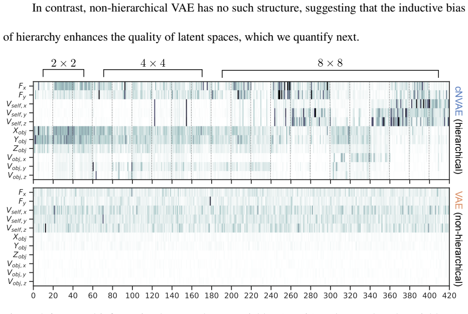
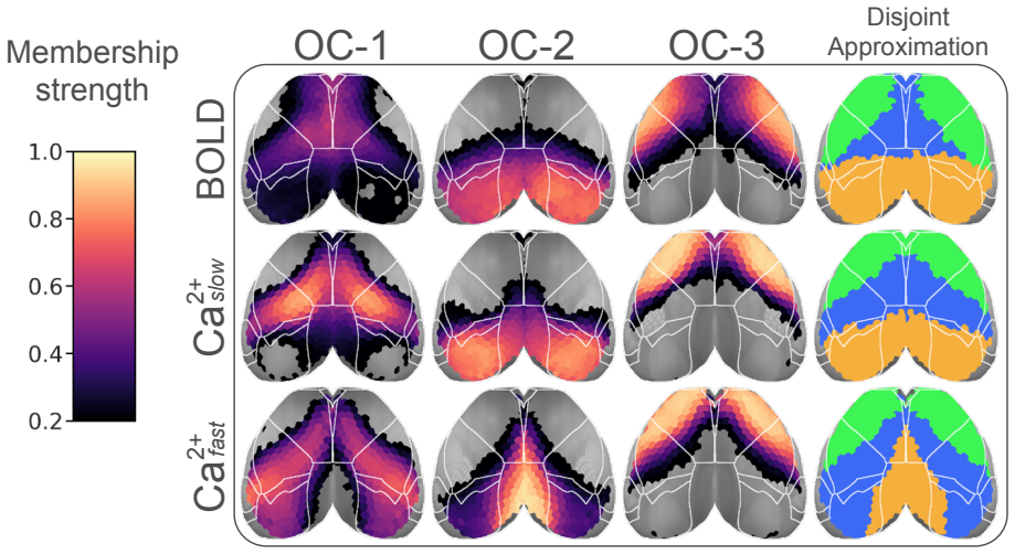
In the author's terms, the dissertation demonstrates that optic flow parsing is possible if a neural network is (a) structured hierarchically and (b) trained with an objective function based on Helmholtzian inference. The compressed hierarchical VAE (cNVAE) is trained on the ROFL synthetic world, which contains a fixating observer, translational and rotational self-motion, and one independently moving object; without any supervision, the latents untangle object position and velocity from self-motion components more sharply than a comparable flat VAE. Evaluated as an encoding model against macaque MT neuron recordings, the hierarchical VAE surpasses the previous state-of-the-art mechanistic MT model [27] by a gain of over 2x in predictive power. Chapter 5 applies the same variational-inference strategy to spontaneous activity: a mixed-membership stochastic blockmodel decomposes simultaneous fMRI-BOLD and wide-field calcium fluorescence in mice into overlapping communities, and around half of the cortical regions belong to multiple communities.
Load-bearing premise
The load-bearing premise is that the simplified simulated retina—one fixating observer, one fixed-size moving object, and prescribed depth statistics—captures the optic-flow structure that real macaque MT neurons encode, so that what the model learns transfers to biological neurons.
Editorial extensions
If this is right
- Unsupervised, visual-only learning can separate self-motion from object motion when the network is hierarchical and trained with an inference-based loss; no extra-retinal signals are required.
- Hierarchical latent structure is the reason for the improved brain alignment: flat VAEs trained on the same objective and data align less well with MT neurons, so the hierarchy itself carries predictive power.
- MT encoding models no longer need hand-designed motion filters to be competitive; the latent representations of a generative model trained on optic flow predict neural responses better than the previous mechanistic standard.
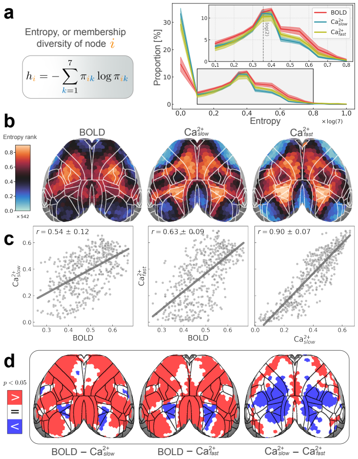
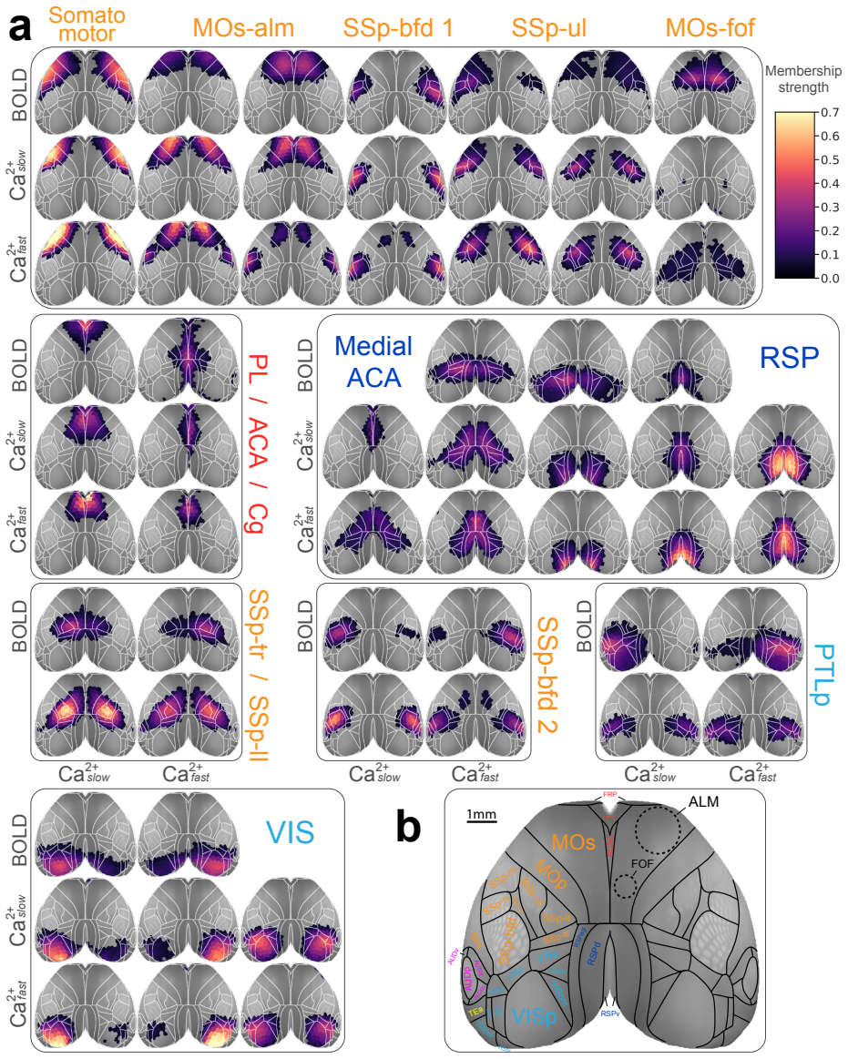
- Around half of mouse cortical regions belong to more than one resting-state network, so disjoint network parcellations misdescribe a substantial fraction of cortex.
- The overlapping organization is not purely a BOLD artifact: wide-field calcium imaging, a more neuron-specific signal, produces largely concordant overlapping communities, with modality-specific differences in degree and diversity metrics.
Reading between the lines
- Taken further than the dissertation states: if the inference objective is the cause of the 2x gain, then training the same model on more ecological optic flow—pursuit eye movements, multiple objects, occlusions—should preserve or increase the gain; if the gain collapses, the result is specific to the simplified stimulus family rather than to hierarchical inference per se.
- A testable extension the author leaves implicit: latent units carrying object-motion information in the model should map onto neurons in area MST, the next cortical stage after MT, predicting that MST-like selectivity for combined optic flow and object motion does not require extra-retinal input.
- On the network side, the overlap result implies that regions with high membership entropy are the most likely to switch community allegiance when the brain changes state, linking static overlap to dynamic circuit reconfiguration in a way that task or arousal manipulations could test.
- Because Ca2+ and fMRI reveal similar principal gradients but different degree and entropy maps, the disparate centrality measures across modalities may index neurovascular rather than purely neural properties; a joint model treating BOLD and calcium as two noisy observations of one shared community latent would test that interpretation.
Editorial analysis
A structured set of objections, weighed in public.
Referee Report
Summary. This manuscript, an arXiv deposit of a PhD dissertation, presents two projects. Project 1 (Chapter 4) develops a hierarchical variational autoencoder, cNVAE, trained on a synthetic optic-flow dataset (ROFL), and claims that its latent representations not only separate self-motion and object-motion causes without extra-retinal signals but also predict macaque MT neuron responses with a gain of over 2x over the previous state of the art (Section 1.8.4). Project 2 (Chapter 5) applies a mixed-membership stochastic blockmodel to simultaneous fMRI and wide-field calcium imaging data in mice and claims that roughly half of cortical regions belong to multiple overlapping communities (abstract and Section 1.13.3). The introduction and background chapters are fully provided, but the two results chapters are truncated in the version under review, so the evidence for these claims is not available in the submitted text.
Significance. If the results hold, Project 1 would provide an important proof-of-concept that unsupervised hierarchical generative models can perform optic-flow parsing and serve as encoding models of primate MT neurons, a domain previously dominated by supervised mechanistic models. The use of external macaque MT recordings to ground the brain-alignment claim is a methodological strength, as is the stated intention to release code and data (Sections 4.8 and 5.5). Project 2 addresses a timely question about overlapping functional organization in the rodent cortex using a rare simultaneous fMRI/Ca2+ dataset. However, because both results chapters are absent from the reviewed text, the actual quantitative support for these claims cannot currently be assessed.
major comments (4)
- [Chapters 4 and 5 (TOC; Sections 1.8.4, 1.13.3)] The central quantitative claims—the over-2x predictive gain of the hierarchical VAE over the Nishimoto-Gallant model and the roughly 50% overlap of mouse cortical regions—are asserted in the introduction and abstract, but Chapters 4 and 5 are not present in the submitted text; only their table-of-contents entries and section headings are available. Consequently, the model architecture, training procedure, evaluation metrics, statistical comparisons, and robustness analyses that would substantiate these claims cannot be inspected. This is a load-bearing incompleteness for the manuscript as submitted.
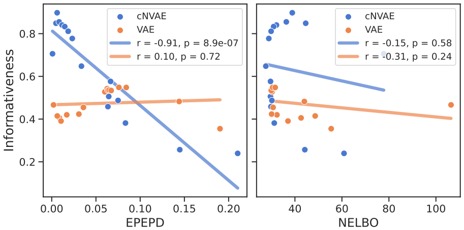
- [§4.9.5; §1.8.4] The over-2x predictive gain claim is potentially confounded by the per-figure β selection disclosed in Section 4.9.5. If β values were chosen after inspecting brain-alignment results (e.g., Fig. 4.16), then the reported gain compares an outcome-selected member of the cNVAE family against a single fixed Nishimoto-Gallant baseline, rather than evaluating a fixed model. Please report the β selection rule, state whether it was blind to the MT recording data, and include a sensitivity analysis of the predictive gain across β values.
- [§4.3, §4.12] The ROFL synthetic dataset contains one fixating observer, one object of fixed size, and known depth distributions. The transfer of representations learned on this simplified stimulus family to real macaque MT responses assumes that its motion statistics capture ecologically relevant structure. The paper should provide evidence about how the disentanglement and MT-alignment results vary with object size, number of objects, presence of pursuit eye movements, and depth variability, or otherwise justify the ecological validity of ROFL.
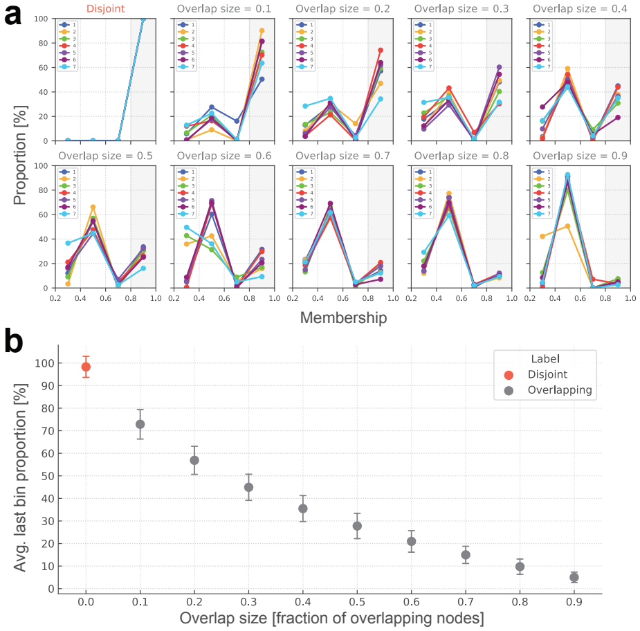
- [§5.4.2, §5.4.3] The Chapter 5 claim that about 50% of cortical regions belong to multiple communities depends on the chosen number of communities K and on the functional connectivity graph threshold. The visible text does not include the robustness analyses that would show this overlap estimate is stable across these choices; the full chapter should report such analyses or qualify the claim accordingly.
minor comments (4)
- [Abstract; §1.8.4] The phrase 'hierarchical inference underlines the brain's understanding' should read 'underlies the brain's understanding'; also, 'V AE' is written with an unusual space throughout, and should be standardized to 'VAE'.
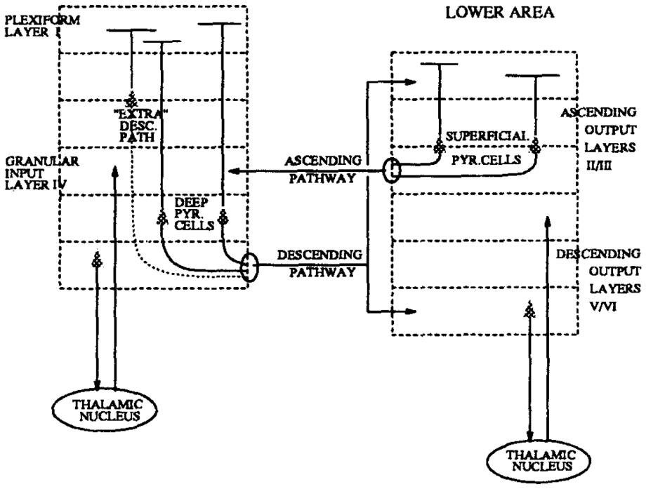
- [Figure 1.11 caption] The caption contains a typo: 'Foodforward (ascending) and feedback' should be 'Feedforward (ascending) and feedback'.
- [§1.8.4] The claim of 'over 2x in predictive power' would be clearer if it specified the evaluation metric (e.g., variance explained, correlation coefficient) and the exact comparison protocol used for the baseline model.
- [Chapter 3 title] The chapter title 'Variational Inference & Variational Autoencoders (V AE)' should use the standard abbreviation 'VAE' consistently, both in the title and in the body text.
Circularity Check
No demonstrated circularity: the MT predictive-power claim is grounded in external macaque recordings; the per-figure β choice is a robustness concern, not an exhibited reduction.
full rationale
The paper's derivation chain is: Helmholtzian inference + cortical hierarchy → cNVAE (Sec. 4.4) → unsupervised training on synthetic ROFL (Sec. 4.3) → evaluation of latent representations against macaque MT spike data (Sec. 4.6.5). The final link is external: the model is scored on predicting real neuronal responses, not on quantities generated by the model or its training distribution. The ROFL disentanglement evaluations (Secs. 4.6.3–4.6.4) use generative factors that are known by construction, but that is a controlled benchmark, not circularity: the model does not receive the factor labels, and the non-hierarchical VAE fails to capture object factors (Sec. 4.10.2), so success is not guaranteed by the architecture or objective. Chapter 5 uses empirical simultaneous fMRI and Ca2+ data (Sec. 5.2) and includes synthetic LFR controls (Sec. 5.4.1). The closest issue to a fitted-input pattern is Sec. 4.9.5 ('Choosing β values for different figures') and Fig. 4.16 ('Alignment scores across β values'), since β controls the reconstruction/disentanglement trade-off and could in principle make the 2x gain an artifact of an outcome-guided hyperparameter search. However, the text available does not state that β was selected using the held-out MT alignment scores, nor does it exhibit the reported gain as the maximum of that search. Without that, the concern remains a validity/robustness risk rather than a demonstrated circular step. No load-bearing self-citation chain was identified.
Assumptions & free parameters
free parameters (4)
- Beta (VAE loss weight) =
not stated in reviewed text
- cNVAE latent dimensionality =
not stated in reviewed text
- Number of communities K (SVINET) =
3, 7, 20 (per figures)
- Functional connectivity graph threshold =
not stated in reviewed text
assumptions (5)
- standard math Subjective Bayesian interpretation of probability; beliefs updated via Bayes' theorem (Equation 3.1)
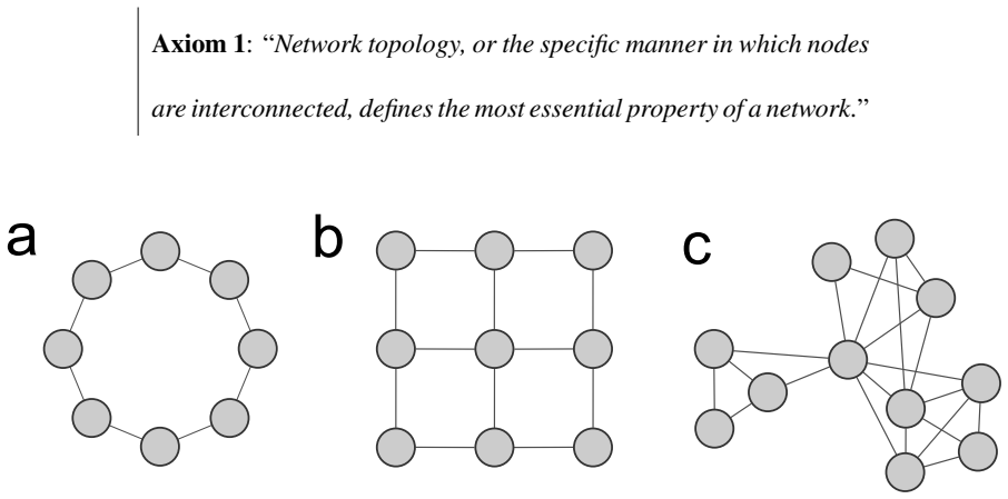
- domain assumption Axiom 1: network topology defines the most essential property of a network
- ad hoc to paper The synthetic ROFL optic flow dataset captures ecologically relevant motion statistics
- domain assumption MT neuron responses can be meaningfully compared to model latents via a linear readout and sparsity alignment metric
- domain assumption BOLD and Ca2+ signals reflect the same underlying neural population activity after preprocessing
invented entities (2)
-
cNVAE (compressed Nouveau VAE)
-
ROFL (Retinal Optic Flow Learning) synthetic dataset
Cite this review
Pith. "Pith review of Unveiling Secrets of Brain Function With Generative Modeling: Motion Perception in Primates & Cortical Network Organization in Mice." pith.science (2026). https://pith.science/paper/LLIXVO5R
@misc{pith2026241219845,
  author       = {Pith},
  title        = {Pith review of: Unveiling Secrets of Brain Function With Generative Modeling: Motion Perception in Primates & Cortical Network Organization in Mice},
  year         = {2026},
  howpublished = {\url{https://pith.science/paper/LLIXVO5R}},
  note         = {Machine review of arXiv:2412.19845}
}
read the original abstract
This Dissertation is comprised of two main projects, addressing questions in neuroscience through applications of generative modeling. Project #1 (Chapter 4) explores how neurons encode features of the external world. I combine Helmholtz's "Perception as Unconscious Inference" -- paralleled by modern generative models like variational autoencoders (VAE) -- with the hierarchical structure of the visual cortex. This combination leads to the development of a hierarchical VAE model, which I test for its ability to mimic neurons from the primate visual cortex in response to motion stimuli. Results show that the hierarchical VAE perceives motion similar to the primate brain. Additionally, the model identifies causal factors of retinal motion inputs, such as object- and self-motion, in a completely unsupervised manner. Collectively, these results suggest that hierarchical inference underlines the brain's understanding of the world, and hierarchical VAEs can effectively model this understanding. Project #2 (Chapter 5) investigates the spatiotemporal structure of spontaneous brain activity and its reflection of brain states like rest. Using simultaneous fMRI and wide-field Ca2+ imaging data, this project demonstrates that the mouse cortex can be decomposed into overlapping communities, with around half of the cortical regions belonging to multiple communities. Comparisons reveal similarities and differences between networks inferred from fMRI and Ca2+ signals. The introduction (Chapter 1) is divided similarly to this abstract: sections 1.1 to 1.8 provide background information about Project #1, and sections 1.9 to 1.13 are related to Project #2. Chapter 2 includes historical background, Chapter 3 provides the necessary mathematical background, and finally, Chapter 6 contains concluding remarks and future directions.
Figures


Figures from the paper (97 more)





















Reference graph
Works this paper leans on
-
[1]
Complexity, entropy and the physics of information
John Wheeler. Complexity, entropy and the physics of information . Ed. by Wojciech H. Zurek. SFI Press, 1990. url: https://www.sfipress.org/books/complexity- entropy-and-the-physics-of-information
1990
-
[2]
Christopher A. Fuchs. “On Participatory Realism”. In: Information and Interaction: Ed- dington, Wheeler, and the Limits of Knowledge. Ed. by Ian T. Durham and Dean Rickles. Springer International Publishing, 2017, pp. 113–134. isbn: 978-3-319-43760-6. doi: 10.1007/978-3-319-43760-6_7
-
[3]
Atomic Theory and the Description of Nature
Niels Bohr. Atomic Theory and the Description of Nature . Cambridge University Press,
-
[4]
Life and scientific work of Peter Guthrie Tait
Cargill Gilston Knott. Quote from undated letter from Maxwell to Tait. “Life and scientific work of Peter Guthrie Tait”, Cambridge University Press, 1911
1911
-
[5]
Irreversibility and heat generation in the computing process
Rolf Landauer. “Irreversibility and heat generation in the computing process”. In: IBM journal of research and development 5.3 (1961), pp. 183–191. doi: 10.1147/rd.53. 0183
-
[6]
Notes on Landauer’s principle, reversible computation, and Maxwell’s Demon
Charles H Bennett. “Notes on Landauer’s principle, reversible computation, and Maxwell’s Demon”. In: Studies In History and Philosophy of Science Part B: Studies In History and Philosophy of Modern Physics 34.3 (2003), pp. 501–510. doi: 10.1016/S1355- 2198(03)00039-X
-
[7]
Colloquium: The physics of Maxwell’s demon and information
Koji Maruyama et al. “Colloquium: The physics of Maxwell’s demon and information”. In: Reviews of Modern Physics 81.1 (2009), p. 1. doi: 10.1103/RevModPhys.81.1
-
[8]
Work and information processing in a solv- able model of Maxwell’s demon
Dibyendu Mandal and Christopher Jarzynski. “Work and information processing in a solv- able model of Maxwell’s demon”. In: Proceedings of the National Academy of Sciences 109.29 (2012), pp. 11641–11645. doi: 10.1073/pnas.12042631
Show all 297 references
-
[9]
Thermodynamics of information
Juan MR Parrondo et al. “Thermodynamics of information”. In: Nature physics 11.2 (2015), pp. 131–139. doi: 10.1038/nphys3230
2015 doi
-
[10]
The analysis of sensations, and the relation of the physical to the psychical
Ernst Mach. The analysis of sensations, and the relation of the physical to the psychical. Open Court Publishing Company, 1914. url: https : / / archive . org / details / analysisofsensat00mach
1914
-
[11]
Handbuch der physiologischen Optik
Hermann Von Helmholtz. Handbuch der physiologischen Optik. Vol. 9. Voss, 1867
-
[12]
Receptive fields of single neurones in the cat’s striate cortex
David H. Hubel and Torsten N. Wiesel. “Receptive fields of single neurones in the cat’s striate cortex”. In: The Journal of Physiology 148 (1959). doi: 10.1113/jphysiol. 1959.sp006308
1959 doi
-
[13]
Receptive fields and functional architecture of monkey striate cortex
David H. Hubel and Torsten N. Wiesel. “Receptive fields and functional architecture of monkey striate cortex”. In: The Journal of Physiology 195 (1968). doi: 10.1113/ jphysiol.1968.sp008455. 272
1968
-
[14]
An introduction to the work of David Hubel and Torsten Wiesel
Eric R Kandel. “An introduction to the work of David Hubel and Torsten Wiesel”. In: The Journal of physiology 587.Pt 12 (2009), p. 2733
2009
-
[15]
Receptive field
J. Alonso and Y. Chen. “Receptive field”. In: Scholarpedia 4.1 (2009). revision #136681, p. 5393. doi: 10.4249/scholarpedia.5393
2009 doi
-
[16]
Functional specialisation in the visual cortex of the rhesus monkey
Semir M Zeki. “Functional specialisation in the visual cortex of the rhesus monkey”. In: Nature 274.5670 (1978), pp. 423–428. doi: 10.1038/274423a0
1978 doi
-
[17]
David H. Hubel. Eye, brain, and vision. Scientific American Library Series. 1988. url: https://www.goodreads.com/book/show/2629579
1988
-
[18]
Laminar origins and terminations of cortical connections of the occipital lobe in the rhesus monkey
Kathleen S Rockland and Deepak N Pandya. “Laminar origins and terminations of cortical connections of the occipital lobe in the rhesus monkey”. In:Brain research179.1 (1979), pp. 3–20. doi: 10.1016/0006-8993(79)90485-2
1979 doi
-
[19]
Neuronal circuits of the neocortex
Rodney J. Douglas and Kevan A. C. Martin. “Neuronal circuits of the neocortex.” In: Annual Review of Neuroscience27 (2004), pp. 419–51.doi: 10.1146/annurev.neuro. 27.070203.144152
2004
-
[20]
The columnar organization of the neocortex
V B Mountcastle. “The columnar organization of the neocortex.” In: Brain 120.4 (Apr. 1997), pp. 701–722. issn: 0006-8950. doi: 10.1093/brain/120.4.701
1997 doi
-
[21]
Distributed hierarchical processing in the primate cerebral cortex
Daniel J Felleman and David C Van Essen. “Distributed hierarchical processing in the primate cerebral cortex”. In: Cerebral Cortex 1.1 (1991), pp. 1–47. doi: 10 . 1093 / CERCOR/1.1.1
1991
-
[22]
Reappraising the functional implications of the primate visual anatomical hierarchy
Jay Hegde and Daniel J Felleman. “Reappraising the functional implications of the primate visual anatomical hierarchy”. In: The Neuroscientist 13.5 (2007), pp. 416–421. doi: 10. 1177/1073858407305201
2007
-
[23]
The Evolving Concept of Cortical Hierarchy
Julien Vezoli et al. “The Evolving Concept of Cortical Hierarchy”. In:The Cerebral Cortex and Thalamus. Ed. by W. Martin Usrey and S. Murray Sherman. Oxford University Press,
-
[24]
Functional organization of a visual area in the posterior bank of the superior temporal sulcus of the rhesus monkey
Semir Zeki. “Functional organization of a visual area in the posterior bank of the superior temporal sulcus of the rhesus monkey”. In: The Journal of Physiology 236 (1974). doi: 10.1113/jphysiol.1974.sp010452
1974 doi
-
[25]
Functional properties of neurons in middle temporal visual area of the macaque monkey. I. Selectivity for stimulus direction, speed, and orientation
John H. R. Maunsell and David C. van Essen. “Functional properties of neurons in middle temporal visual area of the macaque monkey. I. Selectivity for stimulus direction, speed, and orientation.” In: Journal of Neurophysiology 49 5 (1983), pp. 1127–47. doi: 10.1152/JN.1983.49.5.1127
1983 doi
-
[26]
Direction and orientation selectivity of neurons in visual area MT of the macaque
Thomas D. Albright. “Direction and orientation selectivity of neurons in visual area MT of the macaque.” In: Journal of Neurophysiology 52 6 (1984), pp. 1106–30. doi: 10.1152/JN.1984.52.6.1106
1984 doi
-
[27]
A three-dimensional spatiotemporal receptive field model explains responses of area MT neurons to naturalistic movies
Shinji Nishimoto and Jack L Gallant. “A three-dimensional spatiotemporal receptive field model explains responses of area MT neurons to naturalistic movies”. In: Journal of Neuroscience 31.41 (2011), pp. 14551–14564. doi: 10 . 1523 / JNEUROSCI . 6801 - 10.2011
2011
-
[28]
Diverse suppressive influences in area MT and selectivity to complex motion features
Yuwei Cui et al. “Diverse suppressive influences in area MT and selectivity to complex motion features”. In: Journal of Neuroscience 33.42 (2013), pp. 16715–16728. doi: 10.1523/JNEUROSCI.0203-13.2013. 273
2013 doi
-
[29]
Primate Extrastriate Cortical Area MST: A Gateway between Sensation and Cognition
Benedict Wild and Stefan Treue. “Primate Extrastriate Cortical Area MST: A Gateway between Sensation and Cognition.” In: Journal of Neurophysiology (2021). doi: 10 . 1152/jn.00384.2020
2021
-
[30]
Analysis of local and wide-field movements in the superior temporal visual areas of the macaque monkey
K. Tanaka et al. “Analysis of local and wide-field movements in the superior temporal visual areas of the macaque monkey”. In: (1986). doi: 10.1523/JNEUROSCI.06-01- 00134.1986
1986
-
[31]
Sensitivity of MST neurons to optic flow stimuli. I. A continuum of response selectivity to large-field stimuli
Charles Duffy and Robert H. Wurtz. “Sensitivity of MST neurons to optic flow stimuli. I. A continuum of response selectivity to large-field stimuli.” In: Journal of Neurophysiology 65 6 (1991), pp. 1329–45. doi: 10.1152/JN.1991.65.6.1329
1991 doi
-
[32]
Tuning of MST neurons to spiral motions
Mart ´ın Graziano et al. “Tuning of MST neurons to spiral motions”. In: (1994). doi: 10.1523/JNEUROSCI.14-01-00054.1994
1994
-
[33]
Heading representation in MST: sensory interactions and population encoding
William Page and Charles Duffy. “Heading representation in MST: sensory interactions and population encoding.” In: Journal of Neurophysiology 89 4 (2003), pp. 1994–2013. doi: 10.1152/JN.00493.2002
2003
-
[34]
Structure and function of visual area MT
Richard T. Born and David C. Bradley. “Structure and function of visual area MT.” In: Annual Review of Neuroscience28 (2005), pp. 157–89.doi: 10.1146/annurev.neuro. 26.041002.131052
2005
-
[35]
Larry R. Squire. The History of Neuroscience in Autobiography Volume 6 . Oxford Uni- versity Press, 2009. doi: 10.1093/acprof:oso/9780195380101.001.0001
2009
-
[36]
Visual properties of neurons in inferotemporal cortex of the macaque
Charles G Gross et al. “Visual properties of neurons in inferotemporal cortex of the macaque”. In: Journal of Neurophysiology 35.1 (1972), pp. 96–111. doi: 10.1152/jn. 1972.35.1.96
1972 doi
-
[37]
Visual properties of neurons in a polysensory area in superior temporal sulcus of the macaque
Charles Bruce et al. “Visual properties of neurons in a polysensory area in superior temporal sulcus of the macaque”. In: Journal of Neurophysiology 46.2 (1981), pp. 369–
1981
-
[38]
Stimulus-selective properties of inferior temporal neurons in the macaque
Robert Desimone et al. “Stimulus-selective properties of inferior temporal neurons in the macaque”. In: Journal of Neuroscience 4.8 (1984), pp. 2051–2062. doi: 10.1523/ JNEUROSCI.04-08-02051.1984
1984
-
[39]
The fusiform face area: a module in human extrastriate cortex specialized for face perception
Nancy Kanwisher et al. “The fusiform face area: a module in human extrastriate cortex specialized for face perception”. In: Journal of Neuroscience 17.11 (1997), pp. 4302–
1997
-
[40]
Faces and objects in macaque Cerebral Cortex
Doris Y Tsao et al. “Faces and objects in macaque Cerebral Cortex”. In: Nature Neuro- science 6.9 (2003), pp. 989–995. doi: 10.1038/nn1111
2003 doi
-
[41]
A cortical region consisting entirely of face-selective cells
Doris Y Tsao et al. “A cortical region consisting entirely of face-selective cells”. In: Science 311.5761 (2006), pp. 670–674. doi: 10.1126/science.1119983
2006 doi
-
[42]
The code for facial identity in the primate brain
Le Chang and Doris Y Tsao. “The code for facial identity in the primate brain”. In: Cell 169.6 (2017), pp. 1013–1028. doi: 10.1016/j.cell.2017.05.011
2017 doi
-
[43]
Two cortical visual systems
Leslie Ungerleider and Mortimer Mishkin. “Two cortical visual systems”. In: Analysis of Visual Behavior (1982), pp. 549–586. url: https://www.cns.nyu.edu/˜tony/vns/ readings/ungerleider-mishkin-1982.pdf
1982
-
[44]
Separate visual pathways for perception and action
Melvyn A Goodale and A David Milner. “Separate visual pathways for perception and action”. In: Trends in Neurosciences 15.1 (1992), pp. 20–25. doi: 10 . 1016 / 0166 - 2236(92)90344-8
1992
-
[45]
A new neural framework for visuospatial processing
Dwight J Kravitz et al. “A new neural framework for visuospatial processing”. In: Nature Reviews Neuroscience 12.4 (2011), pp. 217–230. doi: 10.1038/nrn3008. 274
2011 doi
-
[46]
Temporal asymmetries and interactions between dorsal and ventral visual pathways during object recognition
Vladislav Ayzenberg et al. “Temporal asymmetries and interactions between dorsal and ventral visual pathways during object recognition”. In:Cerebral Cortex Communications 4.1 (2023), tgad003. doi: 10.1093/texcom/tgad003
2023 doi
-
[47]
Object-selective responses in the human motion area MT/MST
Zoe Kourtzi et al. “Object-selective responses in the human motion area MT/MST”. In: Nature Neuroscience 5.1 (2002), pp. 17–18. doi: 10.1038/nn780
2002 doi
-
[48]
Does the brain’s ventral visual pathway compute object shape?
Vladislav Ayzenberg and Marlene Behrmann. “Does the brain’s ventral visual pathway compute object shape?” In: Trends in Cognitive Sciences 26.12 (2022), pp. 1119–1132. doi: 10.1016/j.tics.2022.09.019
2022 doi
-
[49]
Shape perception does not require dorsal stream processing
Melvyn A Goodale and A David Milner. “Shape perception does not require dorsal stream processing”. In: Trends in Cognitive Sciences(2023). doi: 10.1016/j.tics.2022.12. 007
2023 doi
-
[50]
Patients with dorsal-stream lesions can perceive global shape
Melvyn A Goodale and A David Milner. “Patients with dorsal-stream lesions can perceive global shape”. In: Trends in Cognitive Sciences (2023). doi: 10.1016/j.tics.2023. 03.007
2023 doi
-
[51]
What and where pathways
L. G. Ungerleider and L. Pessoa. “What and where pathways”. In: Scholarpedia 3.11 (2008). revision #91940, p. 5342. doi: 10.4249/scholarpedia.5342
2008 doi
-
[52]
Two visual pathways in primates based on sampling of space: exploitation and exploration of visual information
Bhavin R Sheth and Ryan Young. “Two visual pathways in primates based on sampling of space: exploitation and exploration of visual information”. In: Frontiers in integrative neuroscience 10 (2016), p. 37. doi: 10.3389/fnint.2016.00037
2016
-
[53]
Rise and fall of the two visual systems theory
Yves Rossetti et al. “Rise and fall of the two visual systems theory”. In: Annals of physical and rehabilitation medicine 60.3 (2017), pp. 130–140. doi: 10.1016/j.rehab.2017. 02.002
2017 doi
-
[54]
A feedforward architecture accounts for rapid categorization
Thomas Serre et al. “A feedforward architecture accounts for rapid categorization”. In: Proceedings of the National Academy of Sciences 104.15 (2007), pp. 6424–6429. doi: 10.1073/pnas.0700622104
2007 doi
-
[55]
Performance-optimized hierarchical models predict neural responses in higher visual cortex
Daniel LK Yamins et al. “Performance-optimized hierarchical models predict neural responses in higher visual cortex”. In: Proceedings of the National Academy of Sciences 111.23 (2014), pp. 8619–8624. doi: 10.1073/pnas.1403112111
2014 doi
-
[56]
Using goal-driven deep learning models to understand sensory cortex
Daniel LK Yamins and James J DiCarlo. “Using goal-driven deep learning models to understand sensory cortex”. In: Nature Neuroscience 19.3 (2016), pp. 356–365. doi: 10.1038/nn.4244
2016 doi
-
[57]
Hierarchical processing of complex motion along the primate dorsal visual pathway
Patrick J Mineault et al. “Hierarchical processing of complex motion along the primate dorsal visual pathway”. In: Proceedings of the National Academy of Sciences 109.16 (2012), E972–E980. doi: 10.1073/pnas.1115685109
2012 doi
-
[58]
The macaque face patch system: a turtle’s underbelly for the brain
Janis K Hesse and Doris Y Tsao. “The macaque face patch system: a turtle’s underbelly for the brain”. In: Nature Reviews Neuroscience 21.12 (2020), pp. 695–716. doi: 10. 1038/s41583-020-00393-w
2020
-
[59]
Unsupervised deep learning identifies semantic disentanglement in single inferotemporal face patch neurons
Irina Higgins et al. “Unsupervised deep learning identifies semantic disentanglement in single inferotemporal face patch neurons”. In: Nature Communications 12.1 (2021), p. 6456. doi: 10.1038/s41467-021-26751-5
2021 doi
-
[60]
Efficient inverse graphics in biological face processing
Ilker Yildirim et al. “Efficient inverse graphics in biological face processing”. In: Science Advances 6.10 (2020), eaax5979. doi: 10.1126/sciadv.aax5979
2020 doi
-
[61]
Controversial stimuli: Pitting neural networks against each other as models of human cognition
Tal Golan et al. “Controversial stimuli: Pitting neural networks against each other as models of human cognition”. In:Proceedings of the National Academy of Sciences117.47 (2020), pp. 29330–29337. doi: 10.1073/pnas.1912334117. 275
2020 doi
-
[62]
Unsupervised learning predicts human perception and mis- perception of gloss
Katherine R Storrs et al. “Unsupervised learning predicts human perception and mis- perception of gloss”. In: Nature Human Behaviour 5.10 (2021), pp. 1402–1417. doi: 10.1038/s41562-021-01097-6
2021 doi
-
[63]
Anatomy of hierarchy: feedforward and feedback pathways in macaque visual cortex
Nikola T Markov et al. “Anatomy of hierarchy: feedforward and feedback pathways in macaque visual cortex”. In: Journal of Comparative Neurology 522.1 (2014), pp. 225–
2014
-
[64]
Theory of cortical function
David J Heeger. “Theory of cortical function”. In: Proceedings of the National Academy of Sciences 114.8 (2017), pp. 1773–1782. doi: 10.1073/pnas.1619788114
2017 doi
-
[65]
Some informational aspects of visual perception
Fred Attneave. “Some informational aspects of visual perception.” In: Psychological review 61.3 (1954), p. 183. doi: 10.1037/h0054663
1954 doi
-
[66]
Possible principles underlying the transformation of sensory mes- sages
Horace B. Barlow. “Possible principles underlying the transformation of sensory mes- sages”. In:Sensory communication 1.01 (1961), pp. 217–233.url: https://www.cnbc. cmu.edu/˜tai/microns_papers/Barlow-SensoryCommunication-1961.pdf
1961
-
[67]
Could information theory provide an ecological theory of sensory pro- cessing?
Joseph J Atick. “Could information theory provide an ecological theory of sensory pro- cessing?” In: Network: Computation in neural systems 3.2 (1992), pp. 213–251. doi: 10.1088/0954-898X_3_2_009
1992 doi
-
[68]
A mathematical theory of communication
Claude Elwood Shannon. “A mathematical theory of communication”. In: The Bell system technical journal 27.3 (1948), pp. 379–423. doi: 10 . 1002 / j . 1538 - 7305 . 1948 . tb01338.x
1948
-
[69]
Efficiency and ambiguity in an adaptive neural code
Adrienne L Fairhall et al. “Efficiency and ambiguity in an adaptive neural code”. In: Nature 412.6849 (2001), pp. 787–792. doi: 10.1038/35090500
2001 doi
-
[71]
Adaptive coding of visual information in neural populations
Diego A Gutnisky and Valentin Dragoi. “Adaptive coding of visual information in neural populations”. In: Nature 452.7184 (2008), pp. 220–224. doi: 10.1038/nature06563
2008 doi
-
[72]
The adaptive trade-off between detection and discrimination in cortical representations and behavior
Douglas R Ollerenshaw et al. “The adaptive trade-off between detection and discrimination in cortical representations and behavior”. In: Neuron 81.5 (2014), pp. 1152–1164. doi: 10.1016/j.neuron.2014.01.025
2014 doi
-
[73]
Natural image statistics and neural repre- sentation
Eero P. Simoncelli and Bruno A. Olshausen. “Natural image statistics and neural repre- sentation.” In: Annual Review of Neuroscience 24 (2001), pp. 1193–216. doi: 10.1146/ annurev.neuro.24.1.1193
2001
-
[74]
Visual perception and the statistical properties of natural scenes
Wilson S Geisler. “Visual perception and the statistical properties of natural scenes”. In: Annu. Rev. Psychol. 59 (2008), pp. 167–192. doi: 10.1146/annurev.psych.58. 110405.085632
2008
-
[75]
Efficient coding of natural sounds
Michael S Lewicki. “Efficient coding of natural sounds”. In: Nature Neuroscience 5.4 (2002), pp. 356–363. doi: 10.1038/nn831
2002 doi
-
[76]
Decorrelated neuronal firing in cortical microcircuits
Alexander S Ecker et al. “Decorrelated neuronal firing in cortical microcircuits”. In: Science 327.5965 (2010), pp. 584–587. doi: 10.1126/science.1179867
2010 doi
-
[77]
Decorrelation and efficient coding by retinal ganglion cells
Xaq Pitkow and Markus Meister. “Decorrelation and efficient coding by retinal ganglion cells”. In: Nature Neuroscience 15.4 (2012), pp. 628–635. doi: 10.1038/nn.3064
2012 doi
-
[78]
The asynchronous state in cortical circuits
Alfonso Renart et al. “The asynchronous state in cortical circuits”. In: Science 327.5965 (2010), pp. 587–590. doi: 10.1126/science.1179850
2010 doi
-
[79]
Efficient coding of subjective value
Rafael Polania et al. “Efficient coding of subjective value”. In: Nature Neuroscience 22.1 (2019), pp. 134–142. doi: 10.1038/s41593-018-0292-0. 276
2019 doi
-
[80]
Efficient and adaptive sensory codes
Wiktor F M lynarski and Ann M Hermundstad. “Efficient and adaptive sensory codes”. In: Nature Neuroscience24.7 (2021), pp. 998–1009.doi: 10.1038/s41593-021-00846-0
2021 doi
-
[81]
Efficient coding of natural images in the mouse visual cortex
Federico Bola ˜nos et al. “Efficient coding of natural images in the mouse visual cortex”. In: bioRxiv (2022). doi: 10.1101/2022.09.14.507893
2022 doi
-
[82]
Understanding vision: theory, models, and data
Zhaoping Li. Understanding vision: theory, models, and data . Oxford University Press (UK), 2014. url: https://global.oup.com/academic/product/understanding- vision-9780198829362
2014
-
[83]
Redundancy reduction revisited
Horace Barlow. “Redundancy reduction revisited”. In: Network: computation in neural systems 12.3 (2001), p. 241. doi: 10 . 1088 / 0954 - 898X / 12 / 3 / 301. url: https : //www.researchgate.net/profile/Horace-Barlow/publication/11783583_ Redundancy_reduction_revisited/links/0d...
2001
-
[84]
Sparse and powerful cortical spikes
Jason Wolfe et al. “Sparse and powerful cortical spikes”. In: Current Opinion in Neuro- biology 20.3 (2010), pp. 306–312. doi: 10.1016/j.conb.2010.03.006
2010 doi
-
[85]
Emergence of simple-cell receptive field properties by learning a sparse code for natural images
Bruno A Olshausen and David J Field. “Emergence of simple-cell receptive field properties by learning a sparse code for natural images”. In: Nature 381.6583 (1996), pp. 607–609. doi: 10.1038/381607a0
1996 doi
-
[86]
Sparse coding with an overcomplete basis set: A strategy employed by V1?
Bruno A Olshausen and David J Field. “Sparse coding with an overcomplete basis set: A strategy employed by V1?” In: Vision Research 37.23 (1997), pp. 3311–3325. doi: 10.1016/S0042-6989(97)00169-7
1997 doi
-
[87]
Beyond ℓ1 sparse coding in V1
Ilias Rentzeperis et al. “Beyond ℓ1 sparse coding in V1”. In:PLoS Computational Biology 19.9 (2023), e1011459. doi: 10.1371/journal.pcbi.1011459
2023 doi
-
[88]
The metabolic cost of neural information
Simon B Laughlin et al. “The metabolic cost of neural information”. In: Nature Neuro- science 1.1 (1998), pp. 36–41. doi: 10.1038/236
1998 doi
-
[89]
Metabolic cost as a unifying principle governing neuronal bio- physics
Andrea Hasenstaub et al. “Metabolic cost as a unifying principle governing neuronal bio- physics”. In:Proceedings of the National Academy of Sciences107.27 (2010), pp. 12329– 12334. doi: 10.1073/pnas.132309911
2010 doi
-
[90]
Sparse coding via thresholding and local competition in neural circuits
Christopher J Rozell et al. “Sparse coding via thresholding and local competition in neural circuits”. In: Neural Computation 20.10 (2008), pp. 2526–2563. doi: 10.1162/neco. 2008.03-07-486
2008 doi
-
[91]
Learning and Inference in Sparse Coding Models With Langevin Dynamics
Michael Y-S Fang et al. “Learning and Inference in Sparse Coding Models With Langevin Dynamics”. In: Neural Computation 34.8 (2022), pp. 1676–1700. doi: 10.1162/neco_ a_01505
2022 doi
-
[92]
Sparse coding and decorrelation in primary visual cortex during natural vision
William E Vinje and Jack L Gallant. “Sparse coding and decorrelation in primary visual cortex during natural vision”. In: Science 287.5456 (2000), pp. 1273–1276. doi: 10 . 1126/science.287.5456.1273
2000
-
[93]
Sparse population coding of faces in the in- ferotemporal cortex
Malcolm P Young and Shigeru Yamane. “Sparse population coding of faces in the in- ferotemporal cortex”. In: Science 256.5061 (1992), pp. 1327–1331. doi: 10 . 1126 / science.1598577
1992
-
[94]
Natural stimulation of the nonclassical receptive field increases information transmission efficiency in V1
William E Vinje and Jack L Gallant. “Natural stimulation of the nonclassical receptive field increases information transmission efficiency in V1”. In: Journal of Neuroscience 22.7 (2002), pp. 2904–2915. doi: 10.1523/JNEUROSCI.22-07-02904.2002
2002
-
[95]
Sparse but not ’Grandmother-cell’ coding in the medial temporal lobe
R Quian Quiroga et al. “Sparse but not ’Grandmother-cell’ coding in the medial temporal lobe”. In: Trends in Cognitive Sciences 12.3 (2008), pp. 87–91. doi: 10.1016/j.tics. 2007.12.003. 277
2008 doi
-
[96]
Synaptic and network mechanisms of sparse and reliable visual cortical activity during nonclassical receptive field stimulation
Bilal Haider et al. “Synaptic and network mechanisms of sparse and reliable visual cortical activity during nonclassical receptive field stimulation”. In:Neuron 65.1 (2010), pp. 107–
2010
-
[97]
Population code in mouse V1 facilitates readout of natural scenes through increased sparseness
Emmanouil Froudarakis et al. “Population code in mouse V1 facilitates readout of natural scenes through increased sparseness”. In:Nature Neuroscience17.6 (2014), pp. 851–857. doi: 10.1038/nn.3707
2014 doi
-
[98]
A sparse object coding scheme in area V4
Eric T Carlson et al. “A sparse object coding scheme in area V4”. In: Current Biology 21.4 (2011), pp. 288–293. doi: 10.1016/j.cub.2011.01.013
2011 doi
-
[99]
Sparse coding in striate and extrastriate visual cortex
Ben DB Willmore et al. “Sparse coding in striate and extrastriate visual cortex”. In:Journal of Neurophysiology 105.6 (2011), pp. 2907–2919. doi: 10.1152/jn.00594.2010
2011
-
[100]
Surround suppression and sparse coding in visual and barrel cortices
Robert NS Sachdev et al. “Surround suppression and sparse coding in visual and barrel cortices”. In: Frontiers in neural circuits 6 (2012), p. 43. doi: 10.3389/fncir.2012. 00043
2012 doi
-
[101]
Neural correlates of sparse coding and dimensionality reduction
Michael Beyeler et al. “Neural correlates of sparse coding and dimensionality reduction”. In: PLoS Computational Biology15.6 (2019), e1006908.doi: 10.1371/journal.pcbi. 1006908
2019 doi
-
[102]
Hierarchical sparse coding in the sensory system of Caenorhabditis elegans
Alon Zaslaver et al. “Hierarchical sparse coding in the sensory system of Caenorhabditis elegans”. In: Proceedings of the National Academy of Sciences 112.4 (2015), pp. 1185–
2015
-
[103]
Unlocking the brain: volume 1: coding
Georg Northoff. Unlocking the brain: volume 1: coding. Oxford University Press, 2013. doi: 10.1093/acprof:oso/9780199826988.001.0001
2013
-
[104]
Sparse coding of sensory inputs
Bruno A Olshausen and David J Field. “Sparse coding of sensory inputs”. In: Current Opinion in Neurobiology 14.4 (2004), pp. 481–487. doi: 10.1016/j.conb.2004.07. 007
2004 doi
-
[105]
The Far-Reaching Influence of Sparse Coding in V1
Patrick Mineault. The Far-Reaching Influence of Sparse Coding in V1. Accessed: 2023- 11-28. June 2011. url: https://xcorr.net/2011/06/15/the- far- reaching- influence-of-sparse-coding-in-v1/
2023
-
[106]
Whatever next? Predictive brains, situated agents, and the future of cognitive science
Andy Clark. “Whatever next? Predictive brains, situated agents, and the future of cognitive science”. In: Behavioral and brain sciences 36.3 (2013), pp. 181–204. doi: 10.1017/ S0140525X12000477
2013
-
[107]
Predictive coding in the visual cortex: a functional interpretation of some extra-classical receptive-field effects
Rajesh PN Rao and Dana H Ballard. “Predictive coding in the visual cortex: a functional interpretation of some extra-classical receptive-field effects”. In:Nature Neuroscience2.1 (1999), pp. 79–87. doi: 10.1038/4580
1999 doi
-
[108]
Predictive coding: a fresh view of inhibition in the retina
Mandyam Veerambudi Srinivasan et al. “Predictive coding: a fresh view of inhibition in the retina”. In: Proceedings of the Royal Society of London. Series B. Biological Sciences 216.1205 (1982), pp. 427–459. doi: 10.1098/rspb.1982.0085
1982
-
[109]
Predictive coding explains binocular rivalry: An epistemological review
Jakob Hohwy et al. “Predictive coding explains binocular rivalry: An epistemological review”. In: Cognition 108.3 (2008), pp. 687–701. doi: 10.1016/j.cognition.2008. 05.010
2008 doi
-
[110]
The self-evidencing brain
Jakob Hohwy. “The self-evidencing brain”. In: Noˆus 50.2 (2016), pp. 259–285. doi: 10.1111/nous.12062
2016 doi
-
[111]
An internal model for sensorimotor integration
Daniel M Wolpert et al. “An internal model for sensorimotor integration”. In: Science 269.5232 (1995), pp. 1880–1882. doi: 10.1126/science.7569931. 278
1995 doi
-
[112]
Forward models for physiological motor control
R Chris Miall and Daniel M Wolpert. “Forward models for physiological motor control”. In: Neural Networks9.8 (1996), pp. 1265–1279.doi: 10.1016/S0893-6080(96)00035- 4
1996 doi
-
[113]
A unifying computational framework for motor control and social interaction
Daniel M Wolpert et al. “A unifying computational framework for motor control and social interaction”. In: Philosophical Transactions of the Royal Society of London. Series B: Biological Sciences 358.1431 (2003), pp. 593–602. doi: 10.1098/rstb.2002.1238
2003
-
[114]
Why can’t you tickle yourself?
Sarah-Jayne Blakemore et al. “Why can’t you tickle yourself?” In: Neuroreport 11.11 (2000), R11–R16. doi: 10.1097/00001756-200008030-00002
2000 doi
-
[115]
The predictive coding account of psychosis
Philipp Sterzer et al. “The predictive coding account of psychosis”. In: Biological psychi- atry 84.9 (2018), pp. 634–643. doi: 10.1016/j.biopsych.2018.05.015
2018 doi
-
[116]
The computational anatomy of psychosis
Rick A Adams et al. “The computational anatomy of psychosis”. In:Frontiers in psychiatry 4 (2013), p. 47. doi: 10.3389/fpsyt.2013.00047
2013
-
[117]
An interoceptive predictive coding model of conscious presence
Anil K Seth et al. “An interoceptive predictive coding model of conscious presence”. In: Frontiers in psychology2 (2012), p. 395. doi: 10.3389/fpsyg.2011.00395
2012 arXiv
-
[118]
Pavlovian conditioning–induced hallucinations result from over- weighting of perceptual priors
Albert R Powers et al. “Pavlovian conditioning–induced hallucinations result from over- weighting of perceptual priors”. In: Science 357.6351 (2017), pp. 596–600. doi: 10 . 1126/science.aan3458
2017
-
[119]
Hallucinations and strong priors
Philip R Corlett et al. “Hallucinations and strong priors”. In: Trends in Cognitive Sciences 23.2 (2019), pp. 114–127. doi: 10.1016/j.tics.2018.12.001
2019 doi
-
[120]
Limited evidence for sensory prediction error responses in visual cortex of macaques and humans
Selina S Solomon et al. “Limited evidence for sensory prediction error responses in visual cortex of macaques and humans”. In: Cerebral Cortex 31.6 (2021), pp. 3136–3152. doi: 10.1093/cercor/bhab014
2021 doi
-
[121]
doi: 10.1016/j.neuron.2009.12.005
2009 doi
-
[122]
Evaluating the neurophysiological evidence for predictive processing as a model of perception
Kevin S Walsh et al. “Evaluating the neurophysiological evidence for predictive processing as a model of perception”. In:Annals of the new York Academy of Sciences1464.1 (2020), pp. 242–268. doi: 10.1111/nyas.14321
2020 doi
-
[123]
Predictive Coding: Towards a Future of Deep Learning beyond Backpropagation?
Beren Millidge et al. “Predictive Coding: Towards a Future of Deep Learning beyond Backpropagation?” In: International Joint Conference on Artificial Intelligence . 2022. doi: 10.24963/ijcai.2022/774
2022 doi
-
[124]
Where is the error? Hierarchical predictive coding through dendritic error computation
Fabian A Mikulasch et al. “Where is the error? Hierarchical predictive coding through dendritic error computation”. In: Trends in Neurosciences 46.1 (2023), pp. 45–59. doi: 10.1016/j.tins.2022.09.007
2023 doi
-
[125]
The neocortical circuit: themes and vari- ations
Kenneth D Harris and Gordon MG Shepherd. “The neocortical circuit: themes and vari- ations”. In: Nature Neuroscience 18.2 (2015), pp. 170–181. doi: 10.1038/nn.3917
2015 doi
-
[126]
Predictive coding as a model of biased competition in visual attention
Michael W Spratling. “Predictive coding as a model of biased competition in visual attention”. In: Vision Research 48.12 (2008), pp. 1391–1408. doi: 10.1097/00001756- 200008030-00002
2008 doi
-
[127]
On the computational architecture of the neocortex: II The role of cortico-cortical loops
David Mumford. “On the computational architecture of the neocortex: II The role of cortico-cortical loops”. In: Biological Cybernetics 66.3 (1992), pp. 241–251. doi: 10. 1007/BF00198477
1992
-
[128]
Is predictive coding theory articulated enough to be testable?
Naoki Kogo and Chris Trengove. “Is predictive coding theory articulated enough to be testable?” In: Frontiers in computational neuroscience (2015), p. 111. doi: 10.3389/ fncom.2015.00111
2015
-
[130]
Complete functional characterization of sensory neurons by system identification
Michael C-K Wu et al. “Complete functional characterization of sensory neurons by system identification”. In: Annu. Rev. Neurosci. 29 (2006), pp. 477–505. doi: 10.1146/ annurev.neuro.29.051605.113024
2006
-
[131]
Do we know what the early visual system does?
Matteo Carandini et al. “Do we know what the early visual system does?” In: Journal of Neuroscience 25.46 (2005), pp. 10577–10597. doi: 10 . 1523 / JNEUROSCI . 3726 - 05.2005
2005
-
[132]
Inferring nonlinear neuronal computation based on physio- logically plausible inputs
James M McFarland et al. “Inferring nonlinear neuronal computation based on physio- logically plausible inputs”. In: PLoS Computational Biology 9.7 (2013), e1003143. doi: 10.1371/journal.pcbi.1003143
2013 doi
-
[133]
CORnet: Modeling the Neural Mechanisms of Core Object Recog- nition
Jonas Kubilius et al. “CORnet: Modeling the Neural Mechanisms of Core Object Recog- nition”. In: bioRxiv (2018). doi: 10.1101/408385
2018 doi
-
[134]
The functional specialization of visual cortex emerges from train- ing parallel pathways with self-supervised predictive learning
Shahab Bakhtiari et al. “The functional specialization of visual cortex emerges from train- ing parallel pathways with self-supervised predictive learning”. In: Advances in Neural Information Processing Systems. Ed. by M. Ranzato et al. Vol. 34. Curran Associates, Inc., 2021, ...
2021
-
[135]
Hierarchical Bayesian inference in the visual cortex
Tai Sing Lee and David Mumford. “Hierarchical Bayesian inference in the visual cortex”. In: JOSA A 20.7 (2003), pp. 1434–1448. doi: 10.1364/JOSAA.20.001434. 279
2003 doi
-
[136]
Neural system identification for large populations separating “what
David Klindt et al. “Neural system identification for large populations separating “what”and “where””. In: Advances in Neural Information Processing Systems. Ed. by I. Guyon et al. Vol. 30. Curran Associates, Inc., 2017. url: https://proceedings.neurips.cc/ paper/2017/hash/8c2...
2017
-
[137]
Stimulus domain transfer in recurrent models for large scale cor- tical population prediction on video
Fabian Sinz et al. “Stimulus domain transfer in recurrent models for large scale cor- tical population prediction on video”. In: Advances in Neural Information Processing Systems. Ed. by S. Bengio et al. Vol. 31. Curran Associates, Inc., 2018. url: https:// proceedings.neurips...
2018
-
[138]
A rotation-equivariant convolutional neural network model of primary visual cortex
Alexander S. Ecker et al. “A rotation-equivariant convolutional neural network model of primary visual cortex”. In: International Conference on Learning Representations. 2019. url: https://openreview.net/forum?id=H1fU8iAqKX
2019
-
[139]
Data-driven approaches to understanding visual neuron activity
Daniel A Butts. “Data-driven approaches to understanding visual neuron activity”. In: Annual review of vision science 5 (2019), pp. 451–477. doi: 10 . 1146 / annurev - vision-091718-014731
2019
-
[140]
Towards a Foundation Model of the Mouse Visual Cortex
Eric Y. Wang et al. “Towards a Foundation Model of the Mouse Visual Cortex”. In: bioRxiv (2023). doi: 10.1101/2023.03.21.533548
2023 doi
-
[141]
Deep convolutional models improve predictions of macaque V1 responses to natural images
Santiago A Cadena et al. “Deep convolutional models improve predictions of macaque V1 responses to natural images”. In: PLoS Computational Biology 15.4 (2019), e1006897. doi: 10.1371/journal.pcbi.1006897
2019 doi
-
[142]
Deep Learning Models of the Retinal Response to Natural Scenes
Lane McIntosh et al. “Deep Learning Models of the Retinal Response to Natural Scenes”. In: Advances in Neural Information Processing Systems . Ed. by D. Lee et al. Vol. 29. Curran Associates, Inc., 2016. url: https://proceedings.neurips.cc/paper/ 2016/hash/a1d33d0dfec820b41b54...
2016
-
[143]
Mouse visual cortex as a limited resource system that self-learns an ecologically-general representation
Aran Nayebi et al. “Mouse visual cortex as a limited resource system that self-learns an ecologically-general representation”. In: PLoS Computational Biology 19.10 (2023), e1011506. doi: 10.1371/journal.pcbi.1011506
2023 doi
-
[144]
Natural stimulus statistics alter the receptive field structure of v1 neurons
Stephen V David et al. “Natural stimulus statistics alter the receptive field structure of v1 neurons”. In: Journal of Neuroscience 24.31 (2004), pp. 6991–7006. doi: 10.1523/ JNEUROSCI.1422-04.2004
2004
-
[145]
An ecologically motivated image dataset for deep learning yields better models of human vision
Johannes Mehrer et al. “An ecologically motivated image dataset for deep learning yields better models of human vision”. In: Proceedings of the National Academy of Sciences 118.8 (2021), e2011417118. doi: 10.1073/pnas.2011417118
2021 doi
-
[146]
A self-supervised domain-general learning frame- work for human ventral stream representation
Talia Konkle and George A Alvarez. “A self-supervised domain-general learning frame- work for human ventral stream representation”. In: Nature communications 13.1 (2022), p. 491. doi: 10.1038/s41467-022-28091-4
2022 doi
-
[147]
Competitive learning: From interactive activation to adaptive reso- nance
Stephen Grossberg. “Competitive learning: From interactive activation to adaptive reso- nance”. In: Cognitive science 11.1 (1987), pp. 23–63. doi: 10.1016/S0364-0213(87) 80025-3
1987 doi
-
[148]
Backpropagation and the brain
Timothy P Lillicrap et al. “Backpropagation and the brain”. In: Nature Reviews Neuro- science 21.6 (2020), pp. 335–346. doi: 10.1038/s41583-020-0277-3
2020 doi
-
[149]
Unsupervised neural network models of the ventral visual stream
Chengxu Zhuang et al. “Unsupervised neural network models of the ventral visual stream”. In: Proceedings of the National Academy of Sciences 118.3 (2021), e2014196118. doi: 10.1073/pnas.20141961. 280
2021 doi
-
[150]
A deep learning framework for neuroscience
Blake A Richards et al. “A deep learning framework for neuroscience”. In: Nature Neu- roscience 22.11 (2019), pp. 1761–1770. doi: 10.1038/s41593-019-0520-2
2019 doi
-
[151]
Engineering a less artificial intelligence
Fabian H Sinz et al. “Engineering a less artificial intelligence”. In: Neuron 103.6 (2019), pp. 967–979. doi: 10.1016/j.neuron.2019.08.034
2019 doi
-
[152]
Motion picture testing and research
James Jerome Gibson. Motion picture testing and research. 7. US Government Printing Office, 1947. url: https://apps.dtic.mil/sti/citations/AD0651783
1947
-
[153]
The perception of the visual world
James J Gibson. “The perception of the visual world”. In: (1950). url: https : / / psycnet.apa.org/record/1951-04286-000
1950
-
[154]
Direction of self-motion is perceived from optical flow
William H Warren Jr and Daniel J Hannon. “Direction of self-motion is perceived from optical flow”. In: Nature 336.6195 (1988), pp. 162–163. doi: 10.1038/336162A0
1988 doi
-
[155]
The visual perception of objective motion and subjective movement
James J Gibson. “The visual perception of objective motion and subjective movement”. In: Psychological Review 61.5 (1954), p. 304. doi: 10.1037/h0061885
1954 doi
-
[156]
The neuroconnectionist research programme
Adrien Doerig et al. “The neuroconnectionist research programme”. In: Nature Reviews Neuroscience (2023), pp. 1–20. doi: 10.1038/s41583-023-00705-w
2023 doi
-
[157]
Relations between the central nervous system and the peripheral or- gans
Eduard Von Holst. “Relations between the central nervous system and the peripheral or- gans”. In: British Journal of Animal Behaviour(1954). doi: 10.1016/S0950-5601(54) 80044-X
1954 doi
-
[158]
Vestibular facilitation of optic flow parsing
Paul R MacNeilage et al. “Vestibular facilitation of optic flow parsing”. In: PLoS One 7.7 (2012), e40264. doi: 10.1371/journal.pone.0040264
2012 doi
-
[159]
Proprioception and the predictive sensing of active self-motion
Kathleen E Cullen and Omid A Zobeiri. “Proprioception and the predictive sensing of active self-motion”. In: Current Opinion in Physiology 20 (2021), pp. 29–38. doi: 10.1016/j.cophys.2020.12.001
2021 doi
-
[160]
Human heading judgments in the presence of moving objects
Constance S Royden and Ellen C Hildreth. “Human heading judgments in the presence of moving objects”. In: Perception & Psychophysics 58 (1996), pp. 836–856. doi: 10. 3758/BF03205487. 281
1996
-
[161]
Perceiving heading in the presence of moving objects
William H Warren Jr and Jeffrey A Saunders. “Perceiving heading in the presence of moving objects”. In: Perception 24.3 (1995), pp. 315–331. doi: 10.1068/p240315
1995 doi
-
[162]
Walking humans and running mice: perception and neural encoding of optic flow during self-motion
Edward AB Horrocks et al. “Walking humans and running mice: perception and neural encoding of optic flow during self-motion”. In: Philosophical Transactions of the Royal Society B 378.1869 (2023), p. 20210450. doi: 10.1098/rstb.2021.0450
2023
-
[163]
Visual–Vestibular Integration for Self- Motion Perception
Gregory C. DeAngelis and Dora E. Angelaki. “Visual–Vestibular Integration for Self- Motion Perception”. In: The Neural Bases of Multisensory Processes (2012), pp. 629–
2012
-
[164]
Perception of self-motion from visual flow
Markus Lappe et al. “Perception of self-motion from visual flow”. In: Trends in Cognitive Sciences 3.9 (1999), pp. 329–336. doi: 10.1016/S1364-6613(99)01364-9
1999 doi
-
[165]
Moving observers, relative retinal motion and the detection of object movement
Simon K Rushton and Paul A Warren. “Moving observers, relative retinal motion and the detection of object movement”. In: Current Biology 15.14 (2005), R542–R543. doi: 10.1016/j.cub.2005.07.020
2005 doi
-
[166]
Optic flow processing for the assessment of object movement during ego movement
Paul A Warren and Simon K Rushton. “Optic flow processing for the assessment of object movement during ego movement”. In:Current Biology19.18 (2009), pp. 1555–1560.doi: 10.1016/j.cub.2009.07.057
2009 doi
-
[167]
Perception of object trajectory: Parsing retinal motion into self and object movement components
Paul A Warren and Simon K Rushton. “Perception of object trajectory: Parsing retinal motion into self and object movement components”. In: Journal of Vision 7.11 (2007), pp. 2–2. doi: 10.1167/7.11.2
2007 doi
-
[168]
Community structure in social and biological networks
Michelle Girvan and Mark EJ Newman. “Community structure in social and biological networks”. In: Proceedings of the national academy of sciences 99.12 (2002), pp. 7821–
2002
-
[169]
Modularity and community structure in networks
Mark EJ Newman. “Modularity and community structure in networks”. In: Proceedings of the National Academy of Sciences 103.23 (2006), pp. 8577–8582. doi: 10 . 1073 / pnas.0601602103
2006
-
[170]
Networks
Mark Newman. Networks. Oxford university press, 2018. url: https://global.oup. com/academic/product/networks-9780198805090
2018
-
[171]
The optic flow field: The foundation of vision
Denis N Lee. “The optic flow field: The foundation of vision”. In: Philosophical Trans- actions of the Royal Society of London. B, Biological Sciences290.1038 (1980), pp. 169–
1980
-
[172]
Configuring random graph models with fixed degree sequences
Bailey K Fosdick et al. “Configuring random graph models with fixed degree sequences”. In: Siam Review 60.2 (2018), pp. 315–355. doi: 10.1137/16M108717
2018 doi
-
[173]
Null models in network neuroscience
Franti ˇsek V´aˇsa and Bratislav Mi ˇsi´c. “Null models in network neuroscience”. In: Nature Reviews Neuroscience23.8 (2022), pp. 493–504.doi: 10.1038/s41583-022-00601-9
2022 doi
-
[174]
Fast unfolding of communities in large networks
Vincent D Blondel et al. “Fast unfolding of communities in large networks”. In: Journal of statistical mechanics: theory and experiment 2008.10 (2008), P10008. doi: 10.1088/ 1742-5468/2008/10/P10008
2008
-
[175]
From Louvain to Leiden: guaranteeing well-connected commu- nities
Vincent A Traag et al. “From Louvain to Leiden: guaranteeing well-connected commu- nities”. In: Scientific reports 9.1 (2019), p. 5233. doi: 10.1038/s41598-019-41695-z
2019 doi
-
[176]
Performance of modularity maximization in practical contexts
Benjamin H Good et al. “Performance of modularity maximization in practical contexts”. In: Physical review E 81.4 (2010), p. 046106. doi: 10.1103/PhysRevE.81.046106
2010 doi
-
[177]
Descriptive vs
Tiago P Peixoto. Descriptive vs. inferential community detection in networks: Pitfalls, myths and half-truths. Cambridge University Press, 2023.doi: 10.1017/9781009118897
2023 doi
-
[178]
Maps of random walks on complex networks reveal community structure
Martin Rosvall and Carl T Bergstrom. “Maps of random walks on complex networks reveal community structure”. In: Proceedings of the National Academy of Sciences105.4 (2008), pp. 1118–1123. doi: 10.1073/pnas.070685110. 282
2008 doi
-
[179]
doi: 10.1098/rstb.1980.0089
1980
-
[180]
Stochastic blockmodels: First steps
Paul W Holland et al. “Stochastic blockmodels: First steps”. In: Social networks 5.2 (1983), pp. 109–137. doi: 10.1016/0378-8733(83)90021-7
1983 doi
-
[181]
Modular brain networks
Olaf Sporns and Richard F Betzel. “Modular brain networks”. In: Annual review of psychology 67 (2016), pp. 613–640. doi: 10.1146/annurev-psych-122414-033634
2016 doi
-
[182]
Efficient discovery of overlapping communities in massive networks
Prem Gopalan and David M. Blei. “Efficient discovery of overlapping communities in massive networks”. In: Proceedings of the National Academy of Sciences 110 (2013), pp. 14534–14539. doi: 10.1073/pnas.1221839110
2013 doi
-
[183]
Variational Inference: A Review for Statisticians
David M. Blei et al. “Variational Inference: A Review for Statisticians”. In: Journal of the American Statistical Association 112 (2016), pp. 859–877. doi: 10.1080/01621459. 2017.1285773. url: https://arxiv.org/abs/1601.00670
2016
-
[184]
Stochastic variational inference
Matthew D Hoffman et al. “Stochastic variational inference”. In: Journal of Machine Learning Research (2013). url: https : / / jmlr . org / papers / v14 / hoffman13a . html
2013
-
[185]
Community detection in graphs
Santo Fortunato. “Community detection in graphs”. In: Physics reports 486.3-5 (2010), pp. 75–174
2010
-
[186]
20 years of network community detection
Santo Fortunato and Mark EJ Newman. “20 years of network community detection”. In: Nature Physics 18.8 (2022), pp. 848–850. doi: 10.1038/s41567-022-01716-7
2022 doi
-
[187]
Link communities reveal multiscale complexity in networks
Yong-Yeol Ahn et al. “Link communities reveal multiscale complexity in networks”. In: Nature 466.7307 (2010), pp. 761–764. doi: 10.1038/nature09182
2010 doi
-
[188]
Edge-centric functional network representations of human cere- bral cortex reveal overlapping system-level architecture
Joshua Faskowitz et al. “Edge-centric functional network representations of human cere- bral cortex reveal overlapping system-level architecture”. In: Nature Neuroscience 23 (2020), pp. 1644–1654. doi: 10.1038/s41593-020-00719-y
2020 doi
-
[189]
Functional Network Organization of the Human Brain
Jonathan D. Power et al. “Functional Network Organization of the Human Brain”. In: Neuron 72 (2011), pp. 665–678. doi: 10.1016/j.neuron.2011.09.006
2011 doi
-
[190]
Edges in brain networks: Contributions to models of structure and function
Joshua Faskowitz et al. “Edges in brain networks: Contributions to models of structure and function”. In: Network Neuroscience6.1 (Feb. 2022), pp. 1–28.issn: 2472-1751. doi: 10.1162/netn_a_00204
2022 doi
-
[191]
Mixed membership stochastic blockmodels
Edo M Airoldi et al. “Mixed membership stochastic blockmodels”. In: Advances in neural information processing systems21 (2008). url: https://proceedings.neurips.cc/ paper/2008/hash/8613985ec49eb8f757ae6439e879bb2a-Abstract.html
2008
-
[192]
Understanding circuit dynamics using the stomatogastric nervous system of lobsters and crabs
Eve Marder and Dirk Bucher. “Understanding circuit dynamics using the stomatogastric nervous system of lobsters and crabs”. In: Annu. Rev. Physiol. 69 (2007), pp. 291–316. doi: 10.1146/annurev.physiol.69.031905.161516
2007
-
[193]
Neuromodulation of neuronal circuits: back to the future
Eve Marder. “Neuromodulation of neuronal circuits: back to the future”. In: Neuron 76.1 (2012), pp. 1–11. doi: 10.1016/j.neuron.2012.09.010
2012 doi
-
[194]
Neuromodulation of brain states
Seung-Hee Lee and Yang Dan. “Neuromodulation of brain states”. In:Neuron76.1 (2012), pp. 209–222. doi: 10.1016/j.neuron.2012.09.012
2012 doi
-
[195]
Neuromodulation of brain state and behavior
David A McCormick et al. “Neuromodulation of brain state and behavior”. In: Annual Review of Neuroscience 43 (2020), pp. 391–415. doi: 10 . 1146 / annurev - neuro - 100219-105424. 283
2020
-
[196]
Neuromodulatory influences on integration and segregation in the brain
James M Shine. “Neuromodulatory influences on integration and segregation in the brain”. In: Trends in Cognitive Sciences23.7 (2019), pp. 572–583.doi: 10.1016/j.tics.2019. 04.002
2019 doi
-
[197]
Neuromodulatory control of complex adaptive dynamics in the brain
James M Shine. “Neuromodulatory control of complex adaptive dynamics in the brain”. In: Interface Focus 13.3 (2023), p. 20220079
2023
-
[198]
Neurons that form multiple pattern generators: identification and multiple activity patterns of gastric/pyloric neurons in the crab stomatogastric system
James M Weimann et al. “Neurons that form multiple pattern generators: identification and multiple activity patterns of gastric/pyloric neurons in the crab stomatogastric system”. In: Journal of Neurophysiology 65.1 (1991), pp. 111–122. doi: 10.1152/jn.1991.65. 1.111
1991 doi
-
[199]
Living on the edge: network neuroscience beyond nodes
Richard F Betzel et al. “Living on the edge: network neuroscience beyond nodes”. In: Trends in cognitive sciences(2023). doi: 10.1016/j.tics.2023.08.009
2023 doi
-
[200]
From the connectome to brain function
Cornelia I Bargmann and Eve Marder. “From the connectome to brain function”. In: Nature methods 10.6 (2013), pp. 483–490. doi: 10.1038/nmeth.2451
2013 doi
-
[201]
The diversity and multiplexity of edge communities within and between brain systems
Youngheun Jo et al. “The diversity and multiplexity of edge communities within and between brain systems”. In: Cell reports 37.7 (2021). doi: 10.1016/j.celrep.2021. 110032
2021 doi
-
[202]
Appraising the brain’s energy budget
Marcus E Raichle and Debra A Gusnard. “Appraising the brain’s energy budget”. In: Proceedings of the National Academy of Sciences 99.16 (2002), pp. 10237–10239. doi: 10.1073/pnas.172399499
2002 doi
-
[203]
Brain work and brain imaging
Marcus E Raichle and Mark A Mintun. “Brain work and brain imaging”. In: Annu. Rev. Neurosci. 29 (2006), pp. 449–476. doi: 10 . 1146 / annurev . neuro . 29 . 051605 . 112819
2006
-
[204]
Functional connectivity in the motor cortex of resting human brain using echo-planar MRI
Bharat Biswal et al. “Functional connectivity in the motor cortex of resting human brain using echo-planar MRI”. In: Magnetic resonance in medicine 34.4 (1995), pp. 537–541. doi: 10.1002/mrm.1910340409
1995 doi
-
[205]
The human brain is intrinsically organized into dynamic, anticorre- lated functional networks
Michael D Fox et al. “The human brain is intrinsically organized into dynamic, anticorre- lated functional networks”. In: Proceedings of the National Academy of Sciences 102.27 (2005), pp. 9673–9678. doi: 10.1073/pnas.0504136102
2005 doi
-
[206]
A parsimonious description of global functional brain organization in three spatiotemporal patterns
Taylor Bolt et al. “A parsimonious description of global functional brain organization in three spatiotemporal patterns”. In:Nature Neuroscience25.8 (2022), pp. 1093–1103.doi: 10.1038/s41593-022-01118-1
2022 doi
-
[207]
Traveling and standing waves in the brain
Javier Gonzalez-Castillo. “Traveling and standing waves in the brain”. In: Nature Neuro- science 25.8 (2022), pp. 980–981. doi: 10.1038/s41593-022-01119-0
2022 doi
-
[208]
The variable discharge of cortical neu- rons: implications for connectivity, computation, and information coding
Michael N Shadlen and William T Newsome. “The variable discharge of cortical neu- rons: implications for connectivity, computation, and information coding”. In: Journal of Neuroscience 18.10 (1998), pp. 3870–3896. doi: 10 . 1523 / JNEUROSCI . 18 - 10 - 03870.1998
1998
-
[210]
The variability of central neural activity in a sensory system, and its implications for the central reflection of sensory events
Gerhard Werner and Vernon B Mountcastle. “The variability of central neural activity in a sensory system, and its implications for the central reflection of sensory events”. In: Journal of Neurophysiology 26.6 (1963), pp. 958–977. doi: 10.1152/jn.1963.26.6. 958. 284
1963 doi
-
[211]
The brain’s dark energy
Marcus E Raichle. “The brain’s dark energy”. In: Science 314.5803 (2006), pp. 1249–
2006
-
[212]
Neuronal variability: non-stationary responses to identical visual stimuli
George J Tomko and Donald R Crapper. “Neuronal variability: non-stationary responses to identical visual stimuli”. In: Brain research 79.3 (1974), pp. 405–418. doi: 10.1016/ 0006-8993(74)90438-7
1974
-
[213]
Noise in the nervous system
A Aldo Faisal et al. “Noise in the nervous system”. In: Nature reviews neuroscience 9.4 (2008), pp. 292–303. doi: 10.1038/nrn2258
2008 doi
-
[214]
Brain-wide ongoing activity is responsible for significant cross- trial BOLD variability
Qingqing Zhang et al. “Brain-wide ongoing activity is responsible for significant cross- trial BOLD variability”. In: Cerebral Cortex 32.23 (2022), pp. 5311–5329. doi: 10 . 1093/cercor/bhac016
2022
-
[215]
Spontaneous and task-evoked brain activity negatively interact
Biyu J He. “Spontaneous and task-evoked brain activity negatively interact”. In:Journal of Neuroscience 33.11 (2013), pp. 4672–4682.doi: 10.1523/JNEUROSCI.2922-12.2013
2013 doi
-
[216]
Coherent spontaneous activity accounts for trial-to-trial variability in human evoked brain responses
Michael D Fox et al. “Coherent spontaneous activity accounts for trial-to-trial variability in human evoked brain responses”. In: Nature neuroscience 9.1 (2006), pp. 23–25. doi: 10.1038/nn1616
2006 doi
-
[217]
Stimulus onset quenches neural variability: a widespread cortical phenomenon
Mark M Churchland et al. “Stimulus onset quenches neural variability: a widespread cortical phenomenon”. In: Nature Neuroscience 13.3 (2010), pp. 369–378. doi: 10 . 1038/nn.2501
2010
-
[218]
Neuronal variability as a proxy for network state
Ramon Nogueira et al. “Neuronal variability as a proxy for network state”. In: Trends in Neurosciences 41.4 (2018), pp. 170–173. doi: 10.1016/j.tins.2018.02.003
2018 doi
-
[219]
Whither variability?
Adrienne L Fairhall. “Whither variability?” In: Nature Neuroscience22.3 (2019), pp. 329–
2019
-
[220]
The statistical reliability of signals in single neurons in cat and monkey visual cortex
David J Tolhurst et al. “The statistical reliability of signals in single neurons in cat and monkey visual cortex”. In: Vision Research 23.8 (1983), pp. 775–785. doi: 10.1016/ 0042-6989(83)90200-6
1983
-
[221]
Is there a nonadditive interaction between spontaneous and evoked activity? Phase-dependence and its relation to the temporal structure of scale-free brain activity
Zirui Huang et al. “Is there a nonadditive interaction between spontaneous and evoked activity? Phase-dependence and its relation to the temporal structure of scale-free brain activity”. In: Cerebral Cortex (2015), bhv288. doi: 10.1093/cercor/bhv288
2015 doi
-
[222]
Dynamics of ongoing activity: explanation of the large variability in evoked cortical responses
Amos Arieli et al. “Dynamics of ongoing activity: explanation of the large variability in evoked cortical responses”. In:Science 273.5283 (1996), pp. 1868–1871. doi: 10.1126/ science.273.5283.1868
1996
-
[223]
States of mind
Dario L Ringach. “States of mind”. In: Nature 425.6961 (2003), pp. 912–913. doi: 10.1038/425912a
2003 doi
-
[224]
Spontaneous cortical activity reveals hallmarks of an optimal internal model of the environment
Pietro Berkes et al. “Spontaneous cortical activity reveals hallmarks of an optimal internal model of the environment”. In: Science 331.6013 (2011), pp. 83–87. doi: 10 . 1126 / science.1195870
2011
-
[225]
Endogenous sequential cortical activity evoked by visual stimuli
Luis Carrillo-Reid et al. “Endogenous sequential cortical activity evoked by visual stimuli”. In: Journal of Neuroscience 35.23 (2015), pp. 8813–8828. doi: 10 . 1523 / JNEUROSCI.5214-14.2015
2015
-
[226]
Interpreting Neural Response Variability as Monte Carlo Sampling of the Posterior
Patrik Hoyer and Aapo Hyv ¨arinen. “Interpreting Neural Response Variability as Monte Carlo Sampling of the Posterior”. In:Advances in Neural Information Processing Systems. Vol. 15. MIT Press, 2002. url: https://proceedings.neurips.cc/paper_files/ paper/2002/hash/a486cd07e4ac...
2002
-
[227]
Statistically optimal perception and learning: from behavior to neural representations
J ´ozsef Fiser et al. “Statistically optimal perception and learning: from behavior to neural representations”. In: Trends in cognitive sciences 14.3 (2010), pp. 119–130. doi: 10 . 1016/j.tics.2010.01.003
2010
-
[228]
Neural variability and sampling-based probabilistic representations in the visual cortex
Gerg ˝o Orb´an et al. “Neural variability and sampling-based probabilistic representations in the visual cortex”. In: Neuron 92.2 (2016), pp. 530–543. doi: 10.1016/j.neuron. 2016.09.038
2016 doi
-
[229]
Probability by Time
Xaq Pitkow. “Probability by Time”. In: Neuron 92.2 (2016), pp. 275–277.doi: 10.1016/ j.neuron.2016.10.007
2016
-
[230]
Cortical-like dynamics in recurrent circuits optimized for sampling-based probabilistic inference
Rodrigo Echeveste et al. “Cortical-like dynamics in recurrent circuits optimized for sampling-based probabilistic inference”. In: Nature neuroscience 23.9 (2020), pp. 1138–
2020
-
[231]
Taking the neural sampling code very seriously: A data-driven approach for evaluating generative models of the visual system
Suhas Shrinivasan et al. “Taking the neural sampling code very seriously: A data-driven approach for evaluating generative models of the visual system”. In:Thirty-seventh Con- ference on Neural Information Processing Systems. 2023. url: https://openreview. net/forum?id=Pl416tPkNv
2023
-
[232]
Task-driven activity reduces the cortical activity space of the brain: experiment and whole-brain modeling
Adri ´an Ponce-Alvarez et al. “Task-driven activity reduces the cortical activity space of the brain: experiment and whole-brain modeling”. In: PLoS Computational Biology 11.8 (2015), e1004445. doi: 10.1371/journal.pcbi.1004445
2015 doi
-
[233]
Clinical applications of resting state functional connectivity
Michael D Fox and Michael Greicius. “Clinical applications of resting state functional connectivity”. In: Frontiers in systems neuroscience 4 (2010), p. 1443. doi: 10.3389/ fnsys.2010.00019
2010 arXiv
-
[234]
Spontaneously emerging cortical representations of visual attributes
Tal Kenet et al. “Spontaneously emerging cortical representations of visual attributes”. In: Nature 425.6961 (2003), pp. 954–956. doi: 10.1038/nature02078
2003 doi
-
[235]
Resting-state abnormalities in autism spectrum disorders: a meta- analysis
Way KW Lau et al. “Resting-state abnormalities in autism spectrum disorders: a meta- analysis”. In:Scientific reports 9.1 (2019), p. 3892.doi: 10.1038/s41598-019-40427- 7
2019 doi
-
[236]
Georg Northoff and Niall W Duncan. “How do abnormalities in the brain’s spontaneous activity translate into symptoms in schizophrenia? From an overview of resting state activ- ity findings to a proposed spatiotemporal psychopathology”. In:Progress in neurobiology 145 (2016), p...
2016 doi
-
[237]
Brain network dynamics in high-functioning individuals with autism
Takamitsu Watanabe and Geraint Rees. “Brain network dynamics in high-functioning individuals with autism”. In: Nature communications 8.1 (2017), p. 16048. doi: 10 . 1038/ncomms16048
2017
-
[238]
Functional brain networks in the evaluation of patients with neu- rodegenerative disorders
Matej Perovnik et al. “Functional brain networks in the evaluation of patients with neu- rodegenerative disorders”. In: Nature Reviews Neurology 19.2 (2023), pp. 73–90. doi: 10.1038/s41582-022-00753-3
2023 doi
-
[239]
Selective changes of resting-state networks in individuals at risk for Alzheimer’s disease
Christian Sorg et al. “Selective changes of resting-state networks in individuals at risk for Alzheimer’s disease”. In: Proceedings of the National Academy of Sciences 104.47 (2007), pp. 18760–18765. doi: 10.1073/pnas.0708803104
2007 doi
-
[240]
Altered spontaneous brain activity in subjects with different cognitive states of biologically defined Alzheimer’s Disease: a surface-based functional brain imaging study
Zili Zhu et al. “Altered spontaneous brain activity in subjects with different cognitive states of biologically defined Alzheimer’s Disease: a surface-based functional brain imaging study”. In: Frontiers in Aging Neuroscience13 (2021), p. 683783.doi: 10.3389/fnagi. 2021.683783. 286
2021
-
[241]
Spontaneous fluctuations in brain activity observed with functional magnetic resonance imaging
Michael D Fox and Marcus E Raichle. “Spontaneous fluctuations in brain activity observed with functional magnetic resonance imaging”. In:Nature reviews neuroscience8.9 (2007), pp. 700–711. doi: 10.1038/nrn2201
2007 doi
-
[242]
Studying Brain Organization via Spontaneous fMRI Signal
Jonathan D. Power et al. “Studying Brain Organization via Spontaneous fMRI Signal”. In: Neuron 84 (2014), pp. 681–696. doi: 10.1016/j.neuron.2014.09.007
2014 doi
-
[243]
Is the self a higher-order or fundamental function of the brain? The “basis model of self-specificity
Georg Northoff. “Is the self a higher-order or fundamental function of the brain? The “basis model of self-specificity” and its encoding by the brain’s spontaneous activity”. In: Cognitive Neuroscience7.1-4 (2016), pp. 203–222. doi: 10.1080/17588928.2015. 1111868
2016
-
[244]
The Spontaneous Brain: From the Mind–Body to the World–Brain Prob- lem
Georg Northoff. The Spontaneous Brain: From the Mind–Body to the World–Brain Prob- lem. The MIT Press, 2018.url: https://mitpress.mit.edu/9780262038072/the- spontaneous-brain/
2018
-
[245]
Bayesian encoding and decoding as distinct perspectives on neural coding
Richard D Lange et al. “Bayesian encoding and decoding as distinct perspectives on neural coding”. In: Nature Neuroscience(2023), pp. 1–10. doi: 10.1038/s41593-023- 01458-6
2023 doi
-
[246]
Common functional networks in the mouse brain revealed by multi-centre resting-state fMRI analysis
Joanes Grandjean et al. “Common functional networks in the mouse brain revealed by multi-centre resting-state fMRI analysis”. In: NeuroImage 205 (2020). doi: 10.1016/j. neuroimage.2019.116278
2020
-
[247]
Default-mode brain dysfunction in mental disorders: a systematic review
Samantha J Broyd et al. “Default-mode brain dysfunction in mental disorders: a systematic review”. In: Neuroscience & biobehavioral reviews 33.3 (2009), pp. 279–296. doi: 10. 1016/j.neubiorev.2008.09.002
2009
-
[248]
Bring the noise: reconceptualizing spontaneous neural activity
Lucina Q Uddin. “Bring the noise: reconceptualizing spontaneous neural activity”. In: Trends in Cognitive Sciences 24.9 (2020), pp. 734–746. doi: 10.1016/j.tics.2020. 06.003
2020 doi
-
[249]
Intrinsic and task-evoked network architectures of the human brain
Michael W Cole et al. “Intrinsic and task-evoked network architectures of the human brain”. In: Neuron 83.1 (2014), pp. 238–251. doi: 10.1016/j.neuron.2014.05.014
2014 doi
-
[250]
A default mode of brain function
Marcus E. Raichle et al. “A default mode of brain function”. In: Proceedings of the National Academy of Sciences of the United States of America 98 2 (2001), pp. 676–82. doi: 10.1073/pnas.98.2.676
2001 doi
-
[251]
Consistent resting-state networks across healthy subjects
Jessica S Damoiseaux et al. “Consistent resting-state networks across healthy subjects”. In: Proceedings of the national academy of sciences 103.37 (2006), pp. 13848–13853. doi: 10.1073/pnas.0601417103
2006 doi
-
[252]
The organization of the human cerebral cortex estimated by intrinsic functional connectivity
B. T. Thomas Yeo et al. “The organization of the human cerebral cortex estimated by intrinsic functional connectivity”. In:Journal of neurophysiology106 3 (2011), pp. 1125–
2011
-
[253]
Resting-state fMRI in the human connectome project
Stephen M Smith et al. “Resting-state fMRI in the human connectome project”. In: Neuroimage 80 (2013), pp. 144–168. doi: 10.1016/j.neuroimage.2013.05.039
2013 doi
-
[254]
Mapping and comparing fMRI connectivity networks across species
Marco Pagani et al. “Mapping and comparing fMRI connectivity networks across species”. In: Communications Biology 6.1 (2023), p. 1238.doi: 10.1038/s42003-023-05629-w
2023 doi
-
[255]
Animal functional magnetic resonance imaging: trends and path toward standardization
Francesca Mandino et al. “Animal functional magnetic resonance imaging: trends and path toward standardization”. In: Frontiers in neuroinformatics 13 (2020), p. 78. doi: 10.3389/fninf.2019.00078
2020
-
[256]
Functional networks in the anesthetized rat brain revealed by independent component analysis of resting-state FMRI
R Matthew Hutchison et al. “Functional networks in the anesthetized rat brain revealed by independent component analysis of resting-state FMRI”. In: Journal of neurophysiology 103.6 (2010), pp. 3398–3406. doi: 10.1152/jn.00141.2010
2010
-
[257]
Imaging of functional connectivity in the mouse brain
Brian R White et al. “Imaging of functional connectivity in the mouse brain”. In: PloS one 6.1 (2011), e16322. doi: 10.1371/journal.pone.0016322. 287
2011 doi
-
[258]
Decoding cognition from spontaneous neural activity
Yunzhe Liu et al. “Decoding cognition from spontaneous neural activity”. In: Nature Reviews Neuroscience23.4 (2022), pp. 204–214.doi: 10.1038/s41583-022-00570-z
2022 doi
-
[259]
doi: 10.1002/cne.23458
-
[260]
A consensus protocol for functional connectivity analysis in the rat brain
Joanes Grandjean et al. “A consensus protocol for functional connectivity analysis in the rat brain”. In: Nature Neuroscience(2023), pp. 1–9. doi: 10.1038/s41593-023-01286-8
2023 doi
-
[261]
Large-scale topology and the default mode network in the mouse connectome
James M Stafford et al. “Large-scale topology and the default mode network in the mouse connectome”. In:Proceedings of the National Academy of Sciences111.52 (2014), pp. 18745–18750. doi: 10.1073/pnas.1404346111
2014 doi
-
[262]
Distributed BOLD and CBV-weighted resting-state net- works in the mouse brain
Francesco Sforazzini et al. “Distributed BOLD and CBV-weighted resting-state net- works in the mouse brain”. In: NeuroImage 87 (2014), pp. 403–415. doi: 10 . 1016 / j.neuroimage.2013.09.050
2014
-
[263]
Mapping the mouse brain with rs-fMRI: An optimized pipeline for functional network identification
Valerio Zerbi et al. “Mapping the mouse brain with rs-fMRI: An optimized pipeline for functional network identification”. In:NeuroImage123 (2015), pp. 11–21.doi: 10.1016/ j.neuroimage.2015.07.090
2015
-
[264]
Default mode network, motor network, dorsal and ventral basal ganglia networks in the rat brain: comparison to human networks using resting state- fMRI
Adam Sierakowiak et al. “Default mode network, motor network, dorsal and ventral basal ganglia networks in the rat brain: comparison to human networks using resting state- fMRI”. In: PloS one 10.3 (2015), e0120345. doi: 10.1371/journal.pone.0120345
2015 doi
-
[265]
Functional connectivity hubs of the mouse brain
Adam Liska et al. “Functional connectivity hubs of the mouse brain”. In: NeuroImage 115 (2015), pp. 281–291. doi: 10.1016/j.neuroimage.2015.04.033
2015 doi
-
[266]
doi: 10.1152/jn.00338.2011
2011
-
[267]
Network structure of the mouse brain connectome with voxel resolution
Ludovico Coletta et al. “Network structure of the mouse brain connectome with voxel resolution”. In: Science advances 6 51 (2020). doi: 10.1126/sciadv.abb7187
2020 doi
-
[268]
Individual variability in functional connectivity architecture of the mouse brain
Eyal Bergmann et al. “Individual variability in functional connectivity architecture of the mouse brain”. In: Communications biology 3.1 (2020), p. 738. doi: 10.1038/s42003- 020-01472-5
2020 doi
-
[269]
Emerging imaging methods to study whole-brain function in rodent models
Marija Markicevic et al. “Emerging imaging methods to study whole-brain function in rodent models”. In: Translational Psychiatry 11 (2021). doi: 10.1038/s41398- 021- 01575-5
2021 doi
-
[270]
A triple-network organization for the mouse brain
Francesca Mandino et al. “A triple-network organization for the mouse brain”. In: Molec- ular psychiatry 27.2 (2022), pp. 865–872. doi: 10.1038/s41380-021-01298-5
2022 doi
-
[271]
Increased fMRI connectivity upon chemogenetic inhibition of the mouse prefrontal cortex
Federico Rocchi et al. “Increased fMRI connectivity upon chemogenetic inhibition of the mouse prefrontal cortex”. In: Nature Communications 13 (2022), pp. 1–15. doi: 10.1038/s41467-022-28591-3
2022 doi
-
[272]
Functional connectivity fMRI of the rodent brain: comparison of functional connectivity networks in rat and mouse
Elisabeth Jonckers et al. “Functional connectivity fMRI of the rodent brain: comparison of functional connectivity networks in rat and mouse”. In: PloS one 6.4 (2011), e18876. doi: 10.1371/journal.pone.0018876
2011 doi
-
[273]
Robust reproducible resting state networks in the awake rodent brain
Lino Becerra et al. “Robust reproducible resting state networks in the awake rodent brain”. In: PloS one 6.10 (2011), e25701. doi: 10.1371/journal.pone.0025701
2011 doi
-
[274]
High-resolution photoacoustic tomography of resting- state functional connectivity in the mouse brain
Mohammadreza Nasiriavanaki et al. “High-resolution photoacoustic tomography of resting- state functional connectivity in the mouse brain”. In:Proceedings of the National Academy of Sciences 111.1 (2014), pp. 21–26. doi: 10.1073/pnas.1311868111
2014 doi
-
[275]
Where do we stand on fMRI in awake mice?
Francesca Mandino et al. “Where do we stand on fMRI in awake mice?” In: Cerebral Cortex (Dec. 2023), bhad478. issn: 1047-3211. doi: 10.1093/cercor/bhad478
2023 doi
-
[276]
Simultaneous cortex-wide fluorescence Ca2+ imaging and whole- brain fMRI
Evelyn M. R. Lake et al. “Simultaneous cortex-wide fluorescence Ca2+ imaging and whole- brain fMRI”. In: Nature methods 17 (2020), pp. 1262–1271. doi: 10.1038/s41592- 020-00984-6
2020 doi
-
[277]
Uncovering the overlapping community structure of complex net- works in nature and society
Gergely Palla et al. “Uncovering the overlapping community structure of complex net- works in nature and society”. In: Nature 435.7043 (2005), pp. 814–818. doi: 10.1038/ nature03607
2005
-
[278]
Network medicine: a network-based approach to human disease
Albert-L ´aszl´o Barab ´asi et al. “Network medicine: a network-based approach to human disease”. In: Nature reviews genetics12.1 (2011), pp. 56–68. doi: 10.1038/nrg2918
2011 doi
-
[279]
Uncovering disease-disease relationships through the incomplete in- teractome
J ¨org Menche et al. “Uncovering disease-disease relationships through the incomplete in- teractome”. In:Science 347.6224 (2015), p. 1257601.doi: 10.1126/science.1257601
2015 doi
-
[280]
Large-scale functional connectivity networks in the rodent brain
Alessandro Gozzi and Adam J. Schwarz. “Large-scale functional connectivity networks in the rodent brain”. In: NeuroImage 127 (2016), pp. 496–509. doi: 10 . 1016 / j . neuroimage.2015.12.017
2016
-
[281]
Broad domain generality in focal regions of frontal and parietal cortex
Evelina Fedorenko et al. “Broad domain generality in focal regions of frontal and parietal cortex”. In: Proceedings of the National Academy of Sciences110.41 (2013), pp. 16616– 16621. doi: 10.1073/pnas.1315235110
2013 doi
-
[282]
Functional specialization and flexibility in human association cortex
BT Thomas Yeo et al. “Functional specialization and flexibility in human association cortex”. In: Cerebral cortex 25.10 (2015), pp. 3654–3672. doi: 10 . 1093 / cercor / bhv260
2015
-
[283]
Estimates of segregation and overlap of functional connectivity networks in the human cerebral cortex
B. T. Thomas Yeo et al. “Estimates of segregation and overlap of functional connectivity networks in the human cerebral cortex”. In: NeuroImage 88 (2014), pp. 212–227. doi: 10.1016/j.neuroimage.2013.10.046
2014 doi
-
[284]
Overlapping communities reveal rich structure in large-scale brain networks during rest and task conditions
Mahshid Najafi et al. “Overlapping communities reveal rich structure in large-scale brain networks during rest and task conditions”. In: NeuroImage 135 (2016), pp. 92–106. doi: 10.1016/j.neuroimage.2016.04.054
2016 doi
-
[285]
Neurophysiological investigation of the basis of the fMRI signal
Nikos K Logothetis et al. “Neurophysiological investigation of the basis of the fMRI signal”. In: Nature 412.6843 (2001), pp. 150–157. doi: 10.1038/35084005
2001 doi
-
[286]
Infraslow State Fluctuations Govern Spontaneous fMRI Network Dynamics
Daniel Gutierrez-Barragan et al. “Infraslow State Fluctuations Govern Spontaneous fMRI Network Dynamics”. In: Current Biology 29 (2019), 2295–2306.e5. doi: 10.1016/j. cub.2019.06.017
2019 doi
-
[287]
Unique spatiotemporal fMRI dynamics in the awake mouse brain
Daniel Gutierrez-Barragan et al. “Unique spatiotemporal fMRI dynamics in the awake mouse brain”. In: Current Biology 32 (2022), 631–644.e6. doi: 10.1016/j.cub.2021. 12.015. 288
2022 doi
-
[288]
Modular subgraphs in large-scale connectomes underpin spon- taneous co-fluctuation “events
Elisabeth Ragone et al. “Modular subgraphs in large-scale connectomes underpin spon- taneous co-fluctuation “events” in mouse and human brains”. In: bioRxiv (2023). doi: 10.1101/2023.05.17.538593
2023 doi
-
[294]
Multi-task connectivity reveals flexible hubs for adaptive task control
Michael W Cole et al. “Multi-task connectivity reveals flexible hubs for adaptive task control”. In: Nature neuroscience 16.9 (2013), pp. 1348–1355. doi: 10.1038/nn.3470
2013 doi
-
[300]
Interpreting the BOLD signal
Nikos K Logothetis and Brian A Wandell. “Interpreting the BOLD signal”. In: Annu. Rev. Physiol. 66 (2004), pp. 735–769. doi: 10.1146/annurev.physiol.66.082602. 092845
2004 doi
-
[330]
doi: 10.1038/s41593-019-0344-0
-
[384]
doi: 10.1152/jn.1981.46.2.369
1981 doi
-
[644]
doi: 10.1201/9781439812174
-
[1149]
doi: 10.1038/s41593-020-0671-1
-
[1189]
doi: 10.1073/pnas.1423656112
-
[1250]
doi: 10.1126/science.113440
-
[1934]
url: https://www.goodreads.com/book/show/3492109-atomic-theory- and-the-description-of-nature
-
[2023]
Chap. 37, pp. 393–404. url: https://global.oup.com/academic/product/ the-cerebral-cortex-and-thalamus-9780197676158
-
[4311]
doi: 10.1523/JNEUROSCI.17-11-04302.1997
1997
-
[7826]
doi: 10.1073/pnas.1226537
Reviewed August 11, 2026 · model on record in the stance chip above.
Discussion (0). Continue with ORCID to comment.