REVIEW 3 major objections 8 minor 39 references
Simple is what you need for efficient and accurate medical image segmentation
T0 review · 3 major / 8 minor · reviewed 2026-08-15 · deepseek-v4-flash
Pith's one-line read This paper claims that a U-Net reduced to a fixed width with selected skip connections and adaptive feature fusion stays competitive while shrinking to as little as 16 KB, and at 0.67 MB beats far larger models on a breast-lesion benchmark.
desk verdict A simple, well-ablated lightweight U-Net with credible breast and polyp results, but the ISIC numbers are undermined by test-set-as-validation and the abstract overstates dominance. read the letter →
The pith
A machine-rendered reading of the paper's core claim, the machinery that carries it, and where it could break.
The reading
What carries the argument
Three mechanisms carry the argument. The feature-selection skip connection is a $1\times1$ convolution that shrinks shortcut channels to $R\times C$, so the decoder fuses only a representative subset of features. Fixed-width encoding keeps every stage at $C$ channels, converting U-Net's exponential parameter growth into linear growth. Adaptive feature fusion multiplies the selected shortcut and deep features by learnable vectors $\alpha$ and $\beta$ (Hadamard product) before concatenation, adding almost no parameters while letting the network weight channels by importance. Together they yield the closed-form parameter estimate $\sim 6 C^2 k^2$ for the default settings, which is the quantitative fact that supports the claim that width expansion and full skip concatenation are the main sources of U-Net's bulk.
What would settle it
A re-run of ISIC2017/2018 with a validation split disjoint from the test set would settle the issue: if the best-validation-epoch mDice falls below the reported 84.86%/88.77%, the skin-lesion numbers are inflated by the selection procedure.
Extended reading notes
Core claim
The paper's core claim is that the two habits that make U-Net large — stagewise doubling of channel width and concatenating every shortcut feature into the decoder — are not what makes it accurate. In SimpleUNet, each skip connection passes through a $1\times1$ convolution that keeps only $R\times C$ channels, the encoder and decoder keep a constant width $C$ instead of widening, and a fusion step multiplies the selected shortcut and deep features by learned channel weights $\alpha$ and $\beta$ before concatenation. With feature-selection rate $R=0.5$ and one convolution block per stage, the whole network holds roughly $6 C^2 k^2$ parameters, about 65 times fewer than a standard U-Net. The paper reports that the resulting 16 KB model outperforms LBUNet and other lightweight benchmarks on a merged breast-lesion dataset, and that its best 0.67 MB variant reaches mean DSC/IoU of 85.76%/75.60% there, higher than the reported scores of U-Net and TransUNet, with strong mDice on ISIC2017, ISIC2018, and KVASIR-SEG.
Load-bearing premise
The skin-lesion results rest on the assumption that using the test set to pick the best training epoch and then scoring on that same test set does not inflate the reported numbers, because the paper states that when no validation split exists, the test set is used as the validation set.
Editorial extensions
If this is right
- The 16 KB SimpleUNet can act as a practical strong baseline for memory-constrained medical imaging, outperforming other lightweight models on the merged breast dataset.
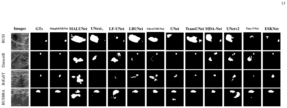
- The 0.67 MB SimpleESKNet gives a parameter-versus-accuracy target: about 140 times fewer parameters than TransUNet with equal or better reported DSC/IoU on the breast-lesion benchmark.
- U-Net's progressive width doubling and full skip concatenation are not essential to accuracy on these tasks; removing them costs little and saves most of the parameters.
- The feature-selection and adaptive-fusion strategies are transferable, since replacing the plain blocks with ESKNet's blocks preserves and extends the gains.
Reading between the lines
- Editorial extension: the same fixed-width recipe should carry over to 3D volumetric segmentation, where memory limits are tighter; the paper reports only 2D experiments.
- Editorial extension: the ablations imply the model's capacity budget is spent on depth and receptive field instead of width, so the gains likely depend on deeper blocks and dilation, not on width.
- Editorial extension: the reported ISIC numbers may be optimistic because the test set is used for best-epoch selection when no validation split exists; re-running with a separate validation split would give an unbiased estimate.
- Editorial extension: replacing plain convolutions with grouped depthwise or spatially reduced operations, which the discussion names but does not implement, should lower the computational cost that currently remains higher than some lightweight rivals.
Signed reviews
Editorial analysis
A structured set of objections, weighed in public.
Referee Report
Summary. This manuscript proposes SimpleUNet, a lightweight U-Net variant built from three strategies: partial channel selection on skip connections, fixed width across all encoder/decoder stages, and a learnable channel-wise weighting of shortcut and deep features. The authors report parameter counts as low as 16 KB and 0.67 MB for their larger variant, with experiments on a merged breast ultrasound dataset (MBD), two skin-lesion datasets (ISIC2017/2018), and a polyp dataset (Kvasir-SEG). Extensive ablations vary width, kernel size, dilation rate, feature-selection rate, number of blocks, and adaptive fusion, and the framework is extended to ESKNet blocks as SimpleESKNet. The central claims are that the 16 KB model outperforms lightweight benchmarks and that the 0.67 MB variant surpasses U-Net and TransUNet on several datasets.
Significance. The architectural idea is attractive and the empirical scope is substantial: every model is trained five times from scratch, all hyperparameters are swept in ablations, and the released code makes the parameter counts directly verifiable. If the reported ISIC numbers were obtained under an independent protocol, the result would be practically important because it would show that extreme parameter compression does not force a large accuracy penalty. The extendability result (replacing convolution blocks with ESKNet blocks) is a useful demonstration that the design pattern is transferable. The main value of the paper, if corrected, would be as a simple, reproducible baseline for efficient medical image segmentation.
major comments (3)
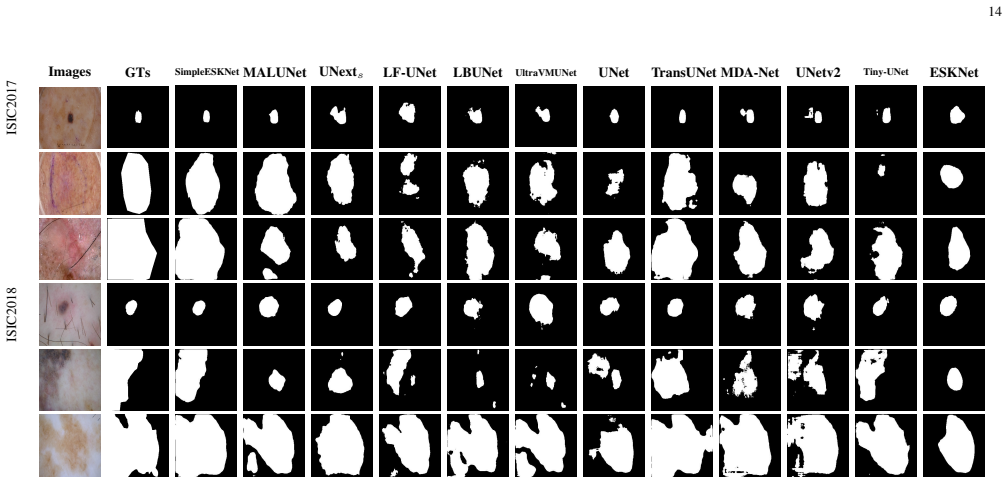
- [Section IV-A and Table II] The protocol sentence 'Otherwise, the testing set is used as the validation set instead' applies directly to ISIC2017 and ISIC2018, because Table II lists only Train and Test for those datasets. Best-epoch weights are therefore selected on the exact test images that are later scored, so the reported ISIC mDice/mIoU values in Table XVI (including the abstract's 84.86% and 88.77%) are in-sample estimates rather than independent test-set evaluations. This is a load-bearing threat to the headline 'consistent dominance' claim on two of the four datasets; the authors must add a proper validation split (or a pre-registered selection rule) and re-evaluate, or remove/qualify the ISIC claims.
- [Abstract and Table XVI] The abstract's statement that the 16 KB configuration 'outperforms LBUNet and other lightweight benchmarks across multiple public datasets' is contradicted by the paper's own Table XVI on ISIC2018: SimpleUNet16 scores 87.57 mDice and 79.98 mIoU, whereas LBUNet scores 87.85 and 80.69. On ISIC2017 the same 16 KB model (83.47 mDice) is also below UNexts (83.79). The headline should be restricted to MBD and Kvasir-SEG, or the claim should be explicitly qualified as 'on some datasets.'
- [Table XVI] Several of the headline superiority claims are based on differences well within one standard deviation, e.g., MBD SimpleESKNet32 85.76±0.49 vs TransUNet 85.32±0.48 and Kvasir-SEG SimpleESKNet32 86.46±0.42 vs ESKNet 86.60±0.83. With M=5 independent runs and no paired significance test, these differences are not established. The authors should report paired tests (e.g., Wilcoxon signed-rank or bootstrap over test images) for at least the abstract's comparison set, and avoid 'dominating' language where differences are not significant.
minor comments (8)
- [Section III-D] The text twice refers to the 'Hardmard product'; this should be 'Hadamard product.'
- [Table I caption] The caption says '(MBT)' but the dataset is called MBD everywhere else; the acronym should be made consistent.
- [References] References [2] and [3] are duplicate entries for the same Mambasam paper and should be merged.
- [Section III-A/III-E] The parameter-count formulas are stated for the case 'N as one,' while Table III experiments use two stacked convolution layers per block; please reconcile the formulas with the exact architectures used in the experiments or explicitly label them as rough estimates for a simplified U-Net.
- [Abstract and Data Availability] The abstract gives a GitHub link, but the Data Availability section states that code will be released after acceptance; these statements should be made consistent.
- [Table XVI notation] The names 'SimpleUNet1_16' and 'SimpleESKNet1+AFF_16' are not explained in a table footnote; the reader cannot tell which subscript/superscript denotes width and which denotes block count.
- [Throughout] Write 'Kvasir-SEG' rather than 'KV ASIR-SEG' for consistency with the dataset's official name.
- [Table XII] The entry 'Para < 0.251' for the model without adaptive feature fusion is imprecise; report the exact parameter count.
Circularity Check
ISIC test-set-as-validation protocol makes headline ISIC mDice values selected on the test set rather than independently predicted; architecture derivation itself is not circular.
-
fitted input called prediction
[Section IV-A, Datasets (KVASIR-SEG paragraph); Table II; Table XVI]
"For datasets containing validation datasets, we only validate the performance of the model during training after each epoch and iteratively save the weights that give the best performance on the validation set. When model training finishes, we reload the weights from the best-performing model for testing on the unseen test set. Otherwise, the testing set is used as the validation set instead."
ISIC2017 and ISIC2018 have no validation split: Table II lists only Train and Test. Under the stated protocol, every model's checkpoint is selected by maximizing the evaluation metric on the exact test images, and the reported mDice/mIoU are then computed on that same test set. The reported ISIC numbers (abstract: mDice 84.86%/88.77%) are therefore best-over-epoch selections on the evaluation set itself, not independent estimates of generalization. This makes the ISIC portion of the headline 'consistent dominance' claim reduce by construction to fitting on the test set.
full rationale
The architectural derivation is self-contained: parameter-count formulas in Section III are arithmetic consequences of layer widths, selection rates, and kernel sizes, not fitted quantities. The ablations are standard hyperparameter tuning, and the central design claim (fixed width plus partial feature selection plus lightweight fusion) does not assume the conclusion. Comparisons on MBD and KVASIR-SEG use proper validation splits, so those results are independent. The partial circularity is confined to ISIC2017/2018, where the lack of a validation split leads the authors to select best checkpoints on the test set and then report metrics on the same test set. This affects the abstract's ISIC mDice claims and the 'consistent dominance' statement; Table XVI also shows SimpleUNet1_16 (87.57) below LBUNet (87.85) on ISIC2018, an internal inconsistency but not itself a circular step. Overall, one load-bearing empirical claim reduces to test-set fitting, while the method and its other evaluations retain independent content, giving a partial-circularity score of 6.
Assumptions & free parameters
free parameters (6)
- Feature selection rate R =
0.5 (default; ablations from 0.25 to 1)
- Model width C =
64 for default SimpleUNet; 48 for best SimpleUNet4+AFF; 32 for SimpleESKNet2+AFF
- Kernel size k =
3
- Dilation rate =
2
- Number of convolution blocks N per stage =
1 for minimal SimpleUNet; 2 for SimpleUNet4 and SimpleESKNet2
- Adaptive feature fusion enabled =
on for the best reported models
assumptions (5)
- standard math The parameter-count estimates in Section III assume square k by k convolutions, a single convolution block per stage, no bias terms, and the stated stage counts.
- domain assumption The public datasets and their annotations are correct and are used with standard train/test splits.
- domain assumption Best-checkpoint selection on the validation set, or on the test set when no validation set exists, yields unbiased performance estimates.
- domain assumption Baseline models such as TransUNet, ESKNet, MALUNet, and LBUNet were re-trained with fair and comparable hyperparameters.
- ad hoc to paper The ESKNet convolution blocks can be treated as drop-in replacements inside the SimpleUNet framework.
Cite this review
Pith. "Pith review of Simple is what you need for efficient and accurate medical image segmentation." pith.science (2026). https://pith.science/paper/MKLJFDXB
@misc{pith2026250613415,
  author       = {Pith},
  title        = {Pith review of: Simple is what you need for efficient and accurate medical image segmentation},
  year         = {2026},
  howpublished = {\url{https://pith.science/paper/MKLJFDXB}},
  note         = {Machine review of arXiv:2506.13415}
}
read the original abstract
While modern segmentation models often prioritize performance over practicality, we advocate a design philosophy prioritizing simplicity and efficiency, and attempted high performance segmentation model design. This paper presents SimpleUNet, a scalable ultra-lightweight medical image segmentation model with three key innovations: (1) A partial feature selection mechanism in skip connections for redundancy reduction while enhancing segmentation performance; (2) A fixed-width architecture that prevents exponential parameter growth across network stages; (3) An adaptive feature fusion module achieving enhanced representation with minimal computational overhead. With a record-breaking 16 KB parameter configuration, SimpleUNet outperforms LBUNet and other lightweight benchmarks across multiple public datasets. The 0.67 MB variant achieves superior efficiency (8.60 GFLOPs) and accuracy, attaining a mean DSC/IoU of 85.76%/75.60% on multi-center breast lesion datasets, surpassing both U-Net and TransUNet. Evaluations on skin lesion datasets (ISIC 2017/2018: mDice 84.86%/88.77%) and endoscopic polyp segmentation (KVASIR-SEG: 86.46%/76.48% mDice/mIoU) confirm consistent dominance over state-of-the-art models. This work demonstrates that extreme model compression need not compromise performance, providing new insights for efficient and accurate medical image segmentation. Codes can be found at https://github.com/Frankyu5666666/SimpleUNet.
Figures


Figures from the paper (7 more)




Reference graph
Works this paper leans on
-
[1]
U-net: Convolutional networks for biomedical image segmentation,
O. Ronneberger, P. Fischer, and T. Brox, “U-net: Convolutional networks for biomedical image segmentation,” in Medical Image Computing and Computer-Assisted Intervention–MICCAI 2015: 18th International Conference, Munich, Germany, October 5-9, 2015, Proceedings, Part III
2015
-
[2]
Mambasam: A visual mamba-adapted sam frame- work for medical image segmentation,
P. Liang, L. Shi, B. Pu, R. Wu, J. Chen, L. Zhou, L. Xu, Z. Chen, Q. Chang, and Y . Li, “Mambasam: A visual mamba-adapted sam frame- work for medical image segmentation,” IEEE Journal of Biomedical and Health Informatics, 2025
work page 2025
-
[3]
Mambasam: A visual mamba-adapted sam framework for med- ical image segmentation,
——, “Mambasam: A visual mamba-adapted sam framework for med- ical image segmentation,” IEEE Journal of Biomedical and Health Informatics, pp. 1–12, 2025
work page 2025
-
[4]
G. Chen, N. Zhu, J. Lin, B. Pu, H. Luo, and K. Li, “Thyfusion: A lightweight attribute enhancement module for thyroid nodule diagnosis using gradient and frequency-domain awareness,” Neurocomputing, vol. 613, p. 128749, 2025. [Online]. Available: https://www.sciencedirect. com/science/article/pii/S0925231224015200
work page 2025
-
[5]
Transunet: Transformers make strong encoders for medical image segmentation,
J. Chen, Y . Lu, Q. Yu, X. Luo, E. Adeli, Y . Wang, L. Lu, A. L. Yuille, and Y . Zhou, “Transunet: Transformers make strong encoders for medical image segmentation,” CoRR, vol. abs/2102.04306, 2021. [Online]. Available: https://arxiv.org/abs/2102.04306
arXiv 2021
-
[6]
Esknet: An enhanced adaptive selection kernel convolution for ultrasound breast tumors segmentation,
G. Chen, L. Zhou, J. Zhang, X. Yin, L. Cui, and Y . Dai, “Esknet: An enhanced adaptive selection kernel convolution for ultrasound breast tumors segmentation,” Expert Systems with Applications, vol. 246, p. 123265, 2024
work page 2024
-
[7]
Ukan: Unbound kolmogorov-arnold network accompanied with accelerated library,
A. Moradzadeh, L. Wawrzyniak, M. Macklin, and S. G. Paliwal, “Ukan: Unbound kolmogorov-arnold network accompanied with accelerated library,” arXiv preprint arXiv:2408.11200, 2024
-
[8]
Mlmseg: a multi-view learning model for ultrasound thyroid nodule segmentation,
G. Chen, G. Tan, M. Duan, B. Pu, H. Luo, S. Li, and K. Li, “Mlmseg: a multi-view learning model for ultrasound thyroid nodule segmentation,” Computers in Biology and Medicine, vol. 169, p. 107898, 2024
work page 2024
Show all 39 references
-
[9]
Mobileunet- fpn: A semantic segmentation model for fetal ultrasound four-chamber segmentation in edge computing environments,
B. Pu, Y . Lu, J. Chen, S. Li, N. Zhu, W. Wei, and K. Li, “Mobileunet- fpn: A semantic segmentation model for fetal ultrasound four-chamber segmentation in edge computing environments,” IEEE Journal of Biomedical and Health Informatics, vol. 26, no. 11, pp. 5540–5550, 2022
2022
-
[10]
Xception: Deep learning with depthwise separable convolu- tions,
F. Chollet, “Xception: Deep learning with depthwise separable convolu- tions,” in Proceedings of the IEEE conference on computer vision and pattern recognition, 2017, pp. 1251–1258. 15
2017
-
[11]
Vm-unet: Vision mamba unet for medical image segmentation,
J. Ruan, J. Li, and S. Xiang, “Vm-unet: Vision mamba unet for medical image segmentation,” arXiv preprint arXiv:2402.02491, 2024
2024 arXiv
-
[12]
Lb-unet: A lightweight boundary-assisted unet for skin lesion segmentation,
J. Xu and L. Tong, “Lb-unet: A lightweight boundary-assisted unet for skin lesion segmentation,” in International Conference on Medical Image Computing and Computer-Assisted Intervention. Springer, 2024, pp. 361–371
2024
-
[13]
Ultralight vm-unet: Parallel vision mamba significantly reduces parameters for skin lesion segmen- tation,
R. Wu, Y . Liu, P. Liang, and Q. Chang, “Ultralight vm-unet: Parallel vision mamba significantly reduces parameters for skin lesion segmen- tation,” arXiv preprint arXiv:2403.20035, 2024
2024 arXiv
-
[14]
Enet: A deep neural network architecture for real-time semantic segmentation,
A. Paszke, A. Chaurasia, S. Kim, and E. Culurciello, “Enet: A deep neural network architecture for real-time semantic segmentation,” arXiv preprint arXiv:1606.02147, 2016
2016 arXiv
-
[15]
Linknet: Exploiting encoder repre- sentations for efficient semantic segmentation,
A. Chaurasia and E. Culurciello, “Linknet: Exploiting encoder repre- sentations for efficient semantic segmentation,” in 2017 IEEE visual communications and image processing (VCIP). IEEE, 2017, pp. 1–4
2017
-
[16]
Unext: Mlp-based rapid medical image segmentation network,
J. M. J. Valanarasu and V . M. Patel, “Unext: Mlp-based rapid medical image segmentation network,” in International conference on medical image computing and computer-assisted intervention. Springer, 2022, pp. 23–33
2022
-
[17]
Lfu-net: a lightweight u-net with full skip connections for medical image segmen- tation,
Y . Deng, H. Wang, Y . Hou, S. Liang, and D. Zeng, “Lfu-net: a lightweight u-net with full skip connections for medical image segmen- tation,” Current Medical Imaging, vol. 19, no. 4, pp. 347–360, 2023
2023
-
[18]
Springer, 2015, pp. 234–241
2015
-
[19]
Malunet: A multi-attention and light-weight unet for skin lesion segmentation,
J. Ruan, S. Xiang, M. Xie, T. Liu, and Y . Fu, “Malunet: A multi-attention and light-weight unet for skin lesion segmentation,” in 2022 IEEE International Conference on Bioinformatics and Biomedicine (BIBM). IEEE, 2022, pp. 1150–1156
2022
-
[20]
Mamba: Linear-time sequence modeling with selective state spaces,
A. Gu and T. Dao, “Mamba: Linear-time sequence modeling with selective state spaces,” arXiv preprint arXiv:2312.00752, 2023
2023 arXiv
-
[21]
Cbam: Convolutional block attention module,
S. Woo, J. Park, J.-Y . Lee, and I. S. Kweon, “Cbam: Convolutional block attention module,” in Proceedings of the European conference on computer vision (ECCV), 2018, pp. 3–19
2018
-
[22]
Atten- tion u-net: Learning where to look for the pancreas,
O. Oktay, J. Schlemper, L. L. Folgoc, M. Lee, M. Heinrich, K. Misawa, K. Mori, S. McDonagh, N. Y . Hammerla, B. Kainz et al., “Atten- tion u-net: Learning where to look for the pancreas,” arXiv preprint arXiv:1804.03999, 2018
2018 arXiv
-
[23]
Dau- net: Dual attention-aided u-net for segmenting tumor in breast ultrasound images,
P. Pramanik, A. Roy, E. Cuevas, M. Perez-Cisneros, and R. Sarkar, “Dau- net: Dual attention-aided u-net for segmenting tumor in breast ultrasound images,” Plos one, vol. 19, no. 5, p. e0303670, 2024
2024
-
[24]
Mda-net: Multiscale dual attention-based network for breast lesion segmentation using ultrasound images,
A. Iqbal and M. Sharif, “Mda-net: Multiscale dual attention-based network for breast lesion segmentation using ultrasound images,”Journal of King Saud University-Computer and Information Sciences, vol. 34, no. 9, pp. 7283–7299, 2022
2022
-
[25]
Unet++: A nested u-net architecture for medical image segmenta- tion,
Z. Zhou, M. M. Rahman Siddiquee, N. Tajbakhsh, and J. Liang, “Unet++: A nested u-net architecture for medical image segmenta- tion,” in Deep Learning in Medical Image Analysis and Multimodal Learning for Clinical Decision Support: 4th International Workshop, DLMIA 2018, and 8t...
2018
-
[26]
Mf-net: Multiple-feature extraction network for breast lesion segmentation in ultrasound images,
J. Wang, G. Liu, D. Liu, and B. Chang, “Mf-net: Multiple-feature extraction network for breast lesion segmentation in ultrasound images,” Expert Systems with Applications, vol. 249, p. 123798, 2024
2024
-
[27]
Unet 3+: A full-scale connected unet for medical image segmentation,
H. Huang, L. Lin, R. Tong, H. Hu, Q. Zhang, Y . Iwamoto, X. Han, Y .-W. Chen, and J. Wu, “Unet 3+: A full-scale connected unet for medical image segmentation,” in ICASSP 2020-2020 IEEE international conference on acoustics, speech and signal processing (ICASSP). IEEE, 2020, pp...
2020
-
[28]
Tinyu-net: Lighter yet better u-net with cascaded multi-receptive fields,
J. Chen, R. Chen, W. Wang, J. Cheng, L. Zhang, and L. Chen, “Tinyu-net: Lighter yet better u-net with cascaded multi-receptive fields,” in International Conference on Medical Image Computing and Computer-Assisted Intervention. Springer, 2024, pp. 626–635
2024
-
[29]
Dataset of breast ultrasound images
W. Al-Dhabyani, M. Gomaa, H. Khaled, and A. Fahmy, “Dataset of breast ultrasound images.” Elsevier, 2020
2020
-
[30]
Bus-set: A benchmark for quantitative evaluation of breast ultrasound segmentation networks with public datasets,
C. Thomas, M. Byra, R. Marti, M. H. Yap, and R. Zwiggelaar, “Bus-set: A benchmark for quantitative evaluation of breast ultrasound segmentation networks with public datasets,” Medical Physics, vol. 50, no. 5, pp. 3223–3243, 2023
2023
-
[31]
Curated benchmark dataset for ultrasound based breast lesion analysis,
A. Pawłowska, A. ´Cwierz-Pie´nkowska, A. Domalik, D. Jagu ´s, P. Kasprzak, R. Matkowski, Ł. Fura, A. Nowicki, and N. ˙Zołek, “Curated benchmark dataset for ultrasound based breast lesion analysis,”Scientific Data, vol. 11, no. 1, p. 148, 2024
2024
-
[32]
Bus-bra: A breast ultrasound dataset for assessing computer-aided diagnosis systems,
W. G ´omez-Flores, M. J. Gregorio-Calas, and W. Coelho de Albu- querque Pereira, “Bus-bra: A breast ultrasound dataset for assessing computer-aided diagnosis systems,” Medical Physics, 2023
2023
-
[33]
Isic 2017-skin lesion analysis towards melanoma detec- tion,
M. Berseth, “Isic 2017-skin lesion analysis towards melanoma detec- tion,” arXiv preprint arXiv:1703.00523, 2017
2017 arXiv
-
[34]
Isic 2018-a method for lesion segmen- tation,
H. Wen, R. Xu, and T. Zhang, “Isic 2018-a method for lesion segmen- tation,” arXiv preprint arXiv:1807.07391, 2018
2018 arXiv
-
[35]
Kvasir-seg: A segmented polyp dataset,
D. Jha, P. H. Smedsrud, M. A. Riegler, P. Halvorsen, T. De Lange, D. Johansen, and H. D. Johansen, “Kvasir-seg: A segmented polyp dataset,” in International conference on multimedia modeling. Springer, 2019, pp. 451–462
2019
-
[36]
S. M. Pizer, “Contrast-limited adaptive histogram equalization: Speed and effectiveness stephen m. pizer, r. eugene johnston, james p. ericksen, bonnie c. yankaskas, keith e. muller medical image display research group,” in Proceedings of the first conference on visualization ...
1990
-
[37]
Decoupled weight decay regularization,
I. Loshchilov, “Decoupled weight decay regularization,” arXiv preprint arXiv:1711.05101, 2017
2017 arXiv
-
[38]
Sgdr: Stochastic gradient descent with warm restarts,
I. Loshchilov and F. Hutter, “Sgdr: Stochastic gradient descent with warm restarts,” arXiv preprint arXiv:1608.03983, 2016
2016 arXiv
-
[39]
U-net v2: Rethinking the skip connections of u-net for medical image segmentation,
Y . Peng, M. Sonka, and D. Z. Chen, “U-net v2: Rethinking the skip connections of u-net for medical image segmentation,” arXiv preprint arXiv:2311.17791, 2023
2023 arXiv
Reviewed August 15, 2026 · model on record in the stance chip above.
Discussion (0). Continue with ORCID to comment.