REVIEW 4 major objections 4 minor 20 references
Toward Accessible Dermatology: Skin Lesion Classification Using Deep Learning Models on Mobile-Acquired Images
T0 review · 4 major / 4 minor · reviewed 2026-08-15 · deepseek-v4-flash
Pith's one-line read Swin Transformer beats CNN baselines on phone-photo skin disease classification, hitting an F1 of 81 percent.
desk verdict The proposed 51-class non-dermoscopic benchmark is a useful idea, but the paper's own numbers and broken dataset link undercut the central claims. read the letter →
The pith
A machine-rendered reading of the paper's core claim, the machinery that carries it, and where it could break.
The reading
What carries the argument
The load-bearing mechanism is the Swin Transformer trained via transfer learning: its hierarchical, shifted-window self-attention, which divides images into patches and computes attention within shifted windows across stages, is what the paper credits for capturing both local lesion detail and global context. The other essential component is the curated non-dermoscopic dataset itself, which supplies the 51-class, real-world mobile-image distribution; both the architecture comparison and the interpretability analysis depend on it.
What would settle it
Sum the per-category image counts in Table I and compare the result with the claimed total of over 27,000 images; then inspect a random sample for phone-camera provenance and have a dermatologist verify a sample of the labels. If the sum falls far short of the claim, or if a meaningful share of sampled images fails the provenance or label check, the reported 81 percent F1 cannot be taken at face value. An independent check would be to run Swin-Base on a separately collected mobile-acquired skin image dataset and see whether accuracy and MCC remain near 80 percent.
Extended reading notes
Core claim
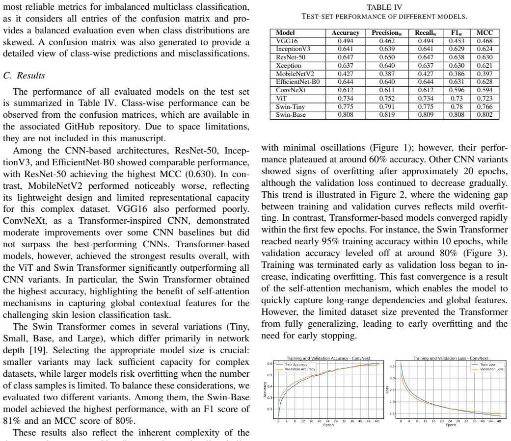
The paper's central claim is that Transformer-based architectures, specifically Swin-Base, outperform CNN baselines for multiclass classification of mobile-acquired, non-dermoscopic skin images, reaching 80.8 percent accuracy, 81 percent F1, and 80.2 percent MCC on a 51-class test set. It further claims that this performance rests on a newly curated dataset of more than 27,000 images across over 50 skin disease categories collected from public online sources, described as one of the largest publicly reported non-dermoscopic collections. The authors argue that self-attention enables Swin to capture global contextual features that CNNs miss, and they use Grad-CAM visualizations to show that the models attend to lesion regions rather than background cues.
Load-bearing premise
The entire result rests on the curated dataset being what it claims: genuine, correctly labeled mobile-phone photos numbering more than 27,000, even though the images were gathered from unverified online sources and the category counts in the paper's own table do not obviously sum to the stated total.
Editorial extensions
If this is right
- If the 81 percent F1 and MCC results hold, a phone-photo-based screening tool is within reach for dozens of common skin disease categories, including in settings with no dermatologist or dermoscopy equipment.
- Transformer architectures become the preferred starting point for non-dermoscopic skin classification over CNN baselines such as ResNet-50 and EfficientNet-B0.
- The released curated dataset gives other researchers a common benchmark for mobile-acquired skin images, reducing the field's reliance on dermoscopic collections.
- Frozen-backbone transfer learning with ImageNet weights is sufficient to obtain strong results on this data scale; fine-tuning backbone layers tends to cause overfitting.
- Grad-CAM heatmaps can serve as a basic trust check, showing whether a prediction is driven by the lesion itself rather than by background or image artifacts.
Reading between the lines
- My reading: if the online-sourced labels are noisy, the reported 81 percent F1 is likely an upper bound on real-world performance, because label noise usually inflates apparent agreement on test data drawn from the same noisy source.
- My reading: the 'global context' explanation for Swin's edge is consistent with the results but not directly proven; an ablation restricting attention windows, or analysis of attention-map statistics, would test it.
- My reading: the dataset's imbalance, with some classes having around 100 images, means per-class reliability varies substantially; deployment should probably be limited to well-represented classes until more data is added.
- A testable extension the paper does not run is evaluating the same models on a held-out set of freshly taken phone photos rather than the curated web collection, which would measure true domain transfer to real screening conditions.
Signed reviews
Editorial analysis
A structured set of objections, weighed in public.
Referee Report
Summary. The manuscript curates a non-dermoscopic skin-image dataset from online sources, claims it contains more than 27,000 images across over 50 skin disease categories captured with mobile devices, and evaluates six CNN and three Transformer-based architectures on a 51-class subset. The authors report that Swin-Base achieves the best test-set performance (accuracy 0.808, F1 0.808, MCC 0.802) and use Grad-CAM to visualize the regions driving predictions. The stated goal is to demonstrate that Transformer models, and Swin in particular, outperform CNN baselines on realistic mobile-acquired images, thereby supporting accessible AI-assisted dermatological screening in low-resource settings.
Significance. If the dataset and evaluation claims are supported, the curated public dataset would be a useful resource for non-dermoscopic skin-image research, and the head-to-head comparison of CNNs and Transformers on such images would be informative. The paper also has strengths: it reports MCC, discusses fine-tuning and data-augmentation experiments, and states limitations honestly. However, the central contributions rest on dataset statistics and provenance that are not yet verified, and the architecture comparison is confounded by input resolution and single-split evaluation. These issues are load-bearing rather than cosmetic, so the claims in their current form are not fully established.
major comments (4)
- [Section III-A and Table I] The text states that the curated dataset contains 'more than 27,000 images,' but Table I lists 68 categories whose counts sum to 24,351. Section IV-A then states that categories with too few images were excluded, leaving 51 classes, so the actual training pool is smaller still. This discrepancy directly affects the paper's primary contribution of providing 'one of the largest publicly reported collections of non-dermoscopic skin lesion images.' Please reconcile the numbers, report the exact total before and after filtering, list which categories were excluded, and provide final per-class counts and train/validation/test sizes.
- [Section III-A and Abstract] The claim that the images are 'captured with mobile devices' is not supported by the evidence in the manuscript. Only PAD-UFES-20 is explicitly described as smartphone-acquired; Dermatology Atlas and the Monkeypox repository are web/atlas collections, and no EXIF, device, or capture-source audit is reported. Section V-E later says all images were 'collected from online sources,' which is a different claim. Please provide verifiable provenance per source, or substantially qualify the mobile-acquired characterization throughout the abstract, introduction, and conclusion.
- [Section III-A and Section V-E] Label correctness and image uniqueness are not established. The sources are unverified online repositories, no expert validation protocol is described, no de-duplication procedure is reported, and the class list contains inconsistent labels such as 'acne' versus 'acne vulgaris.' Because label noise and near-duplicate leakage between training and test sets can alter model rankings, please provide a validation protocol, a deduplication procedure, and a quantitative error analysis. In addition, the repository link in the Acknowledgments is malformed (it contains a space), which prevents independent verification of the dataset and code.
- [Section IV-A, Tables III and IV] The evaluation is based on a single data split, one checkpoint per model, and no error bars or significance tests, so the reported differences between architectures may not be stable. Moreover, Table III shows that CNN inputs are 224x224 or 299x299 while Transformer inputs are 384x384, so image resolution is confounded with architecture. The conclusion that Swin-Base outperforms CNN baselines because of self-attention and global feature capture is therefore not supported by the experiment as designed. Please add repeated runs with confidence intervals, control input resolution across architecture families, or otherwise justify that the observed ranking is not an artifact of resolution or split.
minor comments (4)
- [Section IV-B] The notation 'Precision w', 'Recall w', and 'F1 w' is used without defining that the weights are the class support proportions; please define the weighted averaging convention explicitly.
- [Acknowledgments] The GitHub URL is given as 'https://github.com/newaz-aa/skin lesion classification DL', which contains a space and is not a usable link; please provide the correct URL.
- [Section V-B, Figure 4b] The predicted label 'juvenile xanthogranuloma' does not appear in Table I, so it is unclear whether this class is among the 51 evaluated classes or the figure is mislabeled; please correct this inconsistency.
- [Section V-A, Figures 1-3] The accuracy curves for ConvNeXt and ResNet-50 are plotted as fractions, while the Swin accuracy curve is plotted in percent; please use consistent units and label the axes accordingly.
Circularity Check
No circular reasoning is present: the model rankings come from a held-out test set, and the dataset is described as an external compilation rather than derived from the results.
full rationale
This paper is an empirical benchmarking study. It curates a dataset from external public sources, trains several standard deep learning architectures with ImageNet-pretrained weights, and evaluates them on a held-out test split. The central result, that Swin-Base achieves the highest accuracy and MCC (Table IV), follows from the reported test-set evaluation and is not derived from the dataset definition or from any fitted parameter that is then renamed as a prediction. No equation in the paper defines one result in terms of another, and no fitted input is subsequently presented as a prediction. The interpretive statement that Transformers win by capturing global contextual features is a post hoc explanation and may be untested, but that is an explanatory weakness, not circularity. The paper does not rely on a load-bearing self-citation chain: the cited references are external datasets, architectures, and prior studies, and the authors do not invoke any of their own prior uniqueness theorems or ansatzes to force conclusions. The acknowledged discrepancies in dataset size (Table I sums to 24,351 while the text claims over 27,000) and the unverified mobile-acquisition provenance are serious data-integrity concerns, but they are factual consistency issues rather than circular derivation. Accordingly, no step reduces to its own inputs, and the appropriate circularity score is 0.
Assumptions & free parameters
free parameters (8)
- Learning rate =
1e-4
- Batch size =
32
- Dropout rate =
0.3 or 0.4
- Input image size =
224x224, 299x299, or 384x384 depending on model
- Classification head width and depth =
128 or 256 neurons, one or two fully connected layers
- Early stopping and epochs =
Up to 50 epochs, early stopping; patience unspecified
- Class exclusion threshold =
not stated
- Random seed =
fixed but value not given
assumptions (6)
- domain assumption ImageNet-pretrained weights provide useful generic features for skin images
- domain assumption Online source labels are correct and refer to the same disease definitions
- domain assumption Images are non-dermoscopic and mobile-acquired
- domain assumption Skin diseases can be reliably classified from visual appearance alone
- domain assumption Train/test split is representative and independent
- standard math Standard softmax cross-entropy training with backpropagation
Cite this review
Pith. "Pith review of Toward Accessible Dermatology: Skin Lesion Classification Using Deep Learning Models on Mobile-Acquired Images." pith.science (2026). https://pith.science/paper/OOJI6HXI
@misc{pith2026250904800,
  author       = {Pith},
  title        = {Pith review of: Toward Accessible Dermatology: Skin Lesion Classification Using Deep Learning Models on Mobile-Acquired Images},
  year         = {2026},
  howpublished = {\url{https://pith.science/paper/OOJI6HXI}},
  note         = {Machine review of arXiv:2509.04800}
}
read the original abstract
Skin diseases are among the most prevalent health concerns worldwide, yet conventional diagnostic methods are often costly, complex, and unavailable in low-resource settings. Automated classification using deep learning has emerged as a promising alternative, but existing studies are mostly limited to dermoscopic datasets and a narrow range of disease classes. In this work, we curate a large dataset of over 50 skin disease categories captured with mobile devices, making it more representative of real-world conditions. We evaluate multiple convolutional neural networks and Transformer-based architectures, demonstrating that Transformer models, particularly the Swin Transformer, achieve superior performance by effectively capturing global contextual features. To enhance interpretability, we incorporate Gradient-weighted Class Activation Mapping (Grad-CAM), which highlights clinically relevant regions and provides transparency in model predictions. Our results underscore the potential of Transformer-based approaches for mobile-acquired skin lesion classification, paving the way toward accessible AI-assisted dermatological screening and early diagnosis in resource-limited environments.
Figures


Figures from the paper (2 more)


Reference graph
Works this paper leans on
-
[1]
Hay R. J., et al., “The global burden of skin disease in 2010: An analysis of the prevalence and impact of skin conditions,”Journal of Investigative Dermatology, vol. 134, no. 6, pp. 1527–1534, 2014
work page 2010
-
[2]
Hasan, M. K., Ahamad, M. A., Yap, C. H., & Yang, G. (2023). A survey, review, and future trends of skin lesion segmentation and classification. Computers in Biology and Medicine, 155, 106624
work page 2023
-
[3]
Goyal, M., Knackstedt, T., Yan, S., & Hassanpour, S. (2020). Artifi- cial intelligence-based image classification methods for diagnosis of skin cancer: Challenges and opportunities. Computers in biology and medicine, 127, 104065
work page 2020
-
[4]
https://challenge.isic-archive.com/data/
-
[5]
Tschandl, P., Rosendahl, C., & Kittler, H. (2018). The HAM10000 dataset, a large collection of multi-source dermatoscopic images of common pigmented skin lesions. Scientific data, 5(1), 1-9
work page 2018
-
[6]
https://github.com/jeremykawahara/derm7pt?tab=readme-ov-file
-
[7]
Esteva, A., Kuprel, B., Novoa, R. A., Ko, J., Swetter, S. M., Blau, H. M., & Thrun, S. (2017). Dermatologist-level classification of skin cancer with deep neural networks. nature, 542(7639), 115-118
work page 2017
-
[8]
Dosovitskiy, A., Beyer, L., Kolesnikov, A., Weissenborn, D., Zhai, X., Unterthiner, T., ... & Houlsby, N. (2020). An image is worth 16x16 words: Transformers for image recognition at scale. arXiv preprint arXiv:2010.11929
arXiv 2020
Show all 20 references
-
[9]
G., Lima, G
Pacheco, A. G., Lima, G. R., Salomao, A. S., Krohling, B., Biral, I. P., De Angelo, G. G., ... & de Barros, L. F. (2020). PAD-UFES-20: A skin lesion dataset composed of patient data and clinical images collected from smartphones. Data in brief, 32, 106221
2020
-
[10]
Choudhary, P., Singhai, J., & Yadav, J. S. (2022). Skin lesion detection based on deep neural networks. Chemometrics and Intelligent Labora- tory Systems, 230, 104659
2022
-
[11]
(2019, October)
Young, K., Booth, G., Simpson, B., Dutton, R., & Shrapnel, S. (2019, October). Deep neural network or dermatologist?. In International Work- shop on Multimodal Learning for Clinical Decision Support (pp. 48-55). Cham: Springer International Publishing
2019
-
[12]
K., Jaffery, Z
Arora, G., Dubey, A. K., Jaffery, Z. A., & Rocha, A. (2023). A comparative study of fourteen deep learning networks for multi skin lesion classification (MSLC) on unbalanced data. Neural Computing and Applications, 35(11), 7989-8015
2023
-
[13]
W., & Han, X
Nakai, K., Chen, Y . W., & Han, X. H. (2022). Enhanced deep bottleneck transformer model for skin lesion classification. Biomedical Signal Processing and Control, 78, 103997
2022
-
[14]
Rafay, A., & Hussain, W. (2023). EfficientSkinDis: An EfficientNet- based classification model for a large manually curated dataset of 31 skin diseases. Biomedical Signal Processing and Control, 85, 104869
2023
-
[15]
A., Ahammad, B., & Rahman, M
Sadik, R., Majumder, A., Biswas, A. A., Ahammad, B., & Rahman, M. M. (2023). An in-depth analysis of Convolutional Neural Network architectures with transfer learning for skin disease diagnosis. Healthcare Analytics, 3, 100143
2023
-
[16]
A., Dey, A., & Kabir, M
Hanum, S. A., Dey, A., & Kabir, M. A. (2025). An Attention-Guided Deep Learning Approach for Classifying 39 Skin Lesion Types. arXiv preprint arXiv:2501.05991
2025 arXiv
-
[17]
Monkeypox Skin Images Dataset (MSID)
Bala, Diponkor; Hossain, Md Shamim (2023), “Monkeypox Skin Images Dataset (MSID)”, Mendeley Data, V6, doi: 10.17632/r9bfpnvyxr.6
2023 doi
-
[18]
https://www.atlasdermatologico.com.br/browse.jsf
-
[19]
& Guo, B
Liu, Z., Lin, Y ., Cao, Y ., Hu, H., Wei, Y ., Zhang, Z., ... & Guo, B. (2021). Swin transformer: Hierarchical vision transformer using shifted windows. In Proceedings of the IEEE/CVF international conference on computer vision (pp. 10012-10022)
2021
-
[20]
E., Cosa-Linan, A., Santhanam, N., Jannesari, M., Maros, M
Kim, H. E., Cosa-Linan, A., Santhanam, N., Jannesari, M., Maros, M. E., & Ganslandt, T. (2022). Transfer learning for medical image classification: a literature review. BMC medical imaging, 22(1), 69
2022
Reviewed August 15, 2026 · model on record in the stance chip above.
Discussion (0). Continue with ORCID to comment.