REVIEW 3 major objections 4 minor 57 references
XANES absorption spectra of penta-graphene and penta-SiC2 with different terminations: a computational study
T0 review · 3 major / 4 minor · reviewed 2026-08-06 · deepseek-v4-flash
Pith's one-line read This computational study predicts X-ray absorption fingerprints that distinguish penta-graphene, penta-SiC2, and their hydrogenated and hydroxylated forms.
desk verdict Useful first-pass computational XANES reference for penta-graphene and penta-SiC2, but the 'reliable fingerprints' claim needs an energy calibration before experimentalists should lean on it. read the letter →
The pith
A machine-rendered reading of the paper's core claim, the machinery that carries it, and where it could break.
The reading
What carries the argument
The central object is the K-edge XANES spectrum computed for each inequivalent absorbing site with a core-hole pseudopotential: after X-ray absorption the excited atom's 1s electron is removed and the resulting hole is included in the pseudopotential, so the spectrum reflects the local chemical environment and coordination. The Lanczos chain algorithm evaluates the absorption cross section from the ground-state density without enumerating empty states, and dipole selection rules restrict K-edge transitions to final states with p character or s/p hybrids. Site-resolved spectra are then averaged over the inequivalent sites in the unit cell and broadened with a Lorentzian of 0.8 eV full width at half maximum.
What would settle it
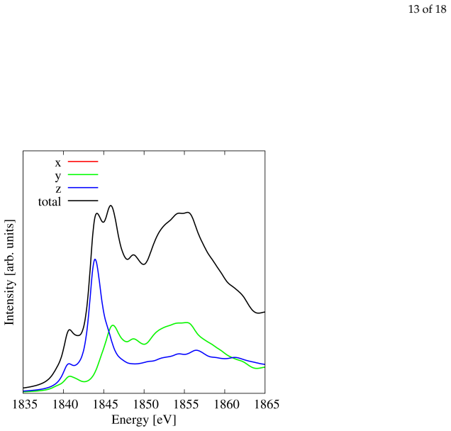
Measure C and Si K-edge XANES on a phase-pure sample of penta-graphene or penta-SiC2; the fingerprint claim fails if the predicted three-plateau C spectrum (283–288, 290–300, and above 305 eV) and the double Si peaks near 1844 and 1857 eV are absent or shifted by more than the several-electronvolt error typical of the uncalibrated scheme.
Extended reading notes
Core claim
On the paper's own terms, the central finding is that pristine, hydrogenated, and hydroxylated penta-graphene, silicon-substituted penta-graphene, and pristine and hydrogenated penta-SiC2 each have distinct C, Si, and O K-edge XANES signatures in peak position, shape, and polarization dependence, so these spectra can serve as reliable fingerprints for experimental identification. Notable discriminators are the 283–288 eV plateau and the site-2 π* peak near 285 eV in pristine penta-graphene, which hydrogenation or hydroxylation removes, and the Si K-edge region around 1840–1860 eV, where coordination site and host lattice change the number, position, and polarization of the peaks.
Load-bearing premise
The fingerprint claim stands on the assumption that the uncalibrated core-hole density functional calculation reproduces absolute XANES peak positions and shapes closely enough for unambiguous identification, yet no comparison to measured spectra is made in the paper.
Editorial extensions
If this is right
- XANES can distinguish pristine penta-graphene from its hydrogenated and hydroxylated forms, because the low-energy plateau at 283–288 eV and the site-2 π* peak near 285 eV vanish on termination.
- The C K-edge spectrum of penta-graphene reproduces graphene's σ* peaks near 293, 298, and 303 eV for the three-coordinated sites, so XANES can recognize the sp2-like sublattice within the pentagonal network.
- Si K-edge spectra separate a single substitutional Si dopant in penta-graphene from true penta-SiC2 by the high-energy peak structure above 1850 eV.
- Polarization-dependent XANES records could reveal the orientation of penta-graphene or penta-SiC2 flakes, since in-plane versus out-of-plane responses are computed to differ strongly in pristine cases.
Reading between the lines
- Beyond the paper's explicit claims: the loss of the low-energy plateau upon hydrogenation or hydroxylation could act as a general marker for conversion of sp2 to sp3 carbon sites in other pentagonal carbon networks.
- The strong site and polarization sensitivity of the Si K-edge suggests a route beyond identification: XANES could locate a single Si dopant within a penta-graphene lattice, provided the computed few-electronvolt energy accuracy holds.
- A testable extension would be to recompute spectra with a self-consistent, many-body treatment of the core hole and compare absolute peak positions to synchrotron measurements, since the current uncalibrated energies are the main uncertainty.
- Because the spectra are computed for isolated 3×3 supercells, an extension to finite temperature or to supported, substrate-bound samples would test whether the fingerprints survive realistic experimental conditions.
Editorial analysis
A structured set of objections, weighed in public.
Referee Report
Summary. The authors present first-principles XANES calculations for penta-graphene in pristine, hydrogenated, and hydroxylated forms, for penta-graphene with a single Si substitution, and for pristine and hydrogenated penta-SiC2. Using the XSpectra code within QUANTUM ESPRESSO with PBE and core-hole pseudopotentials, they compute C, Si, and O K-edge spectra for inequivalent sites and polarizations, and form weighted-average total spectra. They conclude that the calculated peak positions, shapes, and polarization dependencies can serve as reliable fingerprints for experimental identification.
Significance. If the absolute energy scale were validated, the systematic set of computed spectra would be a useful reference for future experimental characterization of these hypothetical materials. The work is methodologically standard and internally consistent: no fitting to experimental data, and the only manually chosen parameter is the Lorentzian broadening. However, the central fingerprint claim is not established because the absolute energy scale of PBE core-hole XANES is not benchmarked against experiment. The paper would be acceptable if the claim is softened to 'predicted spectra' or if a calibration against measured spectra is added.
major comments (3)
- [Section 4 and Section 3.1] The 'reliable fingerprints' claim in the Conclusions requires absolute energy accuracy. The only comparison with experiment is qualitative: site-2 peaks at 293/298/303 eV are said to correspond to graphene's sigma peaks (citing refs [25,55-57]), but no peak-position residuals or calibration shift are reported. Since PBE core-hole XANES can have site- and edge-dependent shifts of several eV, and the sp3 carbon site and all Si K-edge spectra are unbenchmarked, the fingerprints are not validated. Provide a quantitative benchmark against measured C K-edge (e.g., graphene) and Si K-edge (e.g., SiC) spectra, or explicitly restrict the claim to relative spectral features.
- [Section 2.2 and Section 3.1] The 'weighted average' of spectra over inequivalent sites is not defined. It is not stated what weights were used (stoichiometric 1:2 for site 1:site 2, or equal weights). Without this, the total spectra in Figs. 3, 5, 7, 9, 10, 12, and 14 cannot be reproduced, which is a reproducibility problem for the fingerprint claim.
- [Section 2.1] The statement that a 3x3 supercell is 'sufficient for the convergence of the spectra' is not supported by any presented convergence data. Similarly, 'After convergence tests' for cutoff and k-points is asserted but not shown. Because the central claim concerns absolute peak positions, demonstrate supercell-size, cutoff, and k-point convergence of peak energies (e.g., a table of first-peak positions vs supercell size).
minor comments (4)
- [Section 3.6] The first paragraph contains a duplicated sentence: 'We report in Fig. 11 the Si K-edge XANES spectra ... and in Fig. 12 the spectra for the C K-edge.' is immediately followed by 'We show in Fig. 11 the Si K-edge XANES spectra ...' Remove one.
- [Section 2.1] In the sentence 'In addition, penta-SiC and hydrogenated penta-SiC are shown in panels D) and E)', 'penta-SiC' should be 'penta-SiC2' to match the figure and the rest of the manuscript.
- [Figures 2-14] The x- and y-polarized spectra coincide in many figures; the text notes this, but the red and green lines are indistinguishable in print. Consider using dashed/dotted lines for one of the polarizations or stating the coincidence directly in each caption.
- [Section 3.1] The description of the total spectra as 'three plateaus in the ranges 283–288, 290–300 and above 305 eV' is imprecise because the spectra show peaks, not plateaus; rephrase to 'three groups of peaks' or similar.
Circularity Check
No circularity: the XANES fingerprints are computed from first-principles DFT without fitting to measured spectra or importing the target result from self-citations.
full rationale
The paper's central claim is that calculated XANES peak positions, shapes, and polarization dependencies can serve as reliable fingerprints for identifying penta-graphene, penta-SiC2, and their hydrogenated/hydroxylated variants. The spectra are produced by standard DFT-based XANES calculations (XSpectra/Quantum ESPRESSO with ultrasoft core-hole pseudopotentials and the PBE functional). No measured XANES spectrum of these materials enters the calculation, no parameter is fitted to experimental data, and the only manually chosen parameter, the 0.8 eV Lorentzian broadening, affects line shape but does not encode the peak positions or the fingerprint pattern. The comparison with graphene in Section 3.1 is an external qualitative benchmark, not an input to the calculation. The paper does cite prior work by its own authors, but those citations support introductory background and methodological context, not the load-bearing fingerprint derivation. The absence of a quantitative experimental energy-scale validation is a real accuracy concern, but it is a correctness/validation issue, not circularity: the claimed fingerprints are not equivalent to the inputs by construction.
Assumptions & free parameters
free parameters (1)
- Lorentzian broadening FWHM =
0.8 eV
assumptions (4)
- domain assumption PBE functional with core-hole pseudopotential yields XANES spectra accurate enough for fingerprinting
- domain assumption Dipole approximation selects only 1s to p or s/p transitions
- domain assumption 3x3 supercell with 2.0 nm vacuum is converged and isolates the single core-hole interaction
- domain assumption The modeled structures represent the relevant experimental phases of penta-graphene and penta-SiC2
Cite this review
Pith. "Pith review of XANES absorption spectra of penta-graphene and penta-SiC2 with different terminations: a computational study." pith.science (2026). https://pith.science/paper/PL7XWBXY
@misc{pith2026250800704,
  author       = {Pith},
  title        = {Pith review of: XANES absorption spectra of penta-graphene and penta-SiC2 with different terminations: a computational study},
  year         = {2026},
  howpublished = {\url{https://pith.science/paper/PL7XWBXY}},
  note         = {Machine review of arXiv:2508.00704}
}
read the original abstract
In recent research, penta-graphene and penta-SiC2 have emerged as innovative 2D materials consisting exclusively of pentagons. However, there is still a significant gap in the theoretical characterization of these materials, which hinders progress in their synthesis and potential technological applications. This study aims to close this gap by investigating the X-ray absorption near-edge spectroscopy (XANES) of these materials through ab initio calculations. In particular, we analyze the XANES spectra of penta-graphene in its pristine, hydrogenated, and hydroxylated states, and we investigate the effects of substitution by a single silicon in both penta-graphene and pentagraphane. In addition, we calculate the XANES spectra for pristine and hydrogenated penta-SiC2. This work sets the stage for the possible identification of penta-graphene and penta-SiC2 phases by X-ray spectroscopy at the experimental level and lays the foundation for the future engineering of the absorption properties of these materials in optical devices.
Figures


Figures from the paper (11 more)




Reference graph
Works this paper leans on
-
[1]
Geim, A.K.; Novoselov, K.S. The rise of graphene. Nature Materials 2007, 6, 183–191. https://doi.org/10.103 8/nmat1849
work page 2007
-
[2]
Pedrielli, A.; Taioli, S.; Garberoglio, G.; Pugno, N.M. Designing graphene based nanofoams with nonlinear auxetic and anisotropic mechanical properties under tension or compression. Carbon 2017, 111, 796–806. https://doi.org/10.1016/j.carbon.2016.10.034
-
[3]
Honeycomb Carbon: A Review of Graphene
Allen, M.J.; Tung, V .C.; Kaner, R.B. Honeycomb Carbon: A Review of Graphene. Chemical Reviews 2010, 110, 132–145. https://doi.org/10.1021/cr900070d
-
[4]
A decade of graphene research: production, applications and outlook
Randviir, E.P .; Brownson, D.A.; Banks, C.E. A decade of graphene research: production, applications and outlook. Materials Today 2014, 17, 426–432. https://doi.org/10.1016/j.mattod.2014.06.001
-
[5]
Taioli, S. Computational study of graphene growth on copper by first-principles and kinetic Monte Carlo calculations. Journal of Molecular Modeling 2014, 20. https://doi.org/10.1007/s00894-014-2260-2
-
[6]
Azzolini, M.; Morresi, T.; Abrams, K.; Masters, R.; Stehling, N.; Rodenburg, C.; Pugno, N.M.; Taioli, S.; Dapor, M. Anisotropic Approach for Simulating Electron Transport in Layered Materials: Computational and Experimental Study of Highly Oriented Pyrolitic Graphite. The Journal of Physical Chemistry C 2018, 122, 10159–10166. https://doi.org/10.1021/acs....
-
[7]
Production and processing of graphene and related materials
Backes, C.; Abdelkader, A.M.; Alonso, C.; Andrieux-Ledier, A.; Arenal, R.; Azpeitia, J.; Balakrishnan, N.; Banszerus, L.; Barjon, J.; Bartali, R.; et al. Production and processing of graphene and related materials. 2D Materials 2020, 7, 022001. https://doi.org/10.1088/2053-1583/ab1e0a
-
[8]
Lobachevsky crystallography made real through carbon pseudospheres
Taioli, S.; Gabbrielli, R.; Simonucci, S.; Pugno, N.M.; Iorio, A. Lobachevsky crystallography made real through carbon pseudospheres. Journal of Physics: Condensed Matter 2016, 28, 13LT01. https://doi.org/10.1 088/0953-8984/28/13/13lt01
work page 2016
Show all 57 references
-
[9]
Exploring event horizons and Hawking radiation through deformed graphene membranes
Morresi, T.; Binosi, D.; Simonucci, S.; Piergallini, R.; Roche, S.; Pugno, N.M.; Simone, T. Exploring event horizons and Hawking radiation through deformed graphene membranes. 2D Materials 2020, 7, 041006. https://doi.org/10.1088/2053-1583/aba448
2020 doi
-
[10]
Structural, electronic and me- chanical properties of all-sp2 carbon allotropes with density lower than graphene
Morresi, T.; Pedrielli, A.; Beccara, S.a.; Gabbrielli, R.; Pugno, N.M.; Taioli, S. Structural, electronic and me- chanical properties of all-sp2 carbon allotropes with density lower than graphene. Carbon 2020, 159, 512–526. https://doi.org/10.1016/j.carbon.2019.12.024
2020 doi
-
[11]
Mechanical Properties of Twisted Carbon Nanotube Bundles with Carbon Linkers from Molecular Dynamics Simulations
Pedrielli, A.; Dapor, M.; Gkagkas, K.; Taioli, S.; Pugno, N.M. Mechanical Properties of Twisted Carbon Nanotube Bundles with Carbon Linkers from Molecular Dynamics Simulations. International Journal of Molecular Sciences 2023, 24, 2473. https://doi.org/10.3390/ijms24032473
2023 doi
-
[12]
Mechanical and thermal properties of graphene random nanofoams via Molecular Dynamics simulations
Pedrielli, A.; Taioli, S.; Garberoglio, G.; Pugno, N.M. Mechanical and thermal properties of graphene random nanofoams via Molecular Dynamics simulations. Carbon 2018, 132, 766–775. https://doi.org/10.1016/j. carbon.2018.02.081
2018 doi
-
[13]
Computational methods for 2D materials modelling
Carvalho, A.; Trevisanutto, P .E.; Taioli, S.; Castro Neto, A.H. Computational methods for 2D materials modelling. Reports on Progress in Physics 2021, 84, 106501. https://doi.org/10.1088/1361-6633/ac2356
2021 doi
-
[14]
Silicene: Compelling Experimental Evidence for Graphenelike Two-Dimensional Silicon
Vogt, P .; De Padova, P .; Quaresima, C.; Avila, J.; Frantzeskakis, E.; Asensio, M.C.; Resta, A.; Ealet, B.; Le Lay, G. Silicene: Compelling Experimental Evidence for Graphenelike Two-Dimensional Silicon. Phys. Rev. Lett. 2012, 108, 155501. https://doi.org/10.1103/PhysRevLett....
2012 doi
-
[15]
Selective Sputtering and Atomic Resolution Imaging of Atomically Thin Boron Nitride Membranes
Meyer, J.C.; Chuvilin, A.; Algara-Siller, G.; Biskupek, J.; Kaiser, U. Selective Sputtering and Atomic Resolution Imaging of Atomically Thin Boron Nitride Membranes. Nano Letters 2009, 9, 2683–2689. https: //doi.org/10.1021/nl9011497. 16 of 18
2009 doi
-
[16]
The electronic and optical properties of novel germanene and antimonene heterostructures
Chen, X.; Yang, Q.; Meng, R.; Jiang, J.; Liang, Q.; Tan, C.; Sun, X. The electronic and optical properties of novel germanene and antimonene heterostructures. J. Mater. Chem. C 2016, 4, 5434–5441. https: //doi.org/10.1039/C6TC01141A
2016 doi
-
[17]
Massive Dirac quasiparticles in the optical absorbance of graphene, silicene, germanene, and tinene
Matthes, L.; Pulci, O.; Bechstedt, F. Massive Dirac quasiparticles in the optical absorbance of graphene, silicene, germanene, and tinene. Journal of Physics: Condensed Matter 2013, 25, 395305. https://doi.org/10.1 088/0953-8984/25/39/395305
2013
-
[18]
Penta-graphene: A new carbon allotrope
Zhang, S.; Zhou, J.; Wang, Q.; Chen, X.; Kawazoe, Y.; Jena, P . Penta-graphene: A new carbon allotrope. Proceedings of the National Academy of Sciences 2015, 112, 2372–2377. https://doi.org/10.1073/pnas.14165911 12
2015 doi
-
[19]
Computing the band structure and energy gap of penta-graphene by using DFT and G0W0 approximations
Einollahzadeh, H.; Dariani, R.; Fazeli, S. Computing the band structure and energy gap of penta-graphene by using DFT and G0W0 approximations. Solid State Communications 2016, 229, 1–4. https://doi.org/10.101 6/j.ssc.2015.12.012
2016
-
[20]
Electronic properties of extended graphene nanomaterials from GW calculations
Taioli, S.; Umari, P .; De Souza, M.M. Electronic properties of extended graphene nanomaterials from GW calculations. physica status solidi (b) 2009, 246, 2572–2576. https://doi.org/10.1002/pssb.200982339
2009 doi
-
[21]
Communication: Electronic band gaps of semiconducting zig-zag carbon nanotubes from many-body perturbation theory calculations
Umari, P .; Petrenko, O.; Taioli, S.; De Souza, M.M. Communication: Electronic band gaps of semiconducting zig-zag carbon nanotubes from many-body perturbation theory calculations. The Journal of Chemical Physics 2012, 136. https://doi.org/10.1063/1.4716178
2012 doi
-
[22]
Mechanical properties of penta-graphene, hydrogenated penta-graphene, and penta-CN2 sheets
Le, M.Q. Mechanical properties of penta-graphene, hydrogenated penta-graphene, and penta-CN2 sheets. Computational Materials Science 2017, 136, 181 – 190. https://doi.org/https://doi.org/10.1016/j.commatsci. 2017.05.004
2017 doi
-
[23]
Energetic and electronic structure of penta-graphene nanoribbons
Rajbanshi, B.; Sarkar, S.; Mandal, B.; Sarkar, P . Energetic and electronic structure of penta-graphene nanoribbons. Carbon 2016, 100, 118 – 125. https://doi.org/https://doi.org/10.1016/j.carbon.2016.01.014
2016 doi
-
[24]
Electronic structure and magnetic properties of penta-graphene nanoribbons
Yuan, P .F.; Zhang, Z.H.; Fan, Z.Q.; Qiu, M. Electronic structure and magnetic properties of penta-graphene nanoribbons. Phys. Chem. Chem. Phys. 2017, 19, 9528–9536. https://doi.org/10.1039/C7CP00029D
2017 doi
-
[25]
Thermal conductivity of penta-graphene from molecular dynamics study
Xu, W.; Zhang, G.; Li, B. Thermal conductivity of penta-graphene from molecular dynamics study. The Journal of Chemical Physics 2015, 143, 154703. https://doi.org/10.1063/1.4933311
2015 doi
-
[26]
Lattice thermal conductivity of penta-graphene.Carbon 2016, 105, 424–429
Wang, F.Q.; Yu, J.; Wang, Q.; Kawazoe, Y.; Jena, P . Lattice thermal conductivity of penta-graphene.Carbon 2016, 105, 424–429. https://doi.org/10.1016/j.carbon.2016.04.054
2016 doi
-
[27]
Weak interlayer dependence of lattice thermal conductivity on stacking thickness of penta-graphene
Wang, F.Q.; Liu, J.; Li, X.; Wang, Q.; Kawazoe, Y. Weak interlayer dependence of lattice thermal conductivity on stacking thickness of penta-graphene. Applied Physics Letters 2017, 111, 192102. https://doi.org/10.1063/ 1.4996054
2017
-
[28]
Tuning the electronic and magnetic properties of penta-graphene using a hydrogen atom: a theoretical study
Liu, L.L.; Wang, Y.; Chen, C.P .; Yu, H.X.; Zhao, L.S.; Wang, X.C. Tuning the electronic and magnetic properties of penta-graphene using a hydrogen atom: a theoretical study. RSC Adv. 2017, 7, 40200–40207. https://doi.org/10.1039/C7RA06956A
2017 doi
-
[29]
Effect of hydrogen coverage on the buckling of penta-graphene by molecular dynamics simulation
Ebrahimi, S. Effect of hydrogen coverage on the buckling of penta-graphene by molecular dynamics simulation. Molecular Simulation 2016, 42, 1485–1489. https://doi.org/10.1080/08927022.2016.1205191
2016
-
[30]
Tight-binding approach to penta-graphene
Stauber, T.; Beltrán, J.I.; Schliemann, J. Tight-binding approach to penta-graphene. Scientific Reports 2016, 6. https://doi.org/10.1038/srep22672
2016 doi
-
[31]
Studying the electronic and phononic structure of penta- graphane
Einollahzadeh, H.; Fazeli, S.M.; Dariani, R.S. Studying the electronic and phononic structure of penta- graphane. Science and Technology of Advanced Materials 2016, 17, 610–617. https://doi.org/10.1080/14686996 .2016.1219970
2016
-
[32]
Band gap engineering in penta-graphene by substitutional doping: first-principles calculations
Berdiyorov, G.R.; Dixit, G.; Madjet, M.E. Band gap engineering in penta-graphene by substitutional doping: first-principles calculations. Journal of Physics: Condensed Matter 2016, 28, 475001. https://doi.org/10.1088/ 0953-8984/28/47/475001
2016
-
[33]
Tunable Band Gap in Hydrogenated Quasi-Free-Standing Graphene
Haberer, D.; Vyalikh, D.V .; Taioli, S.; Dora, B.; Farjam, M.; Fink, J.; Marchenko, D.; Pichler, T.; Ziegler, K.; Simonucci, S.; et al. Tunable Band Gap in Hydrogenated Quasi-Free-Standing Graphene. Nano Letters 2010, 10, 3360–3366. https://doi.org/10.1021/nl101066m
2010 doi
-
[34]
Direct observation of a dispersionless impurity band in hydrogenated graphene
Haberer, D.; Petaccia, L.; Farjam, M.; Taioli, S.; Jafari, S.A.; Nefedov, A.; Zhang, W.; Calliari, L.; Scarduelli, G.; Dora, B.; et al. Direct observation of a dispersionless impurity band in hydrogenated graphene. Physical Review B 2011, 83. https://doi.org/10.1103/physrevb.83.165433
2011 doi
-
[35]
Tuning the electronic and mechanical properties of penta-graphene via hydrogenation and fluorination
Li, X.; Zhang, S.; Wang, F.Q.; Guo, Y.; Liu, J.; Wang, Q. Tuning the electronic and mechanical properties of penta-graphene via hydrogenation and fluorination. Phys. Chem. Chem. Phys. 2016, 18, 14191–14197. https://doi.org/10.1039/C6CP01092J
2016 doi
-
[36]
Tuning Penta-Graphene Electronic Properties Through Engineered Line Defects
Santos, R.M.d.; Sousa, L.E.d.; Galvão, D.S.; Ribeiro, L.A. Tuning Penta-Graphene Electronic Properties Through Engineered Line Defects. Scientific Reports 2020, 10. https://doi.org/10.1038/s41598-020-64791-x. 17 of 18
2020 doi
-
[37]
Tuning magnetic properties of penta-graphene bilayers through doping with boron, nitrogen, and oxygen
Santos, R.M.d.; da Cunha, W.F.; de Sousa Junior, R.T.; Giozza, W.F.; Ribeiro Junior, L.A. Tuning magnetic properties of penta-graphene bilayers through doping with boron, nitrogen, and oxygen. Scientific Reports 2020, 10. https://doi.org/10.1038/s41598-020-73901-8
2020 doi
-
[38]
Research progress on penta-graphene and its related materials: Properties and applications
Nazir, M.A.; Hassan, A.; Shen, Y.; Wang, Q. Research progress on penta-graphene and its related materials: Properties and applications. Nano Today 2022, 44, 101501. https://doi.org/10.1016/j.nantod.2022.101501
2022
-
[39]
Predicting experimentally stable allotropes: Instability of penta-graphene
Ewels, C.P .; Rocquefelte, X.; Kroto, H.W.; Rayson, M.J.; Briddon, P .R.; Heggie, M.I. Predicting experimentally stable allotropes: Instability of penta-graphene. Proceedings of the National Academy of Sciences2015, 112, 15609– 15612. https://doi.org/10.1073/pnas.1520402112
-
[40]
Unveiling the pentagonal nature of perfectly aligned single-and double-strand Si nano-ribbons on Ag(110)
Cerdá, J.I.; Sławi ´ nska, J.; Le Lay, G.; Marele, A.C.; Gómez-Rodríguez, J.M.; Dávila, M.E. Unveiling the pentagonal nature of perfectly aligned single-and double-strand Si nano-ribbons on Ag(110). Nature Communications 2016, 7. https://doi.org/10.1038/ncomms13076
2016 doi
-
[41]
First-principles study of electronic transport and optical properties of penta-graphene, penta-SiC2 and penta-CN2
Berdiyorov, G.R.; Madjet, M.E.A. First-principles study of electronic transport and optical properties of penta-graphene, penta-SiC2 and penta-CN2. RSC Advances 2016, 6, 50867–50873. https://doi.org/10.1039/ c6ra10376f
2016
-
[42]
Anisotropic ultrahigh hole mobility in two-dimensional penta-SiC2 by strain-engineering: electronic structure and chemical bonding analysis
Xu, Y.; Ning, Z.; Zhang, H.; Ni, G.; Shao, H.; Peng, B.; Zhang, X.; He, X.; Zhu, Y.; Zhu, H. Anisotropic ultrahigh hole mobility in two-dimensional penta-SiC2 by strain-engineering: electronic structure and chemical bonding analysis. RSC Adv. 2017, 7, 45705–45713. https://doi....
2017 doi
-
[43]
Electron spectroscopies and inelastic processes in nanoclusters and solids: Theory and experiment
Taioli, S.; Simonucci, S.; Calliari, L.; Dapor, M. Electron spectroscopies and inelastic processes in nanoclusters and solids: Theory and experiment. Physics Reports 2010, 493, 237–319. https://doi.org/10.1016/j.physrep. 2010.04.003
2010 doi
-
[44]
Advancements in secondary and backscattered electron energy spectra and yields analysis: From theory to applications
Taioli, S.; Dapor, M. Advancements in secondary and backscattered electron energy spectra and yields analysis: From theory to applications. Surface Science Reports 2025, 80, 100646. https://doi.org/10.1016/j. surfrep.2024.100646
2025
-
[45]
X-ray absorption spectroscopy
Yano, J.; Yachandra, V .K. X-ray absorption spectroscopy. Photosynthesis Research 2009, 102, 241–254. https://doi.org/10.1007/s11120-009-9473-8
2009 doi
-
[46]
Enhancement of the core near-band-edge emission induced by an amorphous shell in coaxial one-dimensional nanostructure: the case of SiC/SiO2core/shell self-organized nanowires
Fabbri, F.; Rossi, F.; Attolini, G.; Salviati, G.; Iannotta, S.; Aversa, L.; Verucchi, R.; Nardi, M.; Fukata, N.; Dierre, B.; et al. Enhancement of the core near-band-edge emission induced by an amorphous shell in coaxial one-dimensional nanostructure: the case of SiC/SiO2core...
2010 doi
-
[47]
A novel combined experimental and multiscale theoretical approach to unravel the structure of SiC/SiOx core/shell nanowires for their optimal design
Morresi, T.; Timpel, M.; Pedrielli, A.; Garberoglio, G.; Tatti, R.; Verucchi, R.; Pasquali, L.; Pugno, N.M.; Nardi, M.V .; Taioli, S. A novel combined experimental and multiscale theoretical approach to unravel the structure of SiC/SiOx core/shell nanowires for their optimal d...
2018 doi
-
[48]
Visualization and analysis of atomistic simulation data with OVITO–the Open Visualization Tool
Stukowski, A. Visualization and analysis of atomistic simulation data with OVITO–the Open Visualization Tool. Modelling and Simulation in Materials Science and Engineering 2009, 18, 015012. https://doi.org/10.1088/ 0965-0393/18/1/015012
2009
-
[49]
Computer graphics and graphical user interfaces as tools in simulations of matter at the atomic scale
Kokalj, A. Computer graphics and graphical user interfaces as tools in simulations of matter at the atomic scale. Computational Materials Science 2003, 28, 155–168. https://doi.org/10.1016/s0927-0256(03)00104-6
2003 doi
-
[50]
QUANTUM ESPRESSO: a modular and open-source software project for quantum simulations of materials
Giannozzi, P .; Baroni, S.; Bonini, N.; Calandra, M.; Car, R.; Cavazzoni, C.; Ceresoli, D.; Chiarotti, G.L.; Cococcioni, M.; Dabo, I.; et al. QUANTUM ESPRESSO: a modular and open-source software project for quantum simulations of materials. Journal of Physics: Condensed Matter...
2009 doi
-
[51]
First-principles calculations of x-ray absorption in a scheme based on ultrasoft pseudopotentials: From α-quartz to high-Tc compounds
Gougoussis, C.; Calandra, M.; Seitsonen, A.P .; Mauri, F. First-principles calculations of x-ray absorption in a scheme based on ultrasoft pseudopotentials: From α-quartz to high-Tc compounds. Phys. Rev. B 2009, 80, 075102. https://doi.org/10.1103/PhysRevB.80.075102
2009 doi
-
[52]
http://www.quantum-espresso.org
-
[53]
Generalized Gradient Approximation Made Simple.Phys
Perdew, J.P .; Burke, K.; Ernzerhof, M. Generalized Gradient Approximation Made Simple.Phys. Rev. Lett. 1996, 77, 3865–3868. https://doi.org/10.1103/PhysRevLett.77.3865
1996 doi
-
[54]
Projector augmented-wave method
Blöchl, P .E. Projector augmented-wave method. Phys. Rev. B 1994, 50, 17953–17979. https://doi.org/10.110 3/PhysRevB.50.17953
1994
-
[55]
Near-Edge X-Ray Absorption Fine-Structure Investigation of Graphene
Pacilé, D.; Papagno, M.; Rodríguez, A.F.; Grioni, M.; Papagno, L.; Girit, u.O.; Meyer, J.C.; Begtrup, G.E.; Zettl, A. Near-Edge X-Ray Absorption Fine-Structure Investigation of Graphene. Physical Review Letters 2008, 101. https://doi.org/10.1103/physrevlett.101.066806. 18 of 18
2008 doi
-
[56]
Polarization-dependent C K near-edge X-ray absorption fine-structure of graphene
Papagno, M.; Fraile Rodríguez, A.; Girit, u.; Meyer, J.; Zettl, A.; Pacilé, D. Polarization-dependent C K near-edge X-ray absorption fine-structure of graphene. Chemical Physics Letters 2009, 475, 269–271. https://doi.org/10.1016/j.cplett.2009.05.054
2009 doi
-
[57]
Theory of x-ray absorption spectroscopy: A microscopic Bloch equation approach for two-dimensional solid states
Christiansen, D.; Selig, M.; Biegert, J.; Knorr, A. Theory of x-ray absorption spectroscopy: A microscopic Bloch equation approach for two-dimensional solid states. Physical Review Research 2023, 5. https://doi.org/ 10.1103/physrevresearch.5.023002
2023 doi
Reviewed August 6, 2026 · model on record in the stance chip above.
Discussion (0). Sign in to comment.