REVIEW 2 major objections 6 minor 26 references
Plexus Convolutional Neural Network (PlexusNet): A novel neural network architecture for histologic image analysis
T0 review · 2 major / 6 minor · reviewed 2026-08-14 · deepseek-v4-flash
Pith's one-line read The paper claims that a deliberately small convolutional architecture, PlexusNet, matches much larger networks on histologic cancer classification while using far fewer parameters and achieving better calibration and clinical utility.
desk verdict A useful small-CNN architecture with a real ablation, but the 43.8% slide-reduction headline is an in-sample threshold fit, not a prospective result. read the letter →
The pith
A machine-rendered reading of the paper's core claim, the machinery that carries it, and where it could break.
The reading
What carries the argument
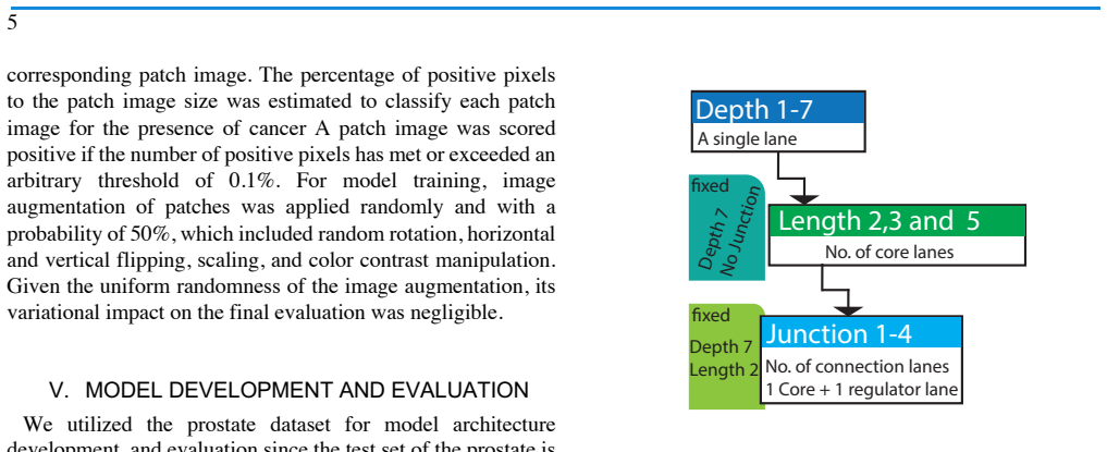
The load-bearing mechanism is the plexus topology: a core lane carries feature maps from input to classification; a regulator lane runs alongside; one or more connection lanes enter and exit at junction points, where a weighted layer $\mathcal{J}_j = \theta_{1j}\mathcal{R}_j + \theta_{2j}\mathcal{C}_j$ fuses regulator features $\mathcal{R}_j$ and core features $\mathcal{C}_j$ at block level $j$. Each lane is a stack of blocks, and each block has a feature-generation segment (plain, residual, or inception-style) followed by a compression segment that reduces channels and controls how much information passes to the global state. The optional normalization section treats color as a vector with learned angle-based weights and rescaling to $[-1,1]$, and the ablation study shows that removing this section lowers slide-level AUROC by 9.3% and worsens the Brier score by 41.9%. The network ends with global pooling, a fully connected layer, and a softmax classification layer.
What would settle it
Fix the 38% threshold on a training or validation set, then evaluate on a held-out set of lymph-node whole-slide images; if the model does not maintain 100% sensitivity and 70% specificity while skipping 43.8% of slides, the clinical-utility claim is refuted.
Extended reading notes
Core claim
The paper's central discovery is that a deliberately condensed architecture can be competitive with large models specifically for histologic image analysis, where data are scarce and overfitting is a practical problem. The PlexusNet architecture combines a feature-extraction section arranged as core lanes, a regulator lane, and connection lanes joined at weighted junction points, with a color-intensity normalization section based on steerable-filter-style angle interpolation. In the prostate cohort, the best PlexusNet model reached a slide-level AUROC of 0.963 with about 177,000 parameters; the comparison models had 23 to 169 times more parameters, yet the PlexusNet model had better calibration and higher net benefit. In the breast lymph-node task, a 57,343-parameter model reached AUROC 0.978, and at a threshold selected by brute-force search on the test set, it captured all positive slides while flagging 70% of negative slides for skipping, corresponding to a 43.8% reduction in slides to review.
Load-bearing premise
The load-bearing premise is that the breast-cancer clinical-utility figures are prospective; the threshold that produces 100% sensitivity and 70% specificity was selected by searching on the same test set, so those numbers are fitting results rather than predicted performance.
Editorial extensions
If this is right
- At 177,382 parameters, the best prostate model is at least 23 times smaller than every comparison network, so training and inference require far less memory and GPU time.
- The breast-metastasis model, with 57,343 parameters, would allow a pathologist to skip roughly 44% of slides if the threshold generalizes, because it flags 70% of negative slides while catching all positives on the test set.
- Color normalization is a necessary component: removing it lowers AUROC by 9.3% and increases Brier score by 41.9% in the ablation study.
- Partitioning of the development set shifts calibration for all architectures, but PlexusNet models achieve similar well-calibrated fits across different partitions, making them less sensitive to how the training/validation split is drawn.
- Larger development sets and ensembling over folds reduce underestimation of cancer probability, so calibration can be tuned by data and ensembling rather than by scaling parameters.
Reading between the lines
- The 43.8% slide-reduction figure is a fitting result: the 38% threshold was chosen by brute-force search on the same test set, so an independent validation is required before expecting the same triage yield prospectively. This is an editorial reading, not the paper's claim.
- The lane-and-junction design is a general inductive bias that could transfer to other dense-image classification problems needing good calibration, such as grading, segmentation, or non-medical imagery; the paper only tests cancer detection.
- Because the architecture starts with two filters and grows channels by a factor of 1.3 to 3 per block, it can be scaled to deeper or wider configurations while remaining far smaller than standard models; the paper does not explore those scaling limits.
Signed reviews
Editorial analysis
A structured set of objections, weighed in public.
Referee Report
Summary. The manuscript introduces PlexusNet, a CNN architecture with a very small parameter count, and evaluates it on two histology tasks: prostate cancer detection on whole-slide images (310 images; development set from TCGA-PRAD, test set from Stanford) and breast cancer lymph node metastasis detection on the Camelyon 2016 dataset (398 slides). The authors report a per-slide AUC of 0.963 for prostate cancer, an AUC of 0.978 on the Camelyon test set, better calibration than several larger comparison architectures, and a clinical-utility claim that a 38% threshold would reduce the number of slides a pathologist must review by 43.8% without missing any metastasis-positive slide. The paper also includes an ablation study of architectural components and an analysis of how development-set partitioning affects model calibration.
Significance. If the results hold, the contribution is useful: a parameter-efficient architecture with competitive discrimination on two histology tasks, reproducible via a public code repository, and with explicit attention to calibration and clinical utility metrics. The ablation study and the finding that fold partitioning affects calibration are informative for practitioners. The main caveat is that the headline clinical-utility result is based on a test-set-optimized threshold and a small number of positive slides, so the 'without consequences' claim is not established by the evidence as presented.
major comments (2)
- [Section VI.E / V.E] The operating point used for the clinical-utility claim is selected by brute-force search on the Camelyon test set itself. The threshold of 38% is chosen to minimize false negatives and false positives on the same 128 slides used to report TPR=100%, TNR=70%, PPV=66.7%, and NPV=100%, so these values are in-sample fit statistics rather than unbiased estimates. Moreover, with only 48 positive slides, an observed 0% false-negative rate does not establish perfect sensitivity; the binomial 95% lower bound is well below 100%. The abstract's 'without consequences' statement and the 43.8% slide-reduction claim should be re-framed as exploratory, and the authors should either fix the threshold on a held-out tuning set or explicitly report the selection procedure and its limitations.
- [Section V and VI (hyperparameter search)] It is unclear whether the PlexusNet architecture hyperparameters (depth D=7, length L=2, junctions J=3, block type) were selected after evaluating multiple configurations on the Stanford test set. The text states the test set was 'utilized once for every experiment,' but the calibration plots in Fig. 3 and the description of the quasi-grid search suggest that test-set results were used to choose the final configuration. If so, the reported AUC of 0.963 and the calibration advantage over comparison models are optimistically biased. The authors should clarify the model-selection protocol and, if the test set was used for selection, report the results with an appropriate correction or a nested evaluation.
minor comments (6)
- [Section III.A] Equation (1) is garbled: the notation for the exponential and the variables omega_1 and omega_2 is incomplete or incorrectly typeset, making the normalization formula hard to verify.
- [Table 2] The column heading 'Greatness (Times)' is unclear; it should be labeled, for example, 'Parameter ratio relative to PlexusNet'.
- [References] Reference [8] misspells Litjens as 'Lijten'; the same reference also appears as [18] and should be consolidated.
- [Section IV.C] 'S.V.S.' should be 'SVS' (Aperio file format).
- [Figure 5 caption] The caption contains 'Net of Bene/f_its', which appears to be an OCR artifact for 'Net Benefit' and should be corrected.
- [Abstract and Section VI.E] The phrase 'without consequences' is not defined; the authors should specify the clinical consequence considered (for example, a missed metastasis at slide level) and the relevant time horizon.
Circularity Check
The clinical-utility claim ('reduce slide number to examine by 43.8% without consequences') is rendered partially circular because the 38% likelihood threshold is chosen on the Camelyon test set by brute-force search to minimize the false-negative rate, and the resulting TPR=100% is then reported as a finding.
-
fitted input called prediction
[Section V.F and Section VI.E (Camelyon 16 evaluation)]
"Then, we estimated the true positive rate (TPR), the true negative rate (TNR), the positive predictive value (PPV), and the negative predictive value (NPV) at the slide level after setting an optimal threshold defined by a brute force search. The optimal threshold is defined by the lowest false-negative rate and the lowest possible false-positive rate. ... We identified that a likelihood threshold of 38% for metastases provided the best false-negative rates (0%) and the lowest possible false positive rate according to a brute force search."
The operational threshold is not fixed a priori or tuned on a separate validation set; it is selected on the same 128-slide Camelyon test set by optimizing precisely the metric that is then reported. The threshold is chosen to minimize the false-negative rate, so reporting a 0% false-negative rate (TPR=100%) at that threshold is the search criterion restated, not an independent prediction. Consequently, the headline clinical-utility claim that pathologists could reduce slide review by 43.8% 'without consequences' is an in-sample fitted operating point, not a prospective estimate. The AUC values remain independent of this threshold choice, but the clinical-utility conclusion is forced by the selection procedure.
full rationale
The paper is an empirical architecture-comparison study, not a first-principles derivation, and there is no load-bearing self-citation chain: the references are external (ImageNet, Camelyon16, standard optimization literature) and the proposed PlexusNet structure is tested against public benchmarks. The central discrimination results (prostate AUC 0.963, Camelyon AUC 0.978) are not circular because they do not depend on a parameter fitted to the evaluation labels. The one significant circularity is the threshold-based clinical-utility claim: the 38% likelihood threshold is found by brute-force search on the Camelyon test set, and the resulting TPR=100%/TNR=70% and the 43.8% slide-reduction figure are then presented as evidence that the model can exclude slides 'without consequences.' Because the threshold was optimized on the very test set used to report sensitivity and specificity, those operating characteristics are fitting results, not predictions. This affects a central advertised benefit of the model, but it does not undermine the architecture's parameter-efficiency or raw discrimination performance, hence a partial-circularity score of 6 rather than a higher score.
Assumptions & free parameters
free parameters (5)
- Architecture hyperparameters (depth D=7, length L=2, junctions J=3) =
D7, L2, J3
- Initial filter size and block compression rate =
2 and 0.5
- Patch positive-pixel threshold =
0.1%
- Camelyon test operating threshold =
38%
- Camelyon oversampling factors =
12x and 4x
assumptions (4)
- domain assumption The steerable-filter-inspired color normalization (Eq. 1) is an appropriate transformation for H&E stain variation.
- domain assumption Pathologist annotations are correct enough to define ground truth.
- domain assumption A patch is cancer-positive if at least 0.1% of its pixels are positive in the annotation mask.
- domain assumption ImageNet-pretrained comparison models trained under the same optimization settings provide a fair baseline for a from-scratch PlexusNet.
Cite this review
Pith. "Pith review of Plexus Convolutional Neural Network (PlexusNet): A novel neural network architecture for histologic image analysis." pith.science (2026). https://pith.science/paper/PPP4KEJT
@misc{pith2026190809067,
  author       = {Pith},
  title        = {Pith review of: Plexus Convolutional Neural Network (PlexusNet): A novel neural network architecture for histologic image analysis},
  year         = {2026},
  howpublished = {\url{https://pith.science/paper/PPP4KEJT}},
  note         = {Machine review of arXiv:1908.09067}
}
read the original abstract
Different convolutional neural network (CNN) models have been tested for their application in histological image analyses. However, these models are prone to overfitting due to their large parameter capacity, requiring more data or valuable computational resources for model training. Given these limitations, we introduced a novel architecture (termed PlexusNet). We utilized 310 Hematoxylin and Eosin stained (H&E) annotated histological images of prostate cancer cases from TCGA-PRAD and Stanford University and 398 H&E whole slides images from the Camelyon 2016 challenge. PlexusNet-architecture -derived models were compared to models derived from several existing "state of the art" architectures. We measured discrimination accuracy, calibration, and clinical utility. An ablation study was conducted to study the effect of each component of PlexusNet on model performance. A well-fitted PlexusNet-based model delivered comparable classification performance (AUC: 0.963) in distinguishing prostate cancer from healthy tissues, although it was at least 23 times smaller, had a better model calibration and clinical utility than the comparison models. A separate smaller PlexusNet model accurately detected slides with breast cancer metastases (AUC: 0.978); it helped reduce the slide number to examine by 43.8% without consequences, although its parameter capacity was 200 times smaller than ResNet18. We found that the partitioning of the development set influences the model calibration for all models. However, with PlexusNet architecture, we could achieve comparable well-calibrated models trained on different partitions. In conclusion, PlexusNet represents a novel model architecture for histological image analysis that achieves classification performance comparable to other models while providing orders-of-magnitude parameter reduction.
Figures

Reference graph
Works this paper leans on
-
[1]
Plexus Convolutional Neural Network (PlexusNet): A novel neural network architecture for histologic image analysis Okyaz Eminaga, Mahmoud Abbas, Christian Kunder, Andreas M. Loening, Jeanne Shen, James D. Brooks, Curtis P. Langlotz, and Daniel L. Rubin Abstract— Different convolutional neural network (CNN) models have been tested for their application in ...
work page 2016
-
[2]
The architecture of the block unit is, by default, Inception. The user can modify the block architecture if needed. IV. HISTOLOGY IMAGES A. Prostate Cancer The study cohort for prostate cancer consisted of 250 whole-slide images randomly selected from the TCGA dataset (TCGA-PRAD, n=250, tumor burden is 45% of the prostatic samples) and the prostate cancer...
-
[3]
E. Arvaniti et al., "Author Correction: Automated Gleason grading of prostate cancer tissue microarrays via deep learning," Sci Rep, vol. 9, no. 1, p. 7668, May 16 2019, doi: 10.1038/s41598-019-43989-8. 10
-
[4]
M. Lucas et al., "Deep learning for automatic Gleason pattern classification for grade group determination of prostate biopsies," Virchows Arch, May 16 2019, doi: 10.1007/s00428-019-02577-x
-
[5]
Automated Gleason grading of prostate cancer tissue microarrays via deep learning,
E. Arvaniti et al., "Automated Gleason grading of prostate cancer tissue microarrays via deep learning," Sci Rep, vol. 8, no. 1, p. 12054, Aug 13 2018, doi: 10.1038/s41598-018-30535-1
-
[6]
Sparse coding of pathology slides compared to transfer learning with deep neural networks,
W. Fischer, S. S. Moudgalya, J. D. Cohn, N. T. T. Nguyen, and G. T. Kenyon, "Sparse coding of pathology slides compared to transfer learning with deep neural networks," B.M.C. Bioinformatics, vol. 19, no. Suppl 18, p. 489, Dec 21 2018, doi: 10.1186/s12859-018-2504-8
-
[7]
Clinical-grade computational pathology using weakly supervised deep learning on whole slide images,
G. Campanella et al., "Clinical-grade computational pathology using weakly supervised deep learning on whole slide images," Nature Medicine, 2019/07/15 2019, doi: 10.1038/s41591-019-0508-1
-
[8]
Y. Jiang, L. Chen, H. Zhang, and X. Xiao, "Breast cancer histopathological image classification using convolutional neural networks with small SE-ResNet module," PLoS One, vol. 14, no. 3, p. e0214587, 2019, doi: 10.1371/journal.pone.0214587
Show all 26 references
-
[9]
U-Net: deep learning for cell counting, detection, and morphometry,
T. Falk et al., "U-Net: deep learning for cell counting, detection, and morphometry," Nat Methods, vol. 16, no. 1, pp. 67-70, Jan 2019, doi: 10.1038/s41592-018-0261-2
2019 doi
-
[10]
Deep learning as a tool for increased accuracy and efficiency of histopathological diagnosis,
G. Litjens et al., "Deep learning as a tool for increased accuracy and efficiency of histopathological diagnosis," Sci Rep, vol. 6, p. 26286, May 23 2016, doi: 10.1038/srep26286
2016 doi
-
[11]
Rethinking the inception architecture for computer vision,
C. Szegedy, V. Vanhoucke, S. Ioffe, J. Shlens, and Z. Wojna, "Rethinking the inception architecture for computer vision," in Proceedings of the IEEE conference on computer vision and pattern recognition, 2016, pp. 2818-2826
2016
-
[12]
Very deep convolutional networks for large-scale image recognition,
K. Simonyan and A. Zisserman, "Very deep convolutional networks for large-scale image recognition," arXiv preprint arXiv:1409.1556,
-
[13]
Densenet: Implementing efficient convnet descriptor pyramids,
F. Iandola, M. Moskewicz, S. Karayev, R. Girshick, T. Darrell, and K. Keutzer, "Densenet: Implementing efficient convnet descriptor pyramids," arXiv preprint arXiv:1404.1869,
-
[14]
Deep residual learning for image recognition,
K. He, X. Zhang, S. Ren, and J. Sun, "Deep residual learning for image recognition," in Proceedings of the IEEE conference on computer vision and pattern recognition, 2016, pp. 770-778
2016
-
[15]
An EM-based semi-supervised deep learning approach for semantic segmentation of histopathological images from radical prostatectomies,
J. Li et al., "An EM-based semi-supervised deep learning approach for semantic segmentation of histopathological images from radical prostatectomies," Comput Med Imaging Graph, vol. 69, pp. 125-133, Nov 2018, doi: 10.1016/j.compmedimag.2018.08.003
2018 doi
-
[16]
Epithelium segmentation using deep learning in H&E-stained prostate specimens with immunohistochemistry as reference standard,
W. Bulten et al., "Epithelium segmentation using deep learning in H&E-stained prostate specimens with immunohistochemistry as reference standard," Sci Rep, vol. 9, no. 1, p. 864, Jan 29 2019, doi: 10.1038/s41598-018-37257-4
2019 doi
-
[17]
Microscopic medical image classification framework via deep learning and shearlet transform,
H. Rezaeilouyeh, A. Mollahosseini, and M. H. Mahoor, "Microscopic medical image classification framework via deep learning and shearlet transform," J Med Imaging (Bellingham), vol. 3, no. 4, p. 044501, Oct 2016, doi: 10.1117/1.JMI.3.4.044501
2016 doi
-
[18]
Deep learning for digital pathology image analysis: A comprehensive tutorial with selected use cases,
A. Janowczyk and A. Madabhushi, "Deep learning for digital pathology image analysis: A comprehensive tutorial with selected use cases," J Pathol Inform, vol. 7, p. 29, 2016, doi: 10.4103/2153-3539.186902
2016
-
[22]
Diagnostic Assessment of Deep Learning Algorithms for Detection of Lymph Node Metastases in Women With Breast Cancer,
B. Ehteshami Bejnordi et al., "Diagnostic Assessment of Deep Learning Algorithms for Detection of Lymph Node Metastases in Women With Breast Cancer," JAMA, vol. 318, no. 22, pp. 2199-2210, Dec 12 2017, doi: 10.1001/jama.2017.14585
2017
-
[23]
Surgical Pathology Criteria for Prostatic Adenocarcinoma
"Surgical Pathology Criteria for Prostatic Adenocarcinoma." Stanford Medical School. http://surgpathcriteria.stanford.edu/prostate/adenocarcinoma/grading.html (accessed 07/29/2019, 2019)
2019
-
[24]
Grading of prostatic adenocarcinoma: current state and prognostic implications,
J. Gordetsky and J. Epstein, "Grading of prostatic adenocarcinoma: current state and prognostic implications," Diagn Pathol, vol. 11, p. 25, Mar 9 2016, doi: 10.1186/s13000-016-0478-2
2016 doi
-
[25]
Adam: A method for stochastic optimization,
D. P. Kingma and J. Ba, "Adam: A method for stochastic optimization," arXiv preprint arXiv:1412.6980,
-
[26]
Decision curve analysis: a novel method for evaluating prediction models,
A. J. Vickers and E. B. Elkin, "Decision curve analysis: a novel method for evaluating prediction models," Med Decis Making, vol. 26, no. 6, pp. 565-74, Nov-Dec 2006, doi: 10.1177/0272989X06295361
2006 doi
-
[28]
Net benefit approaches to the evaluation of prediction models, molecular markers, and diagnostic tests,
A. J. Vickers, B. Van Calster, and E. W. Steyerberg, "Net benefit approaches to the evaluation of prediction models, molecular markers, and diagnostic tests," B.M.J., vol. 352, p. i6, Jan 25 2016, doi: 10.1136/bmj.i6
2016 doi
-
[29]
Decision curve analysis: a technical note,
Z. Zhang et al., "Decision curve analysis: a technical note," Ann Transl Med, vol. 6, no. 15, p. 308, Aug 2018, doi: 10.21037/atm.2018.07.02
2018 doi
-
[2019]
Available: https://www.ncbi.nlm.nih.gov/pubmed/31028058
[Online]. Available: https://www.ncbi.nlm.nih.gov/pubmed/31028058
Reviewed August 14, 2026 · model on record in the stance chip above.
Discussion (0). Continue with ORCID to comment.