REVIEW 3 major objections 5 minor 51 references
Image based cellular contractile force evaluation with small-world network inspired CNN: SW-UNet
T0 review · 3 major / 5 minor · reviewed 2026-08-14 · deepseek-v4-flash
Pith's one-line read A convolutional network called SW-UNet, designed with small-world connectivity, segments cell-generated substrate wrinkles with errors 4.9 times below an FFT-based method and 2.9 times below U-Net, and uses the result to show that…
desk verdict A worthwhile engineering idea undermined by a perimeter-length-only evaluation metric and test-set reuse; fixable, but the KRAS force claim needs more support. read the letter →
The pith
A machine-rendered reading of the paper's core claim, the machinery that carries it, and where it could break.
The reading
What carries the argument
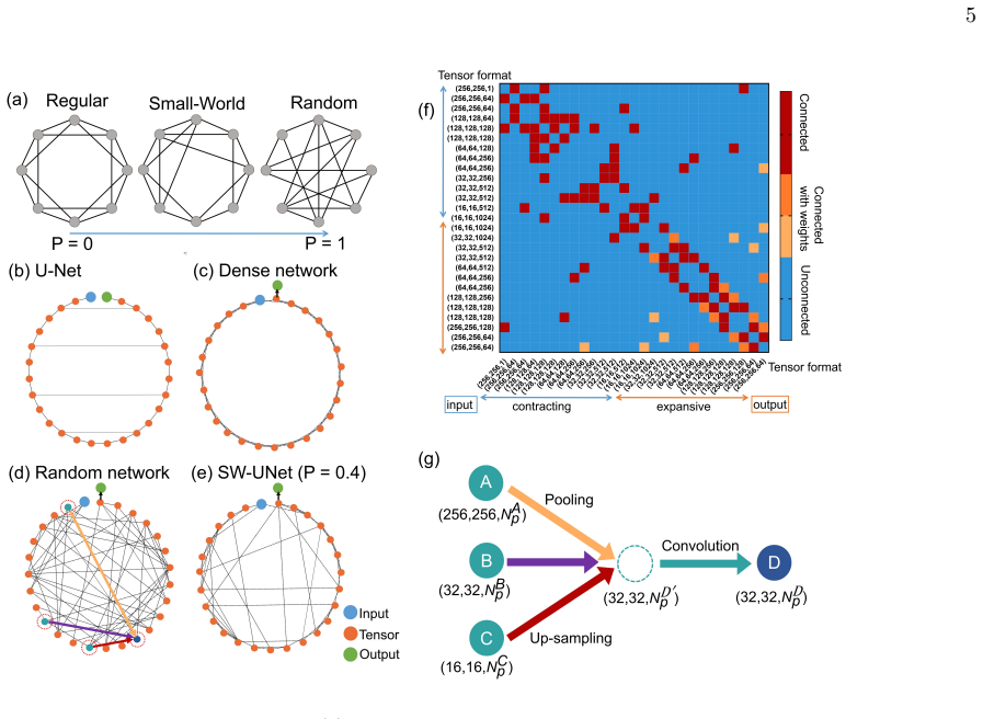
The load-bearing object is SW-UNet, a U-Net-like convolutional architecture whose tensor-flow graph is generated by the Watts-Strogatz small-world network model with N = 27 nodes, K = 4 average neighbor connections, and rewiring probability P. The small-world index (SWI), computed from average path length and clustering coefficient, quantifies how close the topology is to an optimal small-world regime. SW-UNet's job is to map raw phase-contrast images to wrinkle masks; the wrinkles themselves are produced by cells contracting against an oxidized PDMS substrate and are the paper's readout of force magnitude. Segmentation quality is scored by extracting wrinkle perimeter length with the Prewitt operator and measuring Euclidean and cosine distances between network output and human-traced ground truth.
What would settle it
Plate the same U2OS cells on the same wrinkle substrate, treat them with a myosin inhibitor such as blebbistatin, and record SW-UNet wrinkle lengths alongside traction-force-microscopy displacements; the claim fails if wrinkle length does not track the traction force or does not decrease when force is blocked.
Extended reading notes
Core claim
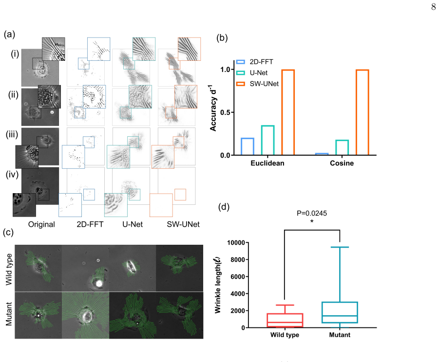
The central discovery is that a U-Net whose internal connections are rewired using the Watts-Strogatz small-world recipe—27 nodes, average degree 4, rewiring probability P—segments substrate wrinkles more accurately than either the classical 2D-FFT bandpass approach or the original U-Net. On 58 test images scored against manual tracings by three researchers, SW-UNet's Euclidean distance error is 4.9 times smaller than 2D-FFT and 2.9 times smaller than U-Net; its cosine-distance accuracy is 36.8 times and 5.5 times better. Segmentation is best near P = 0.2–0.4, where the small-world index SWI peaks, leading the paper to suggest that higher SWI predicts better segmentation performance. Applied to U2OS cells, SW-UNet yields mean wrinkle lengths of 2144 for KRAS G12V-expressing cells versus 901 for wild-type (p = 0.0245), evidence the paper reads as increased contractile force accompanying the oncogenic mutation.
Load-bearing premise
The whole force evaluation rests on the assumption that longer wrinkles always mean stronger contraction; the paper cites earlier studies for this link but never measures actual force itself.
Editorial extensions
If this is right
- Wrinkle-based force assays no longer require manual tracing: SW-UNet outputs wrinkle masks directly from phase-contrast images, so large numbers of cells can be scored automatically with an XY motorized stage.
- The KRAS G12V result directly supports the idea that this common oncogenic mutation raises cellular contractile force, consistent with earlier reports that G12V enhances myosin phosphorylation.
- Because the training labels were generated by image processing (2D-FFT plus curvature filter) rather than hand annotation, the same pipeline can be transferred to other cell types and substrates with modest data-preparation effort.
- The observed link between SWI and segmentation accuracy gives a rough design rule for CNN topology: among randomly rewired variants, those with higher small-world index tend to segment better.
Reading between the lines
- Editorial inference: the 2144-versus-901 ratio suggests roughly a 2.4-fold difference in wrinkle length, but because the paper never calibrates wrinkle length to force units, the number should be read as a relative comparison, not an absolute force ratio.
- Editorial inference: a side-by-side traction-force-microscopy comparison on the same cells would test whether SW-UNet wrinkle length and traction force are monotonically related across cell types; the paper does not report such a calibration.
- Editorial inference: if the SWI-performance correlation generalizes beyond this task, SWI would be a cheap screening statistic for neural architecture search in other segmentation problems; that generalization is not established by the paper.
- Editorial inference: the substrate's fixed stiffness is an untested boundary condition; the method's accuracy on softer or stiffer wrinkle substrates remains unknown.
Signed reviews
Editorial analysis
A structured set of objections, weighed in public.
Referee Report
Summary. The manuscript proposes SW-UNet, a U-Net variant whose inter-node connections are rewired according to the Watts-Strogatz small-world model, for segmenting substrate wrinkles in phase-contrast images of U2OS cells. Training labels are generated by a 2D-FFT bandpass and curvature-filter pipeline, and the network is trained on augmented images. Segmentation is evaluated on 58 test images by Euclidean and cosine distances between total wrinkle perimeter lengths per image and manual ground-truth traces. The authors report that SW-UNet reduces Euclidean error by 4.9 times relative to the 2D-FFT baseline and 2.9 times relative to U-Net, and they demonstrate that KRAS G12V cells show larger wrinkle length than wild-type cells, which they interpret as larger contractile force.
Significance. If the segmentation and force claims held, the work would offer an automated, high-throughput tool for wrinkle quantification in cell contractility studies. The paper has practical strengths: a real biological image dataset, manual ground-truth traces, a clearly described network architecture, and a concrete demonstration on KRAS-mutant versus wild-type cells. However, the headline segmentation accuracy rests on an aggregate perimeter-length metric rather than spatial overlap, and the biological conclusion depends on an uncalibrated wrinkle-length-to-force proxy. The small-world design principle is inferred from the same parameter sweep used to select the network configuration, so it is not independently validated. These issues affect both the technical and biological conclusions, though they appear fixable with additional evaluation and reframing.
major comments (3)
- [Wrinkle evaluation (Eqs. (1)-(2), Fig. 6(b))] The Euclidean and cosine distances are computed from per-image scalar total wrinkle perimeter length, not from pixel-level agreement between predicted and ground-truth masks. As a result, a segmentation that places wrinkles in the wrong locations, or that confuses cell edges with wrinkles, can still achieve a small distance if the total edge-pixel count matches the manual trace. The reported 4.9-fold and 2.9-fold error reductions therefore do not, as stated, demonstrate that SW-UNet segments wrinkles more accurately in space. I request additional pixel-level metrics (e.g., Dice/F1, IoU, precision/recall on wrinkle pixels, and a distance-based measure such as Hausdorff distance) computed on the same test images.
- [Results: Effect of P-value (Fig. 5(b), p. 6)] The reconnection probability P is selected by minimizing dEU on the same Ntest=58 test images that are later used for the final comparison in Fig. 6(b): the text states "From next sections, we will fix the value to P = 0.4 for SW-UNet" immediately after the P-sweep in Fig. 5(b). This is a form of test-set selection, and it makes the headline accuracy ratios optimistically biased. Furthermore, the SWI-performance relationship shown in Fig. 5(c) is inferred from the very same P-sweep used to choose P, so it cannot serve as independent evidence for the small-world design principle. P should be chosen on a validation split (or by cross-validation) and the final metrics reported on a test set not used for any model-selection decision.
- [Demonstration: Effect of KRAS mutation (Fig. 6(d), p. 8)] The conclusion that KRAS G12V cells "exhibit greater forces" rests on the assumption that wrinkle length is a monotonic proxy for contractile force, citing refs [6,9]. The manuscript provides no calibration of the measured aggregate perimeter length to force magnitude, no comparison with traction-force microscopy or another force assay, and no control for potential confounders such as cell size, spreading area, or substrate heterogeneity. Given that the measured quantity is total perimeter length of segmented wrinkles per image, the significant difference (2144 vs 901, p=0.0245) demonstrates a difference in wrinkle length, but not necessarily in contractile force. The authors should either add a calibration/validation experiment or explicitly reframe the conclusion as a difference in wrinkle length.
minor comments (5)
- [Abstract and p. 8] There is a typo in the abstract: "oncogne" should be "oncogene"; also "supplemental meterial" on page 8 should be "supplemental material." Several encoding artifacts appear in the text, such as "10 Îijg/mL" and "Âţg/mL."
- [Eq. (5)] The clustering coefficient formula contains an undefined index h and appears to be misprinted; the standard Watts-Strogatz clustering coefficient involves the number of triangles around node i divided by k_i(k_i-1). Please correct the formula and define all indices.
- [Training dataset preparation (p. 3)] The curvature-filter stopping criterion is given as "200-1000 times until only wrinkles disappear," but "disappear" is not quantified. Please specify an automated stopping rule or a fixed number of iterations with a sensitivity check.
- [Fig. 5(b)] The text states that three randomly generated networks are evaluated for each P value, but Fig. 5(b) shows no error bars or dispersion; please add them or state that the plotted points are means.
- [Fig. 5(c) and conclusion] The inference that "networks with larger SWI might have better performance" is based on a small number of P values from the same sweep used to select P. Please soften the claim or support it with a statistical test and an independent validation.
Circularity Check
Headline error reductions are test-set-selected: P=0.4 is chosen by minimizing dEU on the same 58 test images later used to report the 4.9x/2.9x gains; the central architecture claim otherwise rests on independent manual ground-truth traces.
-
fitted input called prediction
[Results: 'Effect of P-value in SW-UNet' (Fig. 5b) and 'Comparison of different segmentation methods' (Fig. 6b); evaluation via Eqs. (1)-(2)]
"Figure 5(b) shows the distance dEU from the manually tracked ground truth, and the result shows that the segmentation performance is best at P = 0.2−0.4. ... From next sections, we will fix the value to P = 0.4 for SW-UNet. ... SW-UNet has the smallest error compared to 2D-FFT based segmentation and U-Net."
The same Ntest=58 test images are first used to select the network topology parameter P by minimizing the Euclidean distance dEU (Fig. 5b), and the same test images are then used as the basis of the headline comparison (Fig. 6b), where P=0.4 SW-UNet is reported as having 4.9x smaller Euclidean error than 2D-FFT and 2.9x smaller than U-Net. Thus the claimed accuracy is a test-set-selected optimum rather than an independent holdout prediction; the baseline networks were not given the same test-set tuning. The reported improvement is therefore statistically forced by the selection procedure.
full rationale
The core derivation chain--prepare training labels with 2D-FFT and curvature filtering, train SW-UNet, test against manually traced ground truth--is otherwise self-contained and not circular. The CNN is trained on noisy image-processing labels but evaluated against independent human traces, so the central 'SW-UNet beats 2D-FFT/U-Net' claim has real empirical content despite the label-generation overlap. The force-length premise is supported by an external citation [6] in addition to the authors' own [9], so the KRAS wrinkle-length comparison is not a self-citation loop, though the leap from wrinkle length to force magnitude is an uncalibrated assumption. The main circularity is the hyperparameter selection: P=0.4 is fixed after inspecting dEU on the same 58 test images later used for the final error ratios, making the headline factors optimistically selected rather than predicted. The perimeter-length metric (Eqs. 1-2) is also a non-spatial operationalization of 'segmentation accuracy,' which weakens the spatial claim but is not itself a circular derivation; it is a measurement-validity concern.
Assumptions & free parameters
free parameters (7)
- P (reconnection probability) =
0.4
- K (average connection branches) =
4
- N (number of nodes) =
27
- Prewitt threshold =
0.01
- Bandpass wavelength range =
3-6 pixels
- Curvature filter iterations =
200-1000
- Training epochs and steps per epoch =
10 epochs, 300 steps
assumptions (5)
- domain assumption Wrinkle length is positively correlated with contractile force magnitude
- domain assumption Wrinkles in the oxidized PDMS substrate are generated reversibly by cell contraction and reflect force magnitude and direction
- domain assumption Manual tracing by three researchers gives valid ground truth for wrinkle segmentation
- domain assumption Training labels produced by 2D-FFT bandpass and curvature filter are adequate supervision for the CNN
- ad hoc to paper SWI computed from the Watts-Strogatz topology is a meaningful predictor of segmentation performance
Cite this review
Pith. "Pith review of Image based cellular contractile force evaluation with small-world network inspired CNN: SW-UNet." pith.science (2026). https://pith.science/paper/QUTSQLPZ
@misc{pith2026190808631,
  author       = {Pith},
  title        = {Pith review of: Image based cellular contractile force evaluation with small-world network inspired CNN: SW-UNet},
  year         = {2026},
  howpublished = {\url{https://pith.science/paper/QUTSQLPZ}},
  note         = {Machine review of arXiv:1908.08631}
}
read the original abstract
We propose an image-based cellular contractile force evaluation method using a machine learning technique. We use a special substrate that exhibits wrinkles when cells grab the substrate and contract, and the wrinkles can be used to visualize the force magnitude and direction. In order to extract wrinkles from the microscope images, we develop a new CNN (convolutional neural network) architecture SW-UNet (small-world U-Net), which is a CNN that reflects the concept of the small-world network. The SW-UNet shows better performance in wrinkle segmentation task compared to other methods: the error (Euclidean distance) of SW-UNet is 4.9 times smaller than 2D-FFT (fast Fourier transform) based segmentation approach, and is 2.9 times smaller than U-Net. As a demonstration, we compare the contractile force of U2OS (human osteosarcoma) cells and show that cells with a mutation in the KRAS oncogne show larger force compared to the wild-type cells. Our new machine learning based algorithm provides us an efficient, automated and accurate method to evaluate the cell contractile force.
Figures


Figures from the paper (3 more)

Reference graph
Works this paper leans on
-
[1]
at the green node, and the network is designed to extract the desired segmented image at this final tensor. The U-Net mainly consists of two paths, contracting path (left side of Fig. 4(b)) and expansive path (right side). The contracting path is responsible for extracting the feature from the images, while the expansive path is designed to reconstruct the...
-
[2]
W. J. Polacheck and C. S. Chen, Nature methods13, 415 (2016)
work page 2016
-
[3]
S. Munevar, Y.-l. Wang, and M. Dembo, Biophysical journal 80, 1744 (2001)
work page 2001
-
[4]
J. L. Tan, J. Tien, D. M. Pirone, D. S. Gray, K. Bhadri- raju, and C. S. Chen, Proceedings of the National Academy of Sciences100, 1484 (2003)
work page 2003
-
[5]
Z. Liu, J. L. Tan, D. M. Cohen, M. T. Yang, N. J. Sni- adecki, S. A. Ruiz, C. M. Nelson, and C. S. Chen, Pro- ceedings of the National Academy of Sciences107, 9944 (2010)
work page 2010
- [6]
-
[7]
N. Q. Balaban, U. S. Schwarz, D. Riveline, P. Goichberg, G. Tzur, I. Sabanay, D. Mahalu, S. Safran, A. Bershad- sky, L. Addadi,et al., Nature cell biology3, 466 (2001)
work page 2001
-
[8]
S. Yokoyama, T. S. Matsui, and S. Deguchi, Biochem- ical and biophysical research communications482, 975 (2017)
work page 2017
Show all 51 references
-
[9]
Ichikawa, M
T. Ichikawa, M. Kita, T. S. Matsui, A. I. Nagasato, T. Araki, S.-H. Chiang, T. Sezaki, Y. Kimura, K. Ueda, S. Deguchi,et al., J Cell Sci130, 3517 (2017)
2017
-
[10]
S. P. Fukuda, T. S. Matsui, T. Ichikawa, T. Furukawa, N. Kioka, S. Fukushima, and S. Deguchi, Development, growth & differentiation59, 423 (2017)
2017
-
[11]
A. K. Harris, P. Wild, and D. Stopak, Science208, 177 (1980)
1980
-
[12]
Ronneberger, P
O. Ronneberger, P. Fischer, and T. Brox, in Inter- national Conference on Medical image computing and computer-assisted intervention(Springer, 2015) pp. 234– 241
2015
-
[13]
T. Falk, D. Mai, R. Bensch, Ö. Çiçek, A. Abdulkadir, Y. Marrakchi, A. Böhm, J. Deubner, Z. Jäckel, K. Sei- wald, et al., Nature methods16, 67 (2019)
2019
-
[14]
D. A. Van Valen, T. Kudo, K. M. Lane, D. N. Macklin, N. T. Quach, M. M. DeFelice, I. Maayan, Y. Tanouchi, E. A. Ashley, and M. W. Covert, PLoS computational biology 12, e1005177 (2016)
2016
-
[15]
Fabijańska, Artificial intelligence in medicine88, 1 (2018)
A. Fabijańska, Artificial intelligence in medicine88, 1 (2018)
2018
-
[16]
Niioka, S
H. Niioka, S. Asatani, A. Yoshimura, H. Ohigashi, S. Tagawa, and J. Miyake, Human cell31, 87 (2018)
2018
-
[17]
D. J. Watts and S. H. Strogatz, nature393, 440 (1998)
1998
-
[18]
M. D. Humphries and K. Gurney, PloS one3, e0002051 (2008)
2008
-
[19]
Z. P. Neal, Network Science5, 30 (2017)
2017
-
[20]
D. H. Hubel and T. N. Wiesel, The Journal of physiology 195, 215 (1968)
1968
-
[21]
Bullmore and O
E. Bullmore and O. Sporns, Nature reviews neuroscience 10, 186 (2009)
2009
-
[22]
Rubinov and O
M. Rubinov and O. Sporns, Neuroimage52, 1059 (2010)
2010
-
[23]
E. J. Sanz-Arigita, M. M. Schoonheim, J. S. Damoiseaux, S. A. Rombouts, E. Maris, F. Barkhof, P. Scheltens, and C. J. Stam, PloS one5, e13788 (2010)
2010
-
[24]
S. Xie, A. Kirillov, R. Girshick, and K. He, arXiv preprint arXiv:1904.01569 (2019)
2019 arXiv
-
[25]
Javaheripi, B
M. Javaheripi, B. D. Rouhani, and F. Koushanfar, arXiv preprint arXiv:1904.04862 (2019)
2019 arXiv
-
[26]
Gong and I
Y. Gong and I. F. Sbalzarini, IEEE Transactions on Im- age Processing26, 1786 (2017)
2017
-
[27]
R. Wu, S. Yan, Y. Shan, Q. Dang, and G. Sun, arXiv preprint arXiv:1501.02876 (2015)
2015 arXiv
-
[28]
Wang and L
J. Wang and L. Perez, Convolutional Neural Networks Vis. Recognit (2017)
2017
-
[29]
Z. Gao, L. Wang, L. Zhou, and J. Zhang, IEEE journal of biomedical and health informatics21, 416 (2016)
2016
-
[30]
Han and J
Y. Han and J. C. Ye, IEEE transactions on medical imag- ing 37, 1418 (2018)
2018
-
[31]
Kayalibay, G
B. Kayalibay, G. Jensen, and P. van der Smagt, arXiv preprint arXiv:1701.03056 (2017)
2017 arXiv
-
[32]
H. Dong, G. Yang, F. Liu, Y. Mo, and Y. Guo, inannual conference on medical image understanding and analysis (Springer, 2017) pp. 506–517
2017
-
[33]
Kochen,The small world(Ablex Pub., 1989)
M. Kochen,The small world(Ablex Pub., 1989)
1989
-
[34]
Huang, Z
G. Huang, Z. Liu, L. Van Der Maaten, and K. Q. Wein- berger, in Proceedings of the IEEE conference on com- puter vision and pattern recognition (2017) pp. 4700– 4708
2017
-
[35]
Jégou, M
S. Jégou, M. Drozdzal, D. Vazquez, A. Romero, and Y. Bengio, in Proceedings of the IEEE Conference on Computer Vision and Pattern Recognition Workshops (2017) pp. 11–19
2017
-
[36]
S. Li, M. Deng, J. Lee, A. Sinha, and G. Barbastathis, Optica 5, 803 (2018)
2018
-
[37]
D. P. Kingma and J. Ba, arXiv preprint arXiv:1412.6980 (2014)
2014 arXiv
-
[38]
H. R. Roth, L. Lu, A. Seff, K. M. Cherry, J. Hoffman, S. Wang, J. Liu, E. Turkbey, and R. M. Summers, in International conference on medical image computing and computer-assisted intervention(Springer, 2014) pp. 520– 527
2014
-
[39]
Poletti, and H
A.Depeursinge, A.Vargas, A.Platon, A.Geissbuhler, P.- A. Poletti, and H. Müller, Computerized medical imag- 10 ing and graphics36, 227 (2012)
2012
-
[40]
H.-C. Shin, H. R. Roth, M. Gao, L. Lu, Z. Xu, I. Nogues, J. Yao, D. Mollura, and R. M. Summers, IEEE transac- tions on medical imaging35, 1285 (2016)
2016
-
[41]
Zhang, Y
R. Zhang, Y. Zheng, T. W. C. Mak, R. Yu, S. H. Wong, J. Y. Lau, and C. C. Poon, IEEE journal of biomedical and health informatics21, 41 (2016)
2016
-
[42]
W. B. Sampaio, E. M. Diniz, A. C. Silva, A. C. De Paiva, and M. Gattass, Computers in biology and medicine41, 653 (2011)
2011
-
[43]
Loughrey and P
J. Loughrey and P. Cunningham, inInternational Con- ference on Innovative Techniques and Applications of Ar- tificial Intelligence(Springer, 2004) pp. 33–43
2004
-
[44]
Shore and R
J. Shore and R. Johnson, IEEE Transactions on informa- tion theory26, 26 (1980)
1980
-
[45]
Yi-de, L
M. Yi-de, L. Qing, and Q. Zhi-Bai, in Proceedings of 2004 International Symposium on Intelligent Multime- dia, Video and Speech Processing, 2004.(IEEE, 2004) pp. 743–746
2004
-
[46]
Sporns and J
O. Sporns and J. D. Zwi, Neuroinformatics2, 145 (2004)
2004
-
[47]
Tsuchida, A
N. Tsuchida, A. K. Murugan, and M. Grieco, Oncotarget 7, 46717 (2016)
2016
-
[48]
R. G. Amado, M. Wolf, M. Peeters, E. Van Cutsem, S. Siena, D. J. Freeman, T. Juan, R. Sikorski, S. Suggs, R. Radinsky, S. D. Patterson, and D. D. Chang, Journal of Clinical Oncology26, 1626 (2008), pMID: 18316791, https://doi.org/10.1200/JCO.2007.14.7116
2008 doi
-
[49]
J.Son, C.A.Lyssiotis, H.Ying, X.Wang, S.Hua, M.Lig- orio, R. M. Perera, C. R. Ferrone, E. Mullarky, N. Shyh- Chang, et al., Nature496, 101 (2013)
2013
-
[50]
G. J. Riely, J. Marks, and W. Pao, Proceedings of the American Thoracic Society6, 201 (2009)
2009
-
[51]
Hogan, S
C. Hogan, S. Dupré-Crochet, M. Norman, M. Kajita, C. Zimmermann, A. E. Pelling, E. Piddini, L. A. Baena- López, J.-P. Vincent, Y. Itoh,et al., Nature cell biology 11, 460 (2009)
2009
Reviewed August 14, 2026 · model on record in the stance chip above.
Discussion (0). Continue with ORCID to comment.