REVIEW 5 minor 68 references
Efficient Computing for Medical Image Acquisition and Reconstruction
T0 review · 0 major / 5 minor · reviewed 2026-08-02 · deepseek-v4-flash
Pith's one-line read All four major medical imaging modalities reduce to a single linear forward model, Y=AX+E, and the key to fast reconstruction is not faster arithmetic but moving data through the memory hierarchy efficiently.
desk verdict A competent, well-organized book-chapter review of standard CT/MRI/PET/SPECT reconstruction math, with no new results and one thin, self-cited efficiency section that needs tightening. read the letter →
The pith
A machine-rendered reading of the paper's core claim, the machinery that carries it, and where it could break.
The reading
What carries the argument
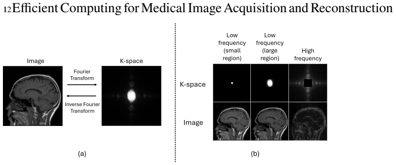
The forward model Y=AX+E (Equation 1.8) is the central object. A is the Radon transform or a ray/voxel/distance-driven system matrix for CT, A=MFS (sampling mask, Fourier transform, coil sensitivities) for MRI, and for PET/SPECT it includes the detector sensitivity C_i(r) times the Beer-Lambert attenuation factor. The chapter's second machine is the super-voxel: a square block of neighboring voxels whose associated measurements form a sinusoidal band in the sinogram, so that cache lines fetched for one voxel are reused for its neighbors. Together these explain both the mathematical unity and the main performance lever (data reuse).
What would settle it
Profile a representative MBIR reconstruction on a current clinical workstation: if the CPU/GPU arithmetic units are busy most of the time (say >80% utilization) and cache misses are low, the memory-bound premise fails. Alternatively, independently reproduce the reported 10x/187x super-voxel speedup on a different scanner geometry (e.g., helical or cone-beam CT); failure to reproduce would show the gain is geometry-specific, not the general principle claimed.
Extended reading notes
Core claim
The central claim, stated the way the authors would state it to a fair reader, is that every reconstruction algorithm discussed—analytical FBP/inverse FFT, algebraic iterative methods, model-based iterative reconstruction, and statistical emission tomography—solves the same inverse problem Y=AX+E. The differences are which quantity X denotes (attenuation coefficient, transverse magnetization, radiotracer activity), how A is built (line integrals, Fourier encoding with coil sensitivities and sampling mask, or emission-detection probability with attenuation), and which statistical model E follows (Gaussian after the log transform in CT, complex Gaussian in MRI, Poisson in PET/SPECT). The chapt
Load-bearing premise
The guidance stands or falls on the claim that reconstruction speed is limited by memory traffic and cache misses rather than by arithmetic throughput; if real clinical workloads are compute-bound, the cache-focused speedup story would not generalize.
Editorial extensions
If this is right
- Algorithms such as OSEM, MBIR, and iterative coordinate descent can be transplanted from one modality to another by swapping the forward operator and the noise model, since all solve the same optimization template.
- Because reconstruction is memory-bound, hardware-aware reorganizations of the computation (like super-voxel blocking) can give order-of-magnitude reductions in reconstruction time without changing image quality.
- The optimization-algorithm choice becomes a co-design decision: gradient-based methods parallelize well but converge slowly; coordinate descent converges fast but is hard to parallelize; grouped coordinate descent interpolates between them.
- Clinical deployment of MBIR is a workstation-computing problem; translating HPC techniques into single-node memory-efficient implementations is what determines whether high-quality reconstruction finishes in minutes rather than hours.
Reading between the lines
- If the memory-bound thesis is right, the next natural lever is acquisition design: reordering views or detector layout so that sinusoidal bands align better with cache lines could extend the super-voxel idea to list-mode PET and non-Cartesian MRI, where access patterns are even more irregular.
- The unification suggests a concrete cross-modality benchmark: running the same optimization algorithm (e.g., OSEM or conjugate gradient) on CT, MRI, PET, and SPECT data of matched size would quantify how much performance transfers and where the forward operator's structure dominates.
- The reported 10x/187x speedups come from a specific implementation on a parallel-beam-style geometry; a testable extension is whether the same cache-reuse gain survives in cone-beam and helical CT, where the sinusoidal band assumption becomes approximate.
Editorial analysis
A structured set of objections, weighed in public.
Referee Report
Summary. This book chapter proposes a unified computational framework for medical image acquisition and reconstruction across CT, MRI, PET, and SPECT. The central claim is that all four modalities, despite differing imaging physics, can be expressed through the forward model Y = AX + E, where the differences lie in the physical meaning of A, the statistical characterization of E, and the resulting reconstruction algorithms. The chapter reviews the physics of data acquisition for each modality, derives analytical (FBP, inverse FFT) and iterative/statistical reconstruction methods from the unified model, and discusses efficient-computing strategies including optimization algorithms, physics-aware forward operators, memory-efficient implementations (e.g., the super-voxel cache-blocking framework), and parallel computing. The stated goal is to provide an organizing framework that connects medical physics, linear algebra, probability, optimization, and high-performance computing, and to enable transfer of computational methods across modalities.
Significance. If the claims hold, the chapter provides a valuable pedagogical and organizational resource. Its main contribution is synthesis: making explicit the common mathematical structure underlying four major imaging modalities and connecting it to practical computational challenges. The mathematics is standard and accurately presented (apart from the typographical issue noted below). The chapter includes useful comparative tables (e.g., Table 1.1 hardware specifications, Table 1.4 reconstruction method comparison) and clear conceptual figures. It does not introduce new algorithms or machine-checked proofs, but as a tutorial/review this is not a weakness. The efficient-computing section raises an important and sometimes underappreciated point about memory bandwidth and cache locality in iterative reconstruction; however, the quantitative support for this point rests on the authors' own prior work and is presented without sufficient experimental context. This should be clarified but does not undermine the central unifying claim.
minor comments (5)
- [§1.4.3.1] The memory-complexity example states 'a system matrix containing approximately254 elements' — the superscript is missing in the rendered text. It should read 'approximately $2^{54}$ elements' (consistent with the subsequent 'tens of petabytes' estimate). Please correct.
- [§1.4.4] The enumeration of the five computational challenges is '(1) Optimization efficiency; (2) Computational complexity; (2) Physics-aware forward system matrix modeling; (4) Data movement and memory hierarchy; (5) Clinical computing constraints.' The duplicate '(2)' should be corrected to '(3)'.
- [§1.4.4] The sentence 'Compared with conventional reconstruction with the super-voxels, the super-voxel framework achieves approximately a 10-fold speedup...' seems to be missing the word 'without'. The intended meaning is likely 'Compared with conventional reconstruction without the super-voxels...' or 'Compared with conventional reconstruction, the super-voxel framework...'.
- [§1.4.4] The super-voxel speedup figures (10-fold on a single core, 187-fold on a 20-core shared-memory system) are cited to [60], but no baseline algorithm, image size, CT geometry, projector type, or hardware details are provided in the text. Since these numbers are the only quantitative support for the memory-bound claim, a brief description of the experimental setting (or an explicit statement that this is an illustrative case study from the authors' own work) would make the claim more credible and avoid overgeneralization to GPU-based clinical systems, where the compute/memory balance may differ.
- [§1.3.2] In the description of Figure 1.6, the phrase 'Protons are finally returned to their alignment with B0 and signal is recorded by receiver coils during that gradual decay' could be rephrased to clarify that the signal is recorded during the decay process, not after the spins have fully returned to equilibrium.
Circularity Check
No significant circularity: the unified forward model is a generalization of independently derived modality equations, and cited efficiency speedups are empirical examples rather than fitted predictions.
full rationale
This chapter is an expository survey rather than a derivation of new predictive results. Its central unification claim is that CT, MRI, PET, and SPECT can each be written in the common form Y = AX + E. That claim is supported by separate physical derivations: the Beer-Lambert line integrals for CT (Eqs. 1.1-1.2), the k-space Fourier relation for MRI (Eq. 1.4), and the attenuated emission integral equations for SPECT/PET (Eqs. 1.6-1.7), whose discretization yields the system-matrix elements in Eq. 1.19. Equation (1.8) is therefore a generalization of independently stated modality-specific forward models, not a definition that presupposes the conclusion. No quantity is fitted and then renamed as a prediction; no uniqueness theorem from the authors' prior work is invoked to force a choice; no ansatz is smuggled in via citation. The super-voxel speedup figures in §1.4.4 are cited empirical results from the authors' earlier paper [60], used as an example of cache-aware reconstruction; they are not derived from the unified framework and therefore cannot be circular in the sense of reducing to their inputs. Whether the memory-bound premise generalizes to clinical cone-beam/helical CT on GPUs is a legitimate external-validity or evidence-quality question, but it is not a circularity. The self-citations are present, but they function as literature pointers to the authors' published implementations rather than as load-bearing premises that reduce the chapter's claims to themselves. Score reflects only minor self-citation prevalence.
Assumptions & free parameters
assumptions (5)
- standard math Beer-Lambert law models X-ray and gamma attenuation as a line integral (Eq. 1.1).
- standard math MRI signal is the Fourier transform of the magnetization under linear gradients (Eq. 1.4).
- domain assumption The unified forward model Y=AX+E (Eq. 1.8) adequately represents all four modalities.
- domain assumption Poisson statistics for PET/SPECT measurements (Eq. 1.20).
- domain assumption The system matrix A for CT/SPECT/PET is approximated by practical projectors (distance-driven, ray-driven, voxel-driven).
Cite this review
Pith. "Pith review of Efficient Computing for Medical Image Acquisition and Reconstruction." pith.science (2026). https://pith.science/paper/QVIRVP2U
@misc{pith2026260713204,
  author       = {Pith},
  title        = {Pith review of: Efficient Computing for Medical Image Acquisition and Reconstruction},
  year         = {2026},
  howpublished = {\url{https://pith.science/paper/QVIRVP2U}},
  note         = {Machine review of arXiv:2607.13204}
}
read the original abstract
Medical imaging systems such as CT, MRI, PET, and SPECT do not directly acquire images. Instead, they measure physical signals that encode anatomical or physiological information, and image reconstruction recovers the underlying image by solving an inverse problem. Although these imaging modalities are governed by different imaging physics, they share a common computational framework that naturally connects medical physics, linear algebra, probability, numerical optimization, and efficient computing. As medical imaging systems acquire increasingly large and higher-dimensional datasets, image reconstruction has become one of the primary computational bottlenecks in modern medical imaging. Advanced reconstruction methods, including analytical reconstruction, iterative optimization, and statistical model-based reconstruction, substantially improve image quality while reducing radiation dose or scan time, but at significantly increased computational cost. Efficient computing has therefore become essential for achieving clinically practical reconstruction times. This chapter presents a unified computational perspective on medical image acquisition and reconstruction across CT, MRI, PET, and SPECT. It first reviews the imaging physics and data acquisition process for each modality and derives a generalized mathematical framework for image reconstruction. Building on this framework, the chapter discusses analytical, iterative, and statistical reconstruction methods together with their computational characteristics. Finally, it examines efficient computing considerations, including optimization algorithms, physics-aware forward operators, memory-efficient implementations, and parallel computing strategies. Together, these topics demonstrate how the integration of imaging physics, mathematical modeling, and efficient computing enables accurate and scalable medical image reconstruction.
Figures


Figures from the paper (20 more)






Reference graph
Works this paper leans on
-
[60]
Medical Physics 48(7), 3595–3613 (2021)
Wang,X.,MacDougall,R.D.,Chen,P.,Bouman,C.A.,Warfield,S.K.:Physics-basediterative reconstruction for dual-source and flying focal spot computed tomography. Medical Physics 48(7), 3595–3613 (2021). DOI 10.1002/mp.14929
-
[1]
WorldAcademyofScience,EngineeringandTechnology(2008)
Aibinu, A.M., Salami, M.J.E., Shafie, A.A., Najeeb, A.R.: MRI reconstruction using discrete Fouriertransform:atutorial. WorldAcademyofScience,EngineeringandTechnology(2008)
2008
-
[2]
Ultrasonic imaging6(1), 81–94 (1984)
Andersen,A.H.,Kak,A.C.:Simultaneousalgebraicreconstructiontechnique(sart):asuperior implementation of the art algorithm. Ultrasonic imaging6(1), 81–94 (1984)
1984
-
[3]
electronic imaging30, 1–1817 (2018)
Balke, T., Majee, S., Buzzard, G.T., Poveromo, S., Howard, P., Groeber, M.A., McClure, J., Bouman, C.A.: Separable models for cone-beam mbir reconstruction. electronic imaging30, 1–1817 (2018)
2018
-
[4]
SIAM(2022)
Bouman,C.A.:Foundationsofcomputationalimaging:amodel-basedapproach. SIAM(2022)
2022
-
[5]
In: 2023 59th annual Allerton conference on communication, control, and computing (Allerton), pp
Bouman, C.A., Buzzard, G.T.: Generative plug and play: Posterior sampling for inverse prob- lems. In: 2023 59th annual Allerton conference on communication, control, and computing (Allerton), pp. 1–7. IEEE (2023)
2023
-
[6]
IEEE Transactions on image processing5(3), 480–492 (1996)
Bouman,C.A.,Sauer,K.:Aunifiedapproachtostatisticaltomographyusingcoordinatedescent optimization. IEEE Transactions on image processing5(3), 480–492 (1996)
1996
-
[7]
John Wiley & Sons (2014)
Brown, R.W., Cheng, Y.C.N., Haacke, E.M., Thompson, M.R., Venkatesan, R.: Magnetic resonance imaging: physical principles and sequence design. John Wiley & Sons (2014)
2014
Show all 68 references
-
[8]
In: Proceedings of the International Conference forHighPerformanceComputing,Networking,StorageandAnalysis,SC’19.Associationfor Computing Machinery, New York, NY, USA (2019)
Chen, P., Wahib, M., Takizawa, S., Takano, R., Matsuoka, S.: ifdk: A scalable framework for instant high-resolution image reconstruction. In: Proceedings of the International Conference forHighPerformanceComputing,Networking,StorageandAnalysis,SC’19.Associationfor Computing Ma...
2019
-
[9]
In: Proceedings of the International Conference for High Performance Computing, Networking, Storage and Analysis, pp
Chen, P., Wahib, M., Wang, X., Hirofuchi, T., Ogawa, H., Biguri, A., Boardman, R., Blu- mensath, T., Matsuoka, S.: Scalable fbp decomposition for cone-beam ct reconstruction. In: Proceedings of the International Conference for High Performance Computing, Networking, Storage an...
2021
-
[10]
In:ProceedingsoftheInternationalConferenceonSupercomputing(ICS’21), pp
Chen, P., Wahib, M., Wang, X., Takizawa, S., Hirofuchi, T., Ogawa, H., Matsuoka, S.: Perfor- mance portable back-projection algorithms on cpus: Agnostic data locality and vectorization optimizations. In:ProceedingsoftheInternationalConferenceonSupercomputing(ICS’21), pp. 1–13 ...
2021
-
[11]
Elsevier Saunders, Philadelphia, PA (2012)
Cherry, S.R., Sorenson, J.A., Phelps, M.E.: Physics in Nuclear Medicine, 4 edn. Elsevier Saunders, Philadelphia, PA (2012). URLhttps://shop.elsevier.com/books/ physics-in-nuclear-medicine/cherry/978-1-4160-5198-5
2012
-
[12]
Physics in Medicine & Biology49(11), 2463–2475 (2004)
De Man, B., Basu, S.: Distance-driven projection and backprojection in three dimensions. Physics in Medicine & Biology49(11), 2463–2475 (2004)
2004
-
[13]
In:IEEENuclearScience Symposium Conference Record, 2005, vol
De Man, B., Basu, S., Thibault, J.B., Hsieh, J., Fessier, J., Bouman, C., Sauer, K.: A study of fourminimizationapproachesforiterativereconstructioninx-rayct. In:IEEENuclearScience Symposium Conference Record, 2005, vol. 5, pp. 2708–2710. IEEE (2005)
2005
-
[14]
Defrise, M., Kinahan, P.E., Michel, C.J.: Image reconstruction algorithms in pet. In: D.L. Bailey, D.W. Townsend, P.E. Valk, M.N. Maisey (eds.) Positron Emission Tomography, pp. 63–91. Springer London, London (2005). DOI 10.1007/1-84628-007-9_4
2005 doi
-
[15]
In: Computational Imaging XIII, vol
Degirmenci,S.,Politte,D.G.,Bosch,C.,Tricha,N.,O’Sullivan,J.A.:Accelerationofiterative image reconstruction for x-ray imaging for security applications. In: Computational Imaging XIII, vol. 9401, pp. 96–110. SPIE (2015)
2015
-
[16]
Journal of Magnetic Resonance Imaging36(1), 55–72 (2012)
Deshmane, A., Gulani, V., Griswold, M.A., Seiberlich, N.: Parallel mr imaging. Journal of Magnetic Resonance Imaging36(1), 55–72 (2012)
2012
-
[17]
CambridgeUniversityPress (2009)
Dougherty,G.:Digitalimageprocessingformedicalapplications. CambridgeUniversityPress (2009)
2009
-
[18]
Journal of Nuclear Medicine Technology30(2), 39–49 (2002)
Fahey, F.H.: Data acquisition in pet imaging. Journal of Nuclear Medicine Technology30(2), 39–49 (2002)
2002
-
[19]
Journal of the Optical Society of America A1(6), 612–619 (1984)
Feldkamp, L.A., Davis, L.C., Kress, J.W.: Practical cone-beam algorithm. Journal of the Optical Society of America A1(6), 612–619 (1984)
1984
-
[20]
Magnetic resonance in medicine72(3), 707–717 (2014)
Feng, L., Grimm, R., Block, K.T., Chandarana, H., Kim, S., Xu, J., Axel, L., Sodickson, D.K.,Otazo,R.:Golden-angleradialsparseparallelmri:combinationofcompressedsensing, 54 Acknowledgements parallel imaging, and golden-angle radial sampling for fast and flexible dynamic volume...
2014
-
[21]
Under preparation (2008)
Fessler, J.A.: Image reconstruction: Algorithms and analysis. Under preparation (2008)
2008
-
[22]
IEEE Signal Processing Magazine 27(4), 81–89 (2010)
Fessler, J.A.: Model-based image reconstruction for mri. IEEE Signal Processing Magazine 27(4), 81–89 (2010). DOI 10.1109/MSP.2010.936726
2010
-
[23]
IEEE transactions on image processing8(5), 688–699 (1999)
Fessler, J.A., Booth, S.D.: Conjugate-gradient preconditioning methods for shift-variant pet image reconstruction. IEEE transactions on image processing8(5), 688–699 (1999)
1999
-
[24]
IEEE transactions on medical imaging16(2), 166–175 (2002)
Fessler,J.A.,Ficaro,E.P.,Clinthorne,N.H.,Lange,K.:Grouped-coordinateascentalgorithms for penalized-likelihood transmission image reconstruction. IEEE transactions on medical imaging16(2), 166–175 (2002)
2002
-
[25]
Fleming, J.: A technique for using ct images in attenuation correction and quantification in spect (1989)
1989
-
[26]
IEEE transactions on medical imaging22(10), 1202–1214 (2003)
Galigekere, R.R., Wiesent, K., Holdsworth, D.W.: Cone-beam reprojection using projection- matrices. IEEE transactions on medical imaging22(10), 1202–1214 (2003)
2003
-
[27]
IEEE Transactions on Nuclear Science21(3), 78–93 (1974)
Gordon, R.: A tutorial on art (algebraic reconstruction techniques). IEEE Transactions on Nuclear Science21(3), 78–93 (1974)
1974
-
[28]
Magnetic Resonance in Medicine47(6), 1202–1210 (2002)
Griswold, M.A., Jakob, P.M., Heidemann, R.M., Nittka, M., Jellus, V., Wang, J., Kiefer, B., Haase, A.: Generalized autocalibrating partially parallel acquisitions (grappa). Magnetic Resonance in Medicine47(6), 1202–1210 (2002). DOI https://doi.org/10.1002/mrm.10171. URLhttps:/...
2002 doi
-
[29]
In:ProceedingsoftheInternationalConferenceforHighPerformanceComputing,Networking, Storage and Analysis, pp
Hidayetoğlu,M.,Biçer,T.,DeGonzalo,S.G.,Ren,B.,Gürsoy,D.,Kettimuthu,R.,Foster,I.T., Hwu,W.m.W.:Memxct:Memory-centricx-rayctreconstructionwithmassiveparallelization. In:ProceedingsoftheInternationalConferenceforHighPerformanceComputing,Networking, Storage and Analysis, pp. 1–56 (2019)
2019
-
[30]
In: SC20: International Conference for High Performance Computing, Networking, Storage and Analysis, pp
Hidayetoğlu, M., Bicer, T., de Gonzalo, S.G., Ren, B., De Andrade, V., Gursoy, D., Ket- timuthu, R., Foster, I.T., Hwu, W.m.W.: Petascale xct: 3d image reconstruction with hi- erarchical communications on multi-gpu nodes. In: SC20: International Conference for High Performance...
2020 arXiv
-
[31]
Medical physics43(3), 1411–1420 (2016)
Hoffman, J., Young, S., Noo, F., McNitt-Gray, M.: Freect_wfbp: A robust, efficient, open- source implementation of weighted filtered backprojection for helical, fan-beam ct. Medical physics43(3), 1411–1420 (2016)
2016
-
[32]
Wikimedia Commons (2010)
Hovden: Shepp Logan Radon. Wikimedia Commons (2010). URLhttps://commons. wikimedia.org/wiki/File:Shepp_logan_radon.png. Radon transform of the Shepp–Logan phantom. Licensed under Creative Commons Attribution-ShareAlike 3.0 Un- ported. Accessed July 8, 2026
2010
-
[33]
SPIEpress (2003)
Hsieh,J.:Computedtomography:principles,design,artifacts,andrecentadvances. SPIEpress (2003)
2003
-
[34]
IEEE transactions on medical imaging13(4), 601–609 (1994)
Hudson, H.M., Larkin, R.S.: Accelerated image reconstruction using ordered subsets of pro- jection data. IEEE transactions on medical imaging13(4), 601–609 (1994)
1994
-
[35]
WikimediaCommons (2006)
Hünniger,D.:GammaCameraSPECTIterativeReconstructionLungs. WikimediaCommons (2006). URLhttps://commons.wikimedia.org/wiki/File:Gamma_Camera_ SPECT_Iterative_Reconstruction_Lungs.png. Public domain. Based on a pub- lic domain image by Kieran Maher. Accessed July 8, 2026
2006
-
[36]
Wikimedia Commons (2024)
Iflaq, P.: MRI Brain T1 Sag (13). Wikimedia Commons (2024). URLhttps:// wikimedia.org. Available under Creative Commons Attribution-ShareAlike 4.0 Inter- national (CC BY-SA 4.0)
2024
-
[37]
IEEE Press and SIAM (1988)
Kak, A.C., Slaney, M.: Principles of Computerized Tomographic Imaging. IEEE Press and SIAM (1988)
1988
-
[38]
SIAM (2001)
Kak, A.C., Slaney, M.: Principles of computerized tomographic imaging. SIAM (2001)
2001
-
[39]
Physics in medicine & Biology51(13), R29– R43 (2006)
Kalender, W.A.: X-ray computed tomography. Physics in medicine & Biology51(13), R29– R43 (2006)
2006
-
[40]
John Wiley & Sons (2011) References 55
Kalender, W.A.: Computed tomography: fundamentals, system technology, image quality, applications. John Wiley & Sons (2011) References 55
2011
-
[41]
IEEE Signal Processing Magazine40(1), 85–97 (2023)
Kamilov, U.S., Bouman, C.A., Buzzard, G.T., Wohlberg, B.: Plug-and-play methods for in- tegrating physical and learned models in computational imaging: Theory, algorithms, and applications. IEEE Signal Processing Magazine40(1), 85–97 (2023)
2023
-
[42]
Radiology273(3), 657–672 (2014)
Mayo-Smith,W.W.,Hara,A.K.,Mahesh,M.,Sahani,D.V.,Pavlicek,W.:Howidoit:managing radiation dose in ct. Radiology273(3), 657–672 (2014)
2014
-
[43]
Turku PET Centre, University of Turku (2021)
Oikonen, V., Liukko, K., Johansson, J., Tolvanen, T.: Image reconstruction. Turku PET Centre, University of Turku (2021). URLhttps://www.turkupetcentre.net/ petanalysis/image_reconstruction.html. Creative Commons Attribution 4.0 License. Accessed July 8, 2026
2021
-
[44]
IEEE transactions on nuclear science28(4), 3641–3647 (1981)
Peters, T.M.: Algorithms for fast back-and re-projection in computed tomography. IEEE transactions on nuclear science28(4), 3641–3647 (1981)
1981
-
[45]
Journal of Magnetic Resonance Imaging35(5), 1038–1054 (2012)
Plewes, D.B., Kucharczyk, W.: Physics of mri: A primer. Journal of Magnetic Resonance Imaging35(5), 1038–1054 (2012). DOI https://doi.org/10.1002/jmri.23642. URLhttps: //onlinelibrary.wiley.com/doi/abs/10.1002/jmri.23642
2012 doi
-
[46]
In: Proceedings of the IEEE/CVF International Conference on Computer Vision Workshops, pp
Pour Yazdanpanah, A., Afacan, O., Warfield, S.: Deep plug-and-play prior for parallel mri reconstruction. In: Proceedings of the IEEE/CVF International Conference on Computer Vision Workshops, pp. 0–0 (2019)
2019
-
[47]
Magnetic Resonance in Medicine42(5), 952–962 (1999)
Pruessmann, K.P., Weiger, M., Scheidegger, M.B., Boesiger, P.: Sense: Sensitivity encoding for fast mri. Magnetic Resonance in Medicine42(5), 952–962 (1999). DOI https://doi.org/10.1002/(SICI)1522-2594(199911)42:5<952::AID-MRM16>3.0.CO;2-S. URLhttps://onlinelibrary.wiley.com/d...
1999 doi
-
[48]
Physics in Medicine and Biology50(20), 4887–4912 (2005)
Rahmim, A., Tang, J., Lodge, M.A., Lashkari, S., Ay, M.R., Lautamaki, R., Tsui, B.M.W.: Statistical dynamic image reconstruction in state-of-the-art high-resolution pet. Physics in Medicine and Biology50(20), 4887–4912 (2005). DOI 10.1088/0031-9155/50/20/010
2005 doi
-
[49]
Wikimedia Commons (2015)
Shepp, L., Logan, B.F., Bitic: Shepp-Logan Phantom. Wikimedia Commons (2015). URLhttps://commons.wikimedia.org/wiki/File:SheppLogan_ Phantom.svg.LicensedunderCreativeCommonsAttribution-ShareAlike4.0International. Accessed July 8, 2026
2015
-
[50]
IEEE transactions on medical imaging1(2), 113–122 (1982)
Shepp, L.A., Vardi, Y.: Maximum likelihood reconstruction for emission tomography. IEEE transactions on medical imaging1(2), 113–122 (1982)
1982
-
[51]
Physics in Medicine & Biology49(11), 2209 (2004)
Stierstorfer, K., Rauscher, A., Boese, J., Bruder, H., Schaller, S., Flohr, T.: Weighted fbp—a simpleapproximate3dfbpalgorithmformultislicespiralctwithgooddoseusageforarbitrary pitch. Physics in Medicine & Biology49(11), 2209 (2004). DOI 10.1088/0031-9155/49/11/
2004 doi
-
[52]
URLhttps://dx.doi.org/10.1088/0031-9155/49/11/007
-
[53]
Journal of chemical education39(7), 333 (1962)
Swinehart, D.F.: The beer-lambert law. Journal of chemical education39(7), 333 (1962)
1962
-
[54]
Radiological physics and technology10(1), 8–22 (2017)
Taguchi, K.: Energy-sensitive photon counting detector-based x-ray computed tomography. Radiological physics and technology10(1), 8–22 (2017)
2017
-
[55]
Physics in Medicine & Biology54(19), 5781–5804 (2009)
Tang, J., Nett, B.E., Chen, G.H.: Performance comparison between total variation (tv)-based compressed sensing and statistical iterative reconstruction algorithms. Physics in Medicine & Biology54(19), 5781–5804 (2009)
2009
-
[56]
Medicalphysics34(11),4526–4544(2007)
Thibault,J.B.,Sauer,K.D.,Bouman,C.A.,Hsieh,J.:Athree-dimensionalstatisticalapproach toimprovedimagequalityformultislicehelicalct. Medicalphysics34(11),4526–4544(2007)
2007
-
[57]
NMR in Biomedicine23(8), 986–994 (2010)
Uecker, M., Zhang, S., Voit, D., Karaus, A., Merboldt, K.D., Frahm, J.: Real-time mri at a resolution of 20 ms. NMR in Biomedicine23(8), 986–994 (2010)
2010
-
[58]
Medical physics42(8), 4796–4813 (2015)
Van Audenhaege, K., Van Holen, R., Vandenberghe, S., Vanhove, C., Metzler, S.D., Moore, S.C.: Review of spect collimator selection, optimization, and fabrication for clinical and preclinical imaging. Medical physics42(8), 4796–4813 (2015)
2015
-
[59]
Wang, X.: High performance tomography. Ph.D. thesis, Purdue University (2017)
2017
-
[61]
ACM SIGPLAN Notices51(8), 1–12 (2016) 56 Acknowledgements
Wang,X.,Sabne,A.,Kisner,S.,Raghunathan,A.,Bouman,C.,Midkiff,S.:Highperformance model based image reconstruction. ACM SIGPLAN Notices51(8), 1–12 (2016) 56 Acknowledgements
2016
-
[62]
In: Proceedings of the International Conference for High Performance Computing, Networking, Storage and Analysis, SC ’17
Wang, X., Sabne, A., Sakdhnagool, P., Kisner, S.J., Bouman, C.A., Midkiff, S.P.: Massively parallel 3d image reconstruction. In: Proceedings of the International Conference for High Performance Computing, Networking, Storage and Analysis, SC ’17. Association for Com- puting Ma...
2017
-
[63]
In:Proceedingsoftheinternationalconference for high performance computing, networking, storage and analysis, pp
Wang, X., Sridhar, V., Ronaghi, Z., Thomas, R., Deslippe, J., Parkinson, D., Buzzard, G.T., Midkiff, S.P., Bouman, C.A., Warfield, S.K.: Consensus equilibrium framework for super- resolutionandextreme-scalectreconstruction. In:Proceedingsoftheinternationalconference for high p...
2019
-
[64]
IEEE Transactions on Medical Imaging40(12), 3337–3348 (2021)
Yoo, J., Jin, K.H., Gupta, H., Yerly, J., Stuber, M., Unser, M.: Time-dependent deep image prior for dynamic mri. IEEE Transactions on Medical Imaging40(12), 3337–3348 (2021)
2021
-
[65]
Wikimedia Commons (2008)
Zereshk: SPECT Sinogram 360. Wikimedia Commons (2008). URLhttps://commons. wikimedia.org/wiki/File:SPECT_Sinogram_360.jpg. Public domain. Ac- cessed July 8, 2026
2008
-
[66]
Physics in Medicine and Biology62(6), 2465–2485 (2017)
Zhang, X., Zhou, J., Cherry, S.R., Badawi, R.D., Qi, J.: Quantitative image reconstruction for total-body pet imaging using the 2-meter long explorer scanner. Physics in Medicine and Biology62(6), 2465–2485 (2017). DOI 10.1088/1361-6560/aa5e46
2017 doi
-
[67]
IEEE Transactions on Image Processing9(10), 1745–1759 (2000)
Zheng, J., Saquib, S.S., Sauer, K., Bouman, C.A.: Parallelizable bayesian tomography algo- rithms with rapid, guaranteed convergence. IEEE Transactions on Image Processing9(10), 1745–1759 (2000)
2000
-
[68]
2044–2049
Ziabari, A., Ye, D.H., Srivastava, S., Sauer, K.D., Thibault, J.B., Bouman, C.A.: 2.5 d deep learningforctimagereconstructionusingamulti-gpuimplementation.In:201852ndAsilomar Conference on Signals, Systems, and Computers, pp. 2044–2049. IEEE (2018)
Reviewed August 2, 2026 · model on record in the stance chip above.
Discussion (0). Sign in to comment.