REVIEW 4 major objections 6 minor 31 references
IntelliCardiac: An Intelligent Platform for Cardiac Image Segmentation and Classification
T0 review · 4 major / 6 minor · reviewed 2026-08-16 · deepseek-v4-flash
Pith's one-line read IntelliCardiac argues that a web-based cardiac MRI platform can segment heart structures at 92.6% mean Dice and classify five disease categories at 98% accuracy, outperforming prior combined segmentation-and-classification systems on the…
desk verdict The platform is real and the segmentation numbers are plausible, but the 98% classification accuracy is not trustworthy as a held-out result because the paper never shows a validation split and describes the SVM's features inconsistently. read the letter →
The pith
A machine-rendered reading of the paper's core claim, the machinery that carries it, and where it could break.
The reading
What carries the argument
The central mechanism is a two-module pipeline. Segmentation uses a 3D residual U-Net with four ResBlock encoder layers, trilinear upsampling, skip connections, and a four-channel output; its loss is a Focal Dice Loss with per-class weights updated each epoch to the inverse of the previous epoch's Dice, so the model concentrates on underperforming classes. An ROI preprocessing step locates the left-ventricle center from temporal intensity variation and circular Hough detection, then crops a fixed 128x128 patch with logical-depth cropping to cover variable temporal depths. From the resulting ED/ES masks, the pipeline extracts 20 features, including volumes, ejection fractions, volume ratios, and myocardial wall thickness statistics. Classification is a two-stage decision: a Random Forest on all 20 features makes the initial five-way call, and an RBF SVM, trained only on the maximum of slice-wise mean wall thickness and the mean of slice-wise standard deviations at end-systole, rewrites predictions that were initially MINF or DCM. That refinement is what lifts accuracy from 92% to 98%.
What would settle it
Re-run the classification module on the same 50-patient ACDC test split while withholding all test labels until after the SVM refinement features are selected on a training or validation split; if final accuracy then fails to exceed the initial Random Forest's 92%, the reported 98% is explained by test-set-guided tuning rather than genuine generalization.
Extended reading notes
Core claim
On the paper's own terms, the discovery is that combining a 3D residual U-Net segmentation module with a two-stage classification pipeline on top of segmentation-derived features yields state-of-the-art results on the ACDC benchmark: an average Dice score of 92.56% (RV 92.27%, Myo 90.33%, LV 95.09%) and a final classification accuracy of 98% across DCM, MINF, HCM, ARV, and normal cases. The classification gain comes from a specific refinement step: a Random Forest first labels each case, and an RBF-kernel SVM re-judges only the cases predicted as MINF or DCM using two myocardial wall-thickness statistics at end-systole, replacing those labels. The paper attributes the segmentation margin to logical-depth cropping, ROI detection via temporal-standard-deviation plus circular Hough voting, dynamically weighted Focal Dice Loss, and largest-connected-component post-processing. It reports these numbers beat the comparison methods listed in Tables II and III, including nnU-Net and nnFormer for segmentation and combined segmentation-classification baselines for accuracy.
Load-bearing premise
The 98% classification accuracy relies on the assumption that the two-stage design, particularly the choice of the two myocardial wall-thickness features and the rule that only initial MINF/DCM predictions get refined, was fixed before the 50-patient test set was examined, with no test information guiding feature selection.
Editorial extensions
If this is right
- A complete MRI upload-to-report run takes about 3 seconds per patient, so the same pipeline can support interactive use rather than offline batch analysis.
- The segmentation module's highest margin over baselines is in the right ventricle and myocardium, the structures whose geometry drives the downstream MINF/DCM distinction.
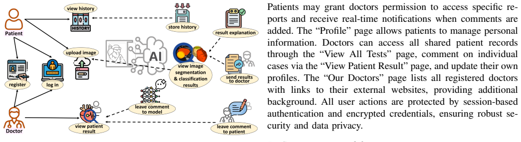
- Combining segmentation and classification in one web platform means the same system can serve patients, doctors, and researchers, with secure sharing and doctor comment workflows.
- The dynamically weighted Focal Dice Loss, which reweights classes by inverse previous-epoch Dice, is proposed as a general fix for class imbalance in 3D medical segmentation.
- On ACDC, the full pipeline reports higher segmentation Dice and higher classification accuracy than the combined-task baselines listed in Table III.
Reading between the lines
- The 98% figure is a single-dataset result on 50 ACDC test patients; before clinical use, the same fixed pipeline would need validation on an external cohort to test whether the two refinement features generalize.
- The refinement recipe is transferable: any diagnostic pair with similar bulk-volume profiles but distinct localized shape statistics could use the same broad-classifier-plus-specialist-SVM pattern.
- If the features were selected after inspecting test confusion, the honest comparison is the initial classifier's 92%, and the platform's real contribution would be integration rather than the reported accuracy gain.
- The doctor-feedback feature could become a data-collection loop for retraining, but the paper does not measure whether that loop improves future models.
Signed reviews
Editorial analysis
A structured set of objections, weighed in public.
Referee Report
Summary. The paper presents IntelliCardiac, a web-based platform for cardiac cine-MRI segmentation and disease classification. It combines a 3D residual U-Net segmentation model with a two-stage classifier (random forest followed by an RBF-SVM expert refinement) and evaluates both tasks on the ACDC dataset (100 training, 50 test patients). The authors report a mean Dice score of 92.56% for segmentation and 98% classification accuracy across five diagnostic categories, and they claim these results outperform prior methods that integrate segmentation and classification. The paper also describes the platform's architecture, user roles, and deployment details.
Significance. If the reported numbers are reproducible, the contribution is valuable: an open-source, web-based tool that addresses segmentation and classification together, with a clearly described interface and a 4D processing pipeline, evaluated on the public ACDC benchmark. The two-stage refinement idea, using clinically motivated myocardial wall thickness features to separate DCM from MINF, is reasonable and potentially useful. However, the scientific claims currently rest on sparse empirical evidence: no error bars or repeated runs, no described validation split for classifier design, and an inconsistent specification of the refinement features. With these added, the paper could provide a credible and useful reference for integrated cardiac MRI analysis platforms.
major comments (4)
- [§III-C2 vs §IV-B] The two myocardial wall thickness (MWT) features used by the expert SVM are described inconsistently. Section III-C2 states the SVM uses the maximum of slice-wise mean wall thickness and the mean of slice-wise standard deviations measured during the end-systolic phase, while Section IV-B and the text around Figure 5 state the features are the mean of slice-wise standard deviations of MWT and the standard deviation of slice-wise mean MWT at end-diastole. This is a load-bearing discrepancy: it makes the classifier design unreproducible and prevents verification of the reported 98% accuracy. The authors must specify the exact feature definitions, the cardiac phase used, and the feature-selection rule that led to these two features.
- [§IV-B (Classification Results)] No validation split or model-selection protocol is reported for the two-stage classifier. The initial classifier is described as obtaining 92% accuracy and the final pipeline 98% on the 50-patient test set; on 50 patients, that is a difference of about three corrected predictions. If the random forest hyperparameters, the choice of the refinement features, the MINF/DCM-only refinement rule, or the SVM hyperparameters (C, gamma) were selected after inspecting the test-set predictions, the 98% figure is not a held-out accuracy. The paper should report the validation procedure, the hyperparameter ranges considered, and the decision rule for selecting the refinement features.
- [Table III and Classification Comparison] The claim of 'outperforming existing state-of-the-art methods' is not supported by the comparison set. Table III compares against Wolterink (2018), Wibowo (2022), Zheng (2019), and Khened (2018), all of which predate many recent ACDC classification results, and no statistical significance tests or confidence intervals are reported for the classification accuracy difference. The paper should either include current state-of-the-art methods that perform both segmentation and classification on ACDC, or temper the claim to 'outperforms the compared methods.'
- [Table II and Segmentation Results] The segmentation results are reported as point estimates without standard deviations, ranges, or patient-level variability. Given that the mean Dice differences between IntelliCardiac and the nearest comparator (nnFormer, EMCAD) are small (0.4–0.5 points), the absence of variability measures makes it difficult to judge whether the improvements are meaningful. The authors should report error bars or per-fold/per-patient statistics and describe how the segmentation model was selected with respect to validation data.
minor comments (6)
- [Abstract/Introduction] The phrase 'bridges the hole between AI research and real-world deployment' should be 'bridges the gap between AI research and real-world deployment.'
- [§III-B2] In the decoder description, 'linked with the unsampled feature maps' should be 'linked with the upsampled feature maps.'
- [§III-B3] The sentence 'We have a contribution lies in the dynamically weighted Focal Dice Loss' should be rewritten, for example as 'Our contribution includes the dynamically weighted Focal Dice Loss.'
- [§III-B3] The sentence 'This immediately address class imbalance and improve segmentation uniformity, this encourages the model to give priority to minority or underperforming buildings' has subject-verb agreement issues and should read 'This addresses class imbalance and improves segmentation uniformity, encouraging the model to give priority to minority or underperforming structures.'
- [§III-C2] The phrase 'avoids minimizes overfitting' is ungrammatical; it should be 'avoids overfitting' or 'minimizes overfitting.'
- [Figure 5] The caption says 'Confusion matrices before (right) and after (left) expert refinement,' but the usual convention is to present the initial state on the left and the refined state on the right; please clarify the ordering or reorder the panels.
Circularity Check
No significant circularity: the reported segmentation and classification accuracies are empirical results measured against the public ACDC benchmark, not derivations that reduce to their inputs.
full rationale
IntelliCardiac's central claims are empirical: a 3D residual U-Net segmentation model and a two-stage random-forest/SVM classifier are trained and evaluated on the publicly available ACDC dataset, with Dice scores and classification accuracies reported against a separate 50-patient test set. The segmentation loss, preprocessing, post-processing, and classifier design are all described as methodological choices rather than as analytical consequences of the target results. The feature set is explicitly inspired by an external reference (Isensee et al. [10]), and the comparisons in Tables II and III are against independent published methods; none of the comparisons rely on a self-citation chain. The two-stage classifier is a legitimate two-step pipeline: the random forest produces initial predictions, and the SVM refines only MINF/DCM predictions using two myocardial wall thickness features. The final 98% accuracy is presented as a measured outcome on ACDC test data, not as a quantity that is definitionally equal to the classifier's training objective or to any input feature. There are no equations in the paper that reduce to one another by construction, and no fitted parameter is renamed as a prediction. The main concerns raised by the paper itself are reproducibility issues rather than circularity: the paper does not describe a validation split or hyperparameter selection procedure, and the description of the SVM's two features is inconsistent (Section III-C2 says end-systolic mean of slice-wise standard deviations and maximum of slice-wise means, while Section IV-B says end-diastolic mean of slice-wise standard deviations and standard deviation of slice-wise means). These inconsistencies create a risk that the 98% figure reflects test-set-guided model selection, but that is a data-leakage or validity concern, not a definitional circularity. Because the core results are benchmarked against independent external data and external methods, the circularity score is 0.
Assumptions & free parameters
free parameters (5)
- ROI patch size =
128 x 128
- Focal Dice exponent beta =
not reported
- Segmentation training schedule =
300 epochs, batch size 4, initial LR 5e-4
- Random Forest hyperparameters =
not reported
- SVM RBF hyperparameters (C, gamma) =
not reported
assumptions (4)
- domain assumption ACDC ground-truth labels are accurate and the 100/50 train/test split is an unbiased sample.
- domain assumption The left ventricle has a roughly circular cross-section in the axial slices used for ROI detection.
- domain assumption Myocardial wall thickness statistics at end-systole separate DCM from MINF.
- standard math Deep learning components (ResBlocks, U-Net, focal Dice loss, LCCA) work as described in the cited literature.
Cite this review
Pith. "Pith review of IntelliCardiac: An Intelligent Platform for Cardiac Image Segmentation and Classification." pith.science (2026). https://pith.science/paper/RMITUZID
@misc{pith2026250503838,
  author       = {Pith},
  title        = {Pith review of: IntelliCardiac: An Intelligent Platform for Cardiac Image Segmentation and Classification},
  year         = {2026},
  howpublished = {\url{https://pith.science/paper/RMITUZID}},
  note         = {Machine review of arXiv:2505.03838}
}
read the original abstract
Precise and effective processing of cardiac imaging data is critical for the identification and management of the cardiovascular diseases. We introduce IntelliCardiac, a comprehensive, web-based medical image processing platform for the automatic segmentation of 4D cardiac images and disease classification, utilizing an AI model trained on the publicly accessible ACDC dataset. The system, intended for patients, cardiologists, and healthcare professionals, offers an intuitive interface and uses deep learning models to identify essential heart structures and categorize cardiac diseases. The system supports analysis of both the right and left ventricles as well as myocardium, and then classifies patient's cardiac images into five diagnostic categories: dilated cardiomyopathy, myocardial infarction, hypertrophic cardiomyopathy, right ventricular abnormality, and no disease. IntelliCardiac combines a deep learning-based segmentation model with a two-step classification pipeline. The segmentation module gains an overall accuracy of 92.6%. The classification module, trained on characteristics taken from segmented heart structures, achieves 98% accuracy in five categories. These results exceed the performance of the existing state-of-the-art methods that integrate both segmentation and classification models. IntelliCardiac, which supports real-time visualization, workflow integration, and AI-assisted diagnostics, has great potential as a scalable, accurate tool for clinical decision assistance in cardiac imaging and diagnosis.
Figures


Figures from the paper (2 more)


Reference graph
Works this paper leans on
-
[1]
Olivier Bernard, Alain Lalande, Clement Zotti, Frederick Cervenan- sky, Xin Yang, Pheng-Ann Heng, Irem Cetin, Karim Lekadir, Oscar Camara, Miguel Angel Gonzalez Ballester, Gerard Sanroma, Sandy Napel, Steffen Petersen, Georgios Tziritas, Elias Grinias, Mahendra Khened, Varghese Alex Kollerathu, Ganapathy Krishnamurthi, Marc- Michel Roh ´e, Xavier Pennec, ...
work page 2018
- [2]
-
[3]
A Computational Approach to Edge Detection
John Canny. A Computational Approach to Edge Detection. IEEE Transactions on Pattern Analysis and Machine Intelligence , PAMI- 8(6):679–698, 1986
work page 1986
-
[4]
Swin-Unet: Unet-Like Pure Transformer for Medical Image Segmentation
Hu Cao, Yueyue Wang, Joy Chen, Dongsheng Jiang, Xiaopeng Zhang, Qi Tian, and Manning Wang. Swin-Unet: Unet-Like Pure Transformer for Medical Image Segmentation. In Computer Vision – ECCV 2022 Workshops, pages 205–218, Cham, 2023. Springer Nature Switzerland
work page 2022
-
[5]
A Cloud-Based System for Automated AI Image Analysis and Reporting
Neil Chatterjee, Jeffrey Duda, James Gee, Ameena Elahi, Kristen Martin, Van Doan, Hannah Liu, Matthew Maclean, Daniel Rader, Arijitt Borthakur, et al. A Cloud-Based System for Automated AI Image Analysis and Reporting. Journal of Imaging Informatics in Medicine , 38(1):368–379, 2025
work page 2025
-
[6]
Jieneng Chen, Yongyi Lu, Qihang Yu, Xiangde Luo, Ehsan Adeli, Yan Wang, Le Lu, Alan L. Yuille, and Yuyin Zhou. TransUNet: Transformers Make Strong Encoders for Medical Image Segmentation. arXiv preprint arXiv:2102.04306, 2021
arXiv 2021
-
[7]
Dillencourt, Hanan Samet, and Markku Tamminen
Michael B. Dillencourt, Hanan Samet, and Markku Tamminen. A general approach to connected-component labeling for arbitrary image representations. J. ACM, 39(2):253–280, Apr 1992
work page 1992
-
[8]
Use of the Hough transformation to detect lines and curves in pictures
Richard O Duda and Peter E Hart. Use of the Hough transformation to detect lines and curves in pictures. Communications of the ACM , 15(1):11–15, 1972
work page 1972
Show all 31 references
-
[9]
Hearst, S.T
M.A. Hearst, S.T. Dumais, E. Osuna, J. Platt, and B. Scholkopf. Support vector machines. IEEE Intelligent Systems and their Applications , 13(4):18–28, 1998
1998
-
[10]
Jaeger, Peter M
Fabian Isensee, Paul F. Jaeger, Peter M. Full, Ivo Wolf, Sandy Engel- hardt, and Klaus H. Maier-Hein. Automatic Cardiac Disease Assess- ment on cine-MRI via Time-Series Segmentation and Domain Specific Features. In Statistical Atlases and Computational Models of the Heart. ACD...
2018
-
[11]
nnU-Net: a self-configuring method for deep learning-based biomedical image segmentation
Fabian Isensee, Paul F Jaeger, Simon A A Kohl, Jens Petersen, and Klaus H Maier-Hein. nnU-Net: a self-configuring method for deep learning-based biomedical image segmentation. Nature methods, 18(2):203—211, Feb 2021
2021
-
[12]
Karamitsos, Jane M
Theodoros D. Karamitsos, Jane M. Francis, Saul Myerson, Joseph B. Selvanayagam, and Stefan Neubauer. The Role of Cardiovascular Magnetic Resonance Imaging in Heart Failure. Journal of the American College of Cardiology, 54(15):1407–1424, 2009
2009
-
[13]
Densely Connected Fully Convolutional Network for Short-Axis Cardiac Cine MR Image Segmentation and Heart Diagnosis Using Random Forest
Mahendra Khened, Varghese Alex, and Ganapathy Krishnamurthi. Densely Connected Fully Convolutional Network for Short-Axis Cardiac Cine MR Image Segmentation and Heart Diagnosis Using Random Forest. In Statistical Atlases and Computational Models of the Heart. ACDC and MMWHS Ch...
2018
-
[14]
3D Slicer: A Platform for Subject-Specific Image Analysis, Visualization, and Clinical Support
Ron Kikinis, Steve D Pieper, and Kirby G V osburgh. 3D Slicer: A Platform for Subject-Specific Image Analysis, Visualization, and Clinical Support. In Intraoperative Imaging and Image-Guided Therapy, pages 277–289. Springer, 2014
2014
-
[15]
Attention U-Net: Learning Where to Look for the Pancreas
Ozan Oktay, Jo Schlemper, Loic Le Folgoc, Matthew Lee, Mattias Heinrich, Kazunari Misawa, Kensaku Mori, Steven McDonagh, Nils Y Hammerla, Bernhard Kainz, et al. Attention U-Net: Learning Where to Look for the Pancreas. arXiv preprint arXiv:1804.03999 , 2018
2018 arXiv
-
[16]
Gernand, Jeffery A
Yimu Pan, Sitao Zhang, Alison D. Gernand, Jeffery A. Goldstein, and James Z. Wang. S2S2: Semantic Stacking for Robust Semantic Segmentation in Medical Imaging. arXiv preprint arXiv:2412.13156 , 2024
2024 arXiv
-
[17]
Circular hough transform
Simon Just Kjeldgaard Pedersen. Circular hough transform. Aalborg University, Vision, Graphics, and Interactive Systems, 123(6):2–3, 2007
2007
-
[18]
SegFormer3D: an Efficient Transformer for 3D Medical Image Segmentation
Shehan Perera, Pouyan Navard, and Alper Yilmaz. SegFormer3D: an Efficient Transformer for 3D Medical Image Segmentation. In 2024 IEEE/CVF Conference on Computer Vision and Pattern Recognition Workshops (CVPRW), pages 4981–4988, Los Alamitos, CA, USA, Jun
2024
-
[19]
EM- CAD: Efficient Multi-Scale Convolutional Attention Decoding for Med- ical Image Segmentation
Md Mostafijur Rahman, Mustafa Munir, and Radu Marculescu. EM- CAD: Efficient Multi-Scale Convolutional Attention Decoding for Med- ical Image Segmentation. In 2024 IEEE/CVF Conference on Computer Vision and Pattern Recognition (CVPR) , pages 11769–11779, 2024
2024
-
[20]
U-Net: Convolu- tional Networks for Biomedical Image Segmentation
Olaf Ronneberger, Philipp Fischer, and Thomas Brox. U-Net: Convolu- tional Networks for Biomedical Image Segmentation. In Medical Image Computing and Computer-Assisted Intervention – MICCAI 2015 , pages 234–241. Springer International Publishing, 2015
2015
-
[21]
Samet and M
H. Samet and M. Tamminen. Efficient component labeling of images of arbitrary dimension represented by linear bintrees. IEEE Transactions on Pattern Analysis and Machine Intelligence , 10(4):579–586, 1988
1988
-
[22]
Spatiotemporal Repre- sentation Learning for Short and Long Medical Image Time Series
Chengzhi Shen, Martin J Menten, Hrvoje Bogunovi ´c, Ursula Schmidt- Erfurth, Hendrik PN Scholl, Sobha Sivaprasad, Andrew Lotery, Daniel Rueckert, Paul Hager, and Robbie Holland. Spatiotemporal Repre- sentation Learning for Short and Long Medical Image Time Series. In Medical I...
2024
-
[23]
Sudre, Wenqi Li, Tom Vercauteren, Sebastien Ourselin, and M
Carole H. Sudre, Wenqi Li, Tom Vercauteren, Sebastien Ourselin, and M. Jorge Cardoso. Generalised Dice Overlap as a Deep Learning Loss Function for Highly Unbalanced Segmentations. In Deep Learning in Medical Image Analysis and Multimodal Learning for Clinical Decision Support...
2017
-
[24]
Pei Wang and Albert C. S. Chung. Focal Dice Loss and Image Dilation for Brain Tumor Segmentation. In Deep Learning in Medical Image Analysis and Multimodal Learning for Clinical Decision Support, Cham, 2018
2018
-
[25]
Cardiac Disease Classification Using Two-Dimensional Thickness and Few-Shot Learning Based on Magnetic Resonance Imaging Image Segmentation
Adi Wibowo, Pandji Triadyaksa, Aris Sugiharto, Eko Adi Sarwoko, Fajar Agung Nugroho, Hideo Arai, and Masateru Kawakubo. Cardiac Disease Classification Using Two-Dimensional Thickness and Few-Shot Learning Based on Magnetic Resonance Imaging Image Segmentation. Journal of Imagi...
2022
-
[26]
Wolterink, Tim Leiner, Max A
Jelmer M. Wolterink, Tim Leiner, Max A. Viergever, and Ivana I ˇsgum. Automatic segmentation and disease classification using cardiac cine MR images. In Statistical Atlases and Computational Models of the Heart, pages 101–110. Springer Verlag, 2018
-
[27]
Cardiovascular diseases (CVDs)
World Health Organization. Cardiovascular diseases (CVDs). World Health Organization Fact Sheets , Jun 2023. Accessed: 2025-04-13
2023
-
[28]
Explainable car- diac pathology classification on cine MRI with motion characterization by semi-supervised learning of apparent flow
Qiao Zheng, Herv ´e Delingette, and Nicholas Ayache. Explainable car- diac pathology classification on cine MRI with motion characterization by semi-supervised learning of apparent flow. Medical Image Analysis , 56:80–95, 2019
2019
-
[29]
nnFormer: V olumetric Medical Image Segmentation via a 3D Transformer
Hong-Yu Zhou, Jiansen Guo, Yinghao Zhang, Xiaoguang Han, Lequan Yu, Liansheng Wang, and Yizhou Yu. nnFormer: V olumetric Medical Image Segmentation via a 3D Transformer. IEEE Transactions on Image Processing, 32:4036–4045, 2023
2023
-
[30]
Open Health Imaging Foundation Viewer: An Extensible Open-Source Framework for Build- ing Web-Based Imaging Applications to Support Cancer Research
Erik Ziegler, Trinity Urban, Danny Brown, James Petts, Steve D Pieper, Rob Lewis, Chris Hafey, and Gordon J Harris. Open Health Imaging Foundation Viewer: An Extensible Open-Source Framework for Build- ing Web-Based Imaging Applications to Support Cancer Research. JCO clinical...
2020
-
[2024]
IEEE Computer Society
Reviewed August 16, 2026 · model on record in the stance chip above.
Discussion (0). Continue with ORCID to comment.