REVIEW 3 major objections 4 minor 1 cited by
UltraGauss: Ultrafast Gaussian Reconstruction of 3D Ultrasound Volumes
T0 review · 3 major / 4 minor · reviewed 2026-08-15 · deepseek-v4-flash
Pith's one-line read An ultrasound-specific Gaussian splatting model that renders by intersecting Gaussians with the probe plane reconstructs 3D fetal brain volumes from 2D scans in minutes, reaching 0.99 SSIM within 20 minutes on a single GPU.
desk verdict Genuine ultrasound-specific Gaussian splatting with strong results, but the rasterizer boundary formula appears mathematically wrong and needs a fix. read the letter →
The pith
A machine-rendered reading of the paper's core claim, the machinery that carries it, and where it could break.
The reading
What carries the argument
The load-bearing object is a mixture of anisotropic 3D Gaussians, each with a mean, a covariance, a colour, and a peak opacity. The rendering mechanism is the probe-plane intersection: a 2D pixel is lifted to the plane $x|0=[x_1,x_2,0]^T$ in the probe frame, and each Gaussian's opacity is the 3D squared Mahalanobis distance $\hat{\alpha}_i(x)=\alpha_i\exp(-\tfrac{1}{2}(x|0-\mu_i^{3D})^T(\Sigma_i^{3D})^{-1}(x|0-\mu_i^{3D}))$. This replaces perspective projection, so there is no depth sorting of occluders and no accumulated transmittance. Efficiency comes from a triangular parametrization $\Sigma^{-1}=LL^T$ that makes inversion and sampling cheap, and from bounding boxes derived from the chi-squared ellipsoid that reject non-intersecting Gaussians and limit pixel iteration to the intersecting 2D box; a buffer-compaction load-balancing pass distributes the remaining work across threads.
What would settle it
Scan a phantom containing a strongly attenuating object placed between the probe and a reflective target, reconstruct the volume with UltraGauss, and compare the recovered intensities behind the object with a no-shadow control scan; if the no-transmittance model is correct, the shadow region should be reconstructed without systematic underestimation, while true attenuation would force the optimizer to absorb the loss into spurious Gaussian opacities. A simpler analytic check is to fit the model's rendering equation to a synthetic volume generated with an exponential attenuation law and measure whether residuals concentrate behind the attenuator.
Extended reading notes
Core claim
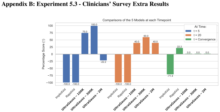
UltraGauss replaces the perspective projection of standard Gaussian splatting with a probe-plane intersection model. Each ultrasound pixel is rendered by evaluating every 3D Gaussian's opacity as a 3D squared Mahalanobis distance at the probe plane, combined through a simple colour equation, with the accumulated transmittance of optical volume rendering omitted because ultrasound does not accumulate intensity along a ray the way light does. The paper's central claim is that this wave-aligned rendering equation, together with a numerically stable lower-triangular covariance parametrization $\Sigma^{-1}=LL^T$ and a two-phase rasterization with chi-squared bounding-box culling, lets a mixture of up to two million anisotropic Gaussians fit a whole fetal brain volume from axial slices in minutes. On twelve clinical volumes it reports average SSIM up to 0.995 at convergence and consistently higher SSIM than two neural baselines at every training time point; on freehand videos it reports 0.91 SSIM on withheld frames. The authors further claim that a survey of ten expert sonographers, averaging 18 years of experience, preferred UltraGauss reconstructions over the baselines at all time points.
Load-bearing premise
The rendering equation drops the accumulated transmittance that optical volume rendering uses, assuming ultrasound images are formed by independent Gaussian reflections at the probe plane with no occlusion or attenuation from overlying tissue; if shadowing is strong in the target anatomy, the model is misspecified, although the flexible mixture may absorb the error.
Editorial extensions
If this is right
- A 3D fetal brain volume can be reconstructed from a sensorless freehand 2D cinesweep, letting clinicians revisit any cross-sectional plane after the scan; the paper reports 0.91 SSIM on frames withheld from end-to-end training.
- Reconstruction quality is nearly saturated when all axial slices are available: average SSIM reaches 0.995 at convergence, and 0.99 within 20 minutes on a single GPU.
- Practical speed-quality trade-offs are controllable by choosing the initial Gaussian count: 100K Gaussians for 5-minute reconstructions, 2M Gaussians for the highest final accuracy.
- Because rasterization is real-time, clinicians could interactively browse reconstructed volumes during a consultation rather than waiting for offline processing.
- The method generalizes across orthogonal views: axial, coronal, and sagittal cross-sections are reconstructed consistently, which is the standard way clinicians inspect 3D volumes.
Reading between the lines
- If the speed holds outside this dataset, 2D ultrasound probes could substitute for 3D probes in routine fetal monitoring, removing a roughly tenfold cost barrier and the need for extra operator training.
- The no-transmittance assumption is the main risk: in anatomies with strong shadowing or attenuation, such as behind ribs or calcified structures, the model may need an explicit attenuation term; a testable extension is to add a learnable transmittance while keeping the plane-intersection rasterizer.
- The same plane-intersection rendering could transfer to other imaging modalities where acquisition is inherently planar rather than projective, such as some tomographic or radar systems.
- The end-to-end cinesweep result mixes pose-estimation error with reconstruction error, so improving the upstream pose predictor should push the 0.91 SSIM higher without changing UltraGauss at all.
Signed reviews
Editorial analysis
A structured set of objections, weighed in public.
Referee Report
Summary. The paper introduces UltraGauss, a Gaussian-splatting framework for reconstructing 3D ultrasound volumes from 2D scans. Unlike standard Gaussian splatting, which projects Gaussians onto a camera image plane, UltraGauss evaluates Gaussian opacities at their intersection with the ultrasound probe plane, motivated by the claim that ultrasound image formation is better modeled by plane intersection than by ray-based transmittance accumulation. The paper contributes a triangular covariance parameterization, a rasterization bounding-box derivation, a load-balancing scheme, and a custom CUDA implementation. Experiments are reported on fetal brain volumes (Dataset A) and freehand cinesweep videos (Dataset B), comparing against ImplicitVol and RapidVol, with quantitative metrics (SSIM, PSNR, LPIPS), a clinician realism survey, and an end-to-end pose-estimation plus reconstruction pipeline.
Significance. If the technical and experimental concerns are resolved, this would be a meaningful contribution: it is, to the best of the authors' knowledge, the first ultrasound-specific Gaussian splatting method, and the reported speed gains (state-of-the-art quality within 5–20 minutes on one GPU) and held-out cinesweep SSIM of 0.91 are strong, concrete results. The paper also ships a reproducible-looking parameterization, evaluates on real clinical data, includes a clinician survey, and commits to releasing the CUDA implementation. These are real strengths. However, the central rasterization derivation contains an internal inconsistency (Eq. 13), and the evaluation protocol partly evaluates training views in the axial direction, so the headline accuracy claims need re-examination before the contribution can be accepted.
major comments (3)
- [Section 5.2] The bounding-box formula in Eq. (13) is not the axis-aligned coordinate extent of the ellipsoid defined by Eq. (12). For an ellipsoid {(x−μ)^T Σ^{−1}(x−μ) ≤ χ²}, the correct coordinate half-widths are sqrt(χ² Σ_kk), obtained by minimizing over the remaining coordinates. Eq. (13) instead uses v_k = Σ_jj Σ_ll − Σ_jl² (the diagonal cofactor) and λ = χ² / det(Σ), giving half-widths sqrt(χ² (Σ^{−1})_kk). The formula is also dimensionally inconsistent: for Σ with units of length², v has units of length⁴ and λ has units of length^{−6}, so sqrt(λ v) has units of length^{−1}, not length. For a concrete example, Σ = diag(100,1,1) and χ² = 7.815 gives a true first-coordinate half-width of 27.96, while Eq. (13) returns 0.28, clipping 99.99% of the Gaussian's probability. Because this box determines which pixels are rasterized, the described algorithm is incorrect for anisotropic covariances. The authors must either correct Eq. (13) to the formula above or, if the shipped CUDA kernel uses the correct bounds, rewrite the derivation in the paper to match the implementation.
- [Section 5.2] The axial evaluation reuses the same 160 linearly spaced axial slices that were used as training input, so the reported axial SSIM partly measures fitting to training views. The coronal and sagittal views are genuinely held out and should be reported separately and prominently; the headline accuracy claims should not rely on the axial or the averaged score without clearly distinguishing training and novel views. The cinesweep held-out-frame results are a cleaner test and are a strength, but the paper's central speed-versus-quality claims would be more credible if this training/test overlap were acknowledged and analyzed.
- [Section 4.1, Eq. (7)] The rendering equation in Eq. (7) drops the accumulated transmittance T_j from Eq. (1), based on the assertion that ultrasound is formed by independent Gaussian reflections at the probe plane with 'no occlusion.' This is a strong modeling assumption: fetal brain scans necessarily include overlying tissue, and attenuation and shadowing are first-order effects in ultrasound. The paper provides no quantitative evidence that the no-transmittance model is sufficient. Since this assumption is the physical motivation for the entire method, the authors should either provide an ablation or a theoretical argument showing that transmittance is negligible in their acquisition geometry, or explicitly discuss the expected failure modes and demonstrate empirically that the flexible Gaussian mixture absorbs the error.
minor comments (4)
- [Section 4.3] The notation χ²_{3,1−p} = 7.815 for p = 95% is inconsistent with standard quantile notation: the 95th percentile of the chi-squared distribution with 3 degrees of freedom is χ²_{3,0.95} = 7.815, i.e., the subscript should be p, not 1−p.
- [Appendix A] The results table in Appendix A is difficult to interpret: rows appear to be repeated (e.g., the UltraGauss-2M entries are duplicated across adjacent blocks), and the PSNR values (around 4.3 dB) are implausibly low for the reported SSIM values near 0.98–1.00. Please reformat the table and double-check the columns and units.
- [Fig. 6] The y-axis label of Fig. 6 says 'Percentage who thought UltraGauss' reconstruction was a real US scan,' while the text says that 70% of experts rated UltraGauss-generated images as 'more realistic than the ground-truth.' These are different statements; please align the wording and clarify what the survey actually asked.
- [Throughout] There are several typos, including 'splattling' in Section 3.1, 'recosntruction' in Section 6.1, and inconsistent hyphenation of 'state-of-the-art.' A careful proofreading pass is recommended.
Circularity Check
No circularity: the derivation is self-contained and the main claims are supported by external benchmarks and held-out evaluations, not by construction or self-citation.
full rationale
The paper's claimed derivation chain maps an explicit ultrasound image-formation assumption (probe-plane intersection, Eq. 8) into a rasterizer boundary formula (Eq. 13), a covariance parameterization (Eq. 11), and a load-balancing scheme (Sec. 4.4). The rendering equation Eq. 7 is an openly stated modeling choice that drops accumulated transmittance; it is not a hidden reuse of the target result. Eq. 13 is asserted to follow from Eq. 12, and the displayed formula appears mathematically inconsistent with the ellipsoid extents, but this is a derivation error rather than circularity: the claimed bound does not reduce to its input equation by construction, it fails to follow from it. Experimental claims are evaluated against held-out coronal and sagittal views, held-out cinesweep frames, and external baselines (RapidVol, ImplicitVol); the present authors' self-citations appear only as baselines or as a pose-estimation component in an end-to-end pipeline, not as the load-bearing justification for the central claim. No fitted parameter is renamed as a prediction, no uniqueness theorem is imported from prior work, and no ansatz is smuggled in via citation. Therefore no circular step is present.
Assumptions & free parameters
free parameters (6)
- Gaussian count =
100k / 300k / 2M
- Initial L_ij range =
uniform [4, 5)
- Initial alpha =
0.731
- Chi-squared cutoff p =
0.95 (7.815)
- Positive-definite regularizer beta =
not reported (beta > 0)
- Learning rates =
0.05 for most parameters, 0.00016 for means
assumptions (4)
- ad hoc to paper Ultrasound rendering is captured by evaluating Gaussian opacities on the probe plane with no accumulated transmittance (Eq. 7)
- domain assumption A Gaussian mixture can represent volumetric ultrasound reflectivity
- domain assumption Known probe poses are available or estimable
- standard math Chi-squared quantile for 3 degrees of freedom gives a valid cutoff ellipsoid
Cite this review
Pith. "Pith review of UltraGauss: Ultrafast Gaussian Reconstruction of 3D Ultrasound Volumes." pith.science (2026). https://pith.science/paper/T57QCUCF
@misc{pith2026250505643,
  author       = {Pith},
  title        = {Pith review of: UltraGauss: Ultrafast Gaussian Reconstruction of 3D Ultrasound Volumes},
  year         = {2026},
  howpublished = {\url{https://pith.science/paper/T57QCUCF}},
  note         = {Machine review of arXiv:2505.05643}
}
read the original abstract
Ultrasound imaging is widely used due to its safety, affordability, and real-time capabilities, but its 2D interpretation is highly operator-dependent, leading to variability and increased cognitive demand. 2D-to-3D reconstruction mitigates these challenges by providing standardized volumetric views, yet existing methods are often computationally expensive, memory-intensive, or incompatible with ultrasound physics. We introduce UltraGauss: the first ultrasound-specific Gaussian Splatting framework, extending view synthesis techniques to ultrasound wave propagation. Unlike conventional perspective-based splatting, UltraGauss models probe-plane intersections in 3D, aligning with acoustic image formation. We derive an efficient rasterization boundary formulation for GPU parallelization and introduce a numerically stable covariance parametrization, improving computational efficiency and reconstruction accuracy. On real clinical ultrasound data, UltraGauss achieves state-of-the-art reconstructions in 5 minutes, and reaching 0.99 SSIM within 20 minutes on a single GPU. A survey of expert clinicians confirms UltraGauss' reconstructions are the most realistic among competing methods. Our CUDA implementation will be released upon publication.
Figures


Figures from the paper (10 more)




Forward citations
Cited by 1 Pith paper
-
K-space Gaussian Representation for Parallel MRI
K-space Gaussian Representation (KGR) fits measured multi-coil k-space with shared Gabor-Gaussian primitives and refines the result with Hankel low-rank projection, improving accelerated MRI reconstruction over baseli...
Reference graph
Works this paper leans on
-
[1]
John E. Aldrich. Basic physics of ultrasound imaging:. Crit- ical Care Medicine, 35(Suppl):S131–S137, 2007
work page 2007
-
[2]
Beryl R. Benacerraf. Three-dimensional fetal sonogra- phy. Journal of Ultrasound in Medicine, 21(10):1063–1067, 2002
work page 2002
-
[3]
Image registration using machine and deep learning
Xiaohuan Cao, Jingfan Fan, Pei Dong, Sahar Ahmad, Pew- Thian Yap, and Dinggang Shen. Image registration using machine and deep learning. In Handbook of Medical Image Computing and Computer Assisted Intervention, pages 319–
-
[4]
Nvidia Corp. Thrust library. https://github.com/ NVIDIA/thrust?tab=readme-ov-file, 2020
work page 2020
-
[5]
Robbersmyr, and Kris- tian Muri Knausg ˚ard
Anurag Dalal, Daniel Hagen, Kjell G. Robbersmyr, and Kris- tian Muri Knausg ˚ard. Gaussian Splatting: 3D Reconstruc- tion and Novel View Synthesis: A Review.IEEE Access, 12: 96797–96820, 2024. Conference Name: IEEE Access
work page 2024
-
[6]
David, Donald Peebles, and Danail Stoyanov
Chiara Di Vece, Maela Le Lous, Brian Dromey, Francisco Vasconcelos, Anna L. David, Donald Peebles, and Danail Stoyanov. Ultrasound Plane Pose Regression: Assessing Generalized Pose Coordinates in the Fetal Brain. IEEE Transactions on Medical Robotics and Bionics , 6(1):41–52,
-
[7]
RapidVol: Rapid Reconstruction of 3D Ultrasound Volumes from Sensorless 2D Scans
Mark C. Eid, Pak-Hei Yeung, Madeleine K. Wyburd, Jo˜ao F. Henriques, and Ana I. L. Namburete. RapidV ol: Rapid Re- construction of 3D Ultrasound V olumes from Sensorless 2D Scans, 2024. arXiv:2404.10766 [cs, eess]
work page Pith review arXiv 2024
-
[8]
Free-SurGS: SfM-Free 3D Gaussian Splatting for Surgical Scene Reconstruction
Jiaxin Guo, Jiangliu Wang, Di Kang, Wenzhen Dong, Went- ing Wang, and Yun-hui Liu. Free-SurGS: SfM-Free 3D Gaussian Splatting for Surgical Scene Reconstruction. In Medical Image Computing and Computer Assisted Inter- vention – MICCAI 2024 , pages 350–360. Springer Nature Switzerland, Cham, 2024. Series Title: Lecture Notes in Computer Science
work page 2024
Show all 35 references
-
[9]
2d gaussian splatting for geometrically accu- rate radiance fields
Binbin Huang, Zehao Yu, Anpei Chen, Andreas Geiger, and Shenghua Gao. 2d gaussian splatting for geometrically accu- rate radiance fields. In SIGGRAPH 2024 Conference Papers. Association for Computing Machinery, 2024
2024
-
[10]
3D Gaussian Splatting for Real-Time Radiance Field Rendering
Bernhard Kerbl, Georgios Kopanas, Thomas Leimkuehler, and George Drettakis. 3D Gaussian Splatting for Real-Time Radiance Field Rendering. ACM Transactions on Graphics, 42(4):1–14, 2023
2023
-
[11]
Merz and C
E. Merz and C. Welter. 2D and 3D Ultrasound in the eval- uation of normal and abnormal fetal anatomy in the second and third trimesters in a level III center. Ultraschall in Der Medizin (Stuttgart, Germany: 1980), 26(1):9–16, 2005
1980
-
[12]
Srinivasan, Matthew Tancik, Jonathan T
Ben Mildenhall, Pratul P. Srinivasan, Matthew Tancik, Jonathan T. Barron, Ravi Ramamoorthi, and Ren Ng. NeRF: Representing Scenes as Neural Radiance Fields for View Synthesis, 2020. arXiv:2003.08934 [cs]
2020 arXiv
-
[13]
Namburete, Richard V
Ana I.L. Namburete, Richard V . Stebbing, Bryn Kemp, Mo- hammad Yaqub, Aris T. Papageorghiou, and J. Alison No- ble. Learning-based prediction of gestational age from ultra- sound images of the fetal brain. Medical Image Analysis, 21 (1):72–86, 2015
2015
-
[14]
Ana I. L. Namburete, Bartłomiej W. Papie ˙z, Michelle Fer- nandes, Madeleine K. Wyburd, Linde S. Hesse, Felipe A. Moser, Leila Cheikh Ismail, Robert B. Gunier, Waney Squier, Eric O. Ohuma, Maria Carvalho, Yasmin Jaffer, Michael Gravett, Qingqing Wu, Ann Lambert, Adele Win- sey...
2023
-
[15]
Nelson and Dolores H
Thomas R. Nelson and Dolores H. Pretorius. Three- dimensional ultrasound imaging. Ultrasound in Medicine and Biology, 24(9):1243–1270, 1998
1998
-
[16]
Powles, David J
Alexander Ej. Powles, David J. Martin, Irving Tp. Wells, and Christopher R. Goodwin. Physics of ultrasound.Anaesthesia & Intensive Care Medicine, 19(4):202–205, 2018
2018
-
[17]
MODELING ILLUMINATION V ARI- ATION WITH SPHERICAL HARMONICS
Ravi Ramamoorthi. MODELING ILLUMINATION V ARI- ATION WITH SPHERICAL HARMONICS. In Face Pro- cessing, pages 385–424. Elsevier, 2006
2006
-
[18]
Jayroop Ramesh, Nicola Dinsdale, Pak-Hei Yeung, and Ana I. L. Namburete. Geometric Transformation Uncertainty for 9 Improving 3D Fetal Brain Pose Prediction from Freehand 2D Ultrasound Videos. In Medical Image Computing and Com- puter Assisted Intervention – MICCAI 2024, pages...
2024
-
[19]
Revising Densification in Gaussian Splatting
Samuel Rota Bul `o, Lorenzo Porzi, and Peter Kontschieder. Revising Densification in Gaussian Splatting. In Computer Vision – ECCV 2024, pages 347–362, Cham, 2025. Springer Nature Switzerland
2024
-
[20]
L. J. Salomon, Z. Alfirevic, V . Berghella, C. M. Bilardo, G. E. Chalouhi, F. Da Silva Costa, E. Hernandez-Andrade, G. Malinger, H. Munoz, D. Paladini, F. Prefumo, A. Sotiri- adis, A. Toi, and W. Lee. ISUOG Practice Guidelines (up- dated): performance of the routine mid-trimes...
2022
-
[21]
Survey of methods and principles in three- dimensional reconstruction from two-dimensional medical images
Mriganka Sarmah, Arambam Neelima, and Heisnam Ro- hen Singh. Survey of methods and principles in three- dimensional reconstruction from two-dimensional medical images. Visual Computing for Industry, Biomedicine, and Art, 6(1):15, 2023
2023
-
[22]
Structure-from-motion revisited
Johannes Lutz Sch ¨onberger and Jan-Michael Frahm. Structure-from-motion revisited. In Conference on computer vision and pattern recognition (CVPR), 2016
2016
-
[23]
Blake, and Toril A
Ole Vegard Solberg, Frank Lindseth, Hans Torp, Richard E. Blake, and Toril A. Nagelhus Hernes. Freehand 3D Ultra- sound Reconstruction Algorithms—A Review. Ultrasound in Medicine & Biology, 33(7):991–1009, 2007
2007
-
[24]
Thomas L. Szabo. Diagnostic ultrasound imaging : inside out. Academic Press, Amsterdam, 2nd ed. edition, 2014. Includes bibliographical references and index
2014
-
[25]
Wang, A.C
Z. Wang, A.C. Bovik, H.R. Sheikh, and E.P. Simoncelli. Im- age Quality Assessment: From Error Visibility to Structural Similarity. IEEE Transactions on Image Processing, 13(4): 600–612, 2004
2004
-
[26]
gsplat: An Open-Source Library for Gaussian Splatting, 2024
Vickie Ye, Ruilong Li, Justin Kerr, Matias Turkulainen, Brent Yi, Zhuoyang Pan, Otto Seiskari, Jianbo Ye, Jef- frey Hu, Matthew Tancik, and Angjoo Kanazawa. gsplat: An Open-Source Library for Gaussian Splatting, 2024. arXiv:2409.06765 [cs]
2024 arXiv
-
[27]
Pak-Hei Yeung, Linde Hesse, Moska Aliasi, Monique Haak, the INTERGROWTH-21st Consortium, Weidi Xie, and Ana I. L. Namburete. ImplicitV ol: Sensorless 3D Ultrasound Reconstruction with Deep Implicit Representation, 2021. arXiv:2109.12108 [cs, eess]
2021 arXiv
-
[28]
Pak-Hei Yeung, Moska Aliasi, Monique Haak, The INTERGROWTH-21st Consortium, Weidi Xie, and Ana I. L. Namburete. Adaptive 3D Localization of 2D Free- hand Ultrasound Brain Images, 2022. arXiv:2209.05477 [cs, eess]
2022 arXiv
-
[29]
Plenox- els: Radiance Fields without Neural Networks, 2021
Alex Yu, Sara Fridovich-Keil, Matthew Tancik, Qinhong Chen, Benjamin Recht, and Angjoo Kanazawa. Plenox- els: Radiance Fields without Neural Networks, 2021. arXiv:2112.05131 [cs]
2021 arXiv
-
[30]
Mip-Splatting: Alias-free 3D Gaussian Splatting, 2023
Zehao Yu, Anpei Chen, Binbin Huang, Torsten Sattler, and Andreas Geiger. Mip-Splatting: Alias-free 3D Gaussian Splatting, 2023. arXiv:2311.16493 [cs]
2023 arXiv
-
[31]
A Review of Deep Learn- ing Techniques for 3D Reconstruction of 2D Images
Anny Yuniarti and Nanik Suciati. A Review of Deep Learn- ing Techniques for 3D Reconstruction of 2D Images. In2019 12th International Conference on Information & Communi- cation Technology and System (ICTS), pages 327–331, 2019
2019
-
[32]
Rˆ2-gaussian: Rectifying radia- tive gaussian splatting for tomographic reconstruction
Ruyi Zha, Tao Jun Lin, Yuanhao Cai, Jiwen Cao, Yanhao Zhang, and Hongdong Li. Rˆ2-gaussian: Rectifying radia- tive gaussian splatting for tomographic reconstruction. In Advances in neural information processing systems , pages 44907–44934. Curran Associates, Inc., 2024
2024
-
[33]
Efros, Eli Shecht- man, and Oliver Wang
Richard Zhang, Phillip Isola, Alexei A. Efros, Eli Shecht- man, and Oliver Wang. The Unreasonable Effectiveness of Deep Features as a Perceptual Metric. In 2018 IEEE/CVF Conference on Computer Vision and Pattern Recognition , pages 586–595, Salt Lake City, UT, 2018. IEEE
2018
-
[34]
UltraGauss - N
M. Zwicker, H. Pfister, J. Van Baar, and M. Gross. EW A volume splatting. In Proceedings Visualization, 2001. VIS ’01., pages 29–538, San Diego, CA, USA, 2001. IEEE. 10 Appendix A: Experiment 5.2 - Results Table SSIM (↑) PSNR (↑) LPIPS (↓) SSIM (↑) PSNR (↑) LPIPS (↓) SSIM (↑) ...
2001
-
[2024]
Conference Name: IEEE Transactions on Medical Robotics and Bionics
Reviewed August 15, 2026 · model on record in the stance chip above.
Discussion (0). Continue with ORCID to comment.