REVIEW 4 major objections 5 minor 31 references
Beyond the First Read: AI-Assisted Perceptual Error Detection in Chest Radiography Accounting for Interobserver Variability
T0 review · 4 major / 5 minor · reviewed 2026-08-07 · deepseek-v4-flash
Pith's one-line read A second-read AI companion, RADAR, catches 78% of simulated missed chest X-ray abnormalities by comparing a detector's candidate boxes against the radiologist's own annotations.
desk verdict A transparent, reproducible post-read CXR miss-detection prototype that is cleverly motivated but evaluated only on a simulated dataset that removes whole consensus-visible boxes—so the headline recall is not yet evidence about real perceptual misses. read the letter →
The pith
A machine-rendered reading of the paper's core claim, the machinery that carries it, and where it could break.
The reading
What carries the argument
The mechanism is a two-module pipeline. The Abnormality Detection Module adapts a fine-tuned single-class YOLOv11 detector to output candidate bounding boxes for any abnormality. The Differential Detection Module then compares each candidate box with the radiologist's boxes, and any candidate whose maximum IoU against all radiologist annotations is zero is flagged as a referral. Non-maximum suppression with an IoU threshold of zero keeps one box per overlapping cluster, reducing redundancy while preserving distinct abnormal regions.
What would settle it
Take a collection of genuine missed chest X-ray findings identified through double reading, eye tracking, or a discrepancy log, run RADAR on those original reads, and compare referral recall and IoU against the simulated figures; if real-miss referral recall falls well below 0.78 or median IoU below 0.78, the central claim would not survive the transition from the simulation.
Extended reading notes
Core claim
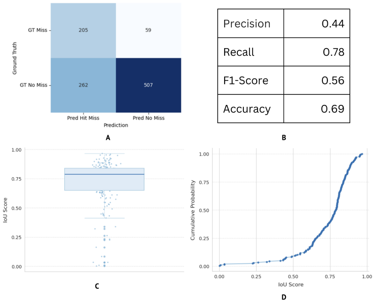
RADAR is designed to work after the radiologist has finalized a reading. Its central claim is that a simple differential rule—flag any region the detector calls abnormal that has zero overlap with any of the radiologist's boxes—recovers most simulated misses while telling the radiologist where to look rather than what to conclude. In the simulated test set it recovered 204 of 263 omitted abnormalities, with more than 90% of its true referrals exceeding 0.5 IoU, so the system does not just name a missed finding but points to the right spot. The authors treat the 0.44 precision as a deliberate design choice: uncertain referrals keep the radiologist in the loop and prevent blind over-reliance on the AI.
Load-bearing premise
The entire evaluation rests on the assumption that deleting one fused radiologist-annotated box from 30% of abnormal cases mimics the way radiologists actually miss abnormalities, and if real misses are partial, ambiguous, or concentrated in low-conspicuity lesions rather than whole omitted regions, the reported recall and IoU figures will not transfer to clinical practice.
Editorial extensions
If this is right
- If RADAR's simulated-dataset performance holds, a practical second-look workflow becomes available at low cost: the system runs in a few seconds on a consumer laptop and in about one second on a modest GPU, so it can be inserted after a normal read without changing the radiologist's workflow.
- Because referrals are regions rather than fixed labels, the system can accommodate interobserver disagreement: a region one radiologist deliberately skips can still be flagged for another without being framed as an error.
- The single-class detection design sidelines inter-class label disagreement, such as the consolidation versus infiltration boundary, making the detector more robust to annotation noise in chest radiography.
- The gating ablation shows that prepending a normal-versus-abnormal classifier improves precision from 0.44 to 0.48 and accuracy from 0.69 to 0.71 but drops recall from 0.78 to 0.69, holding F1 at 0.56, which is why the authors kept the ungated version as the final pipeline.
- The open-source web application and the simulated error dataset give other groups a reusable benchmark for perceptual-error-correction systems in chest X-ray interpretation.
Reading between the lines
- Editorial extension: because the simulation deletes whole fused abnormality boxes at random, real perceptual misses that are partial, subtle, or class-specific could yield lower clinical recall; a study against real misses would calibrate the gap.
- Editorial extension: the differential-detection logic could generalize beyond box geometry to pixel-level anomaly maps, which might improve spatial agreement with human misses and reduce sensitivity to the detector's box shape.
- Editorial extension: with precision at 44%, the practical value of the system depends on how cheaply radiologists can dismiss false referrals, so a prospective workflow study counting adjudication time per referral would test whether the F1 of 0.56 translates into a net clinical benefit.
- Editorial extension: the same post-read comparison strategy could transfer to other double-read imaging tasks, such as CT or mammography, whenever a detector's proposals can be aligned with a reader's annotations.
Signed reviews
Editorial analysis
A structured set of objections, weighed in public.
Referee Report
Summary. This paper presents RADAR, a post-interpretation decision-support system for chest radiography. RADAR consists of an Abnormality Detection Module (ADM) built on a fine-tuned YOLOv11 model and a Differential Detection Module (DDM) that flags ADM predictions with zero Intersection over Union (IoU) against the radiologist's annotations as potential missed findings. The authors construct a simulated perceptual-error dataset by fusing multi-radiologist VinDr-CXR annotations with an IoU threshold of 0.3 and randomly deleting one fused box in 30% of abnormal images. On this dataset they report recall of 0.78, precision of 0.44, F1-score of 0.56, and median IoU of 0.78 for true referrals. They also describe a Flask/ngrok-based web application, latency benchmarks on laptop and GPU platforms, and an ablation study using a DenseNet-121 gating classifier. The paper concludes that RADAR effectively complements radiologist judgment as a second-look tool for perceptual-error detection.
Significance. If the reported performance transferred to real clinical misses, RADAR would be a useful, lightweight second-read aid, and the open-source release of code, data, and a web application is commendable for reproducibility. However, the evaluation rests entirely on a simulated error proxy, and several technical details needed to reproduce the headline numbers are missing. The contribution is therefore best viewed as a proof-of-concept that needs validation on real double-reading or eye-tracking data before clinical claims are warranted.
major comments (4)
- [Section 3.5 and Eq. (2)] The simulated perceptual-error dataset deletes whole fused boxes that are consensus-visible lesions agreed on by multiple radiologists, whereas real perceptual errors are often subtle and reader-dependent. Because the ADM was trained on the same VinDr-CXR box distribution, the DDM task reduces to re-detecting a typical training-label-like box absent from the annotation set. The 0.78 recall therefore measures detection of conspicuous omissions, not genuine perceptual misses; the authors acknowledge this in Section 8, but the Abstract and Section 4.2 conclusions still overstate what has been established.
- [Section 3.2 and Figure 4] No confidence threshold is reported for the ADM predictions that enter the DDM. Section 3.2 states only that NMS with an IoU threshold of zero is applied. Table 1 reports an ADM recall of 0.388, while Figure 4 reports RADAR recall of 0.78 (204/263); these figures may be reconcilable because the denominators differ, but without the operating threshold and a precision-recall curve the headline numbers are not reproducible and could reflect a threshold artifact.
- [Section 3.5] Section 3.5 refers to "the aforementioned test set," but Sections 3.3 and 3.4 describe splitting a balanced training subset (8,788 images) into training/validation and held-out test. It is unclear whether the simulated errors were generated on the held-out test subset of that balanced dataset or on the original 3,000-image VinDr-CXR test set. This distinction matters for possible data leakage and for reproducing the reported 204/263 count.
- [Sections 4.2 and 8] The conclusion that RADAR "effectively complements radiologist judgment" is not supported by the evidence: Section 8 concedes that no user studies or workflow trials were conducted. The system's clinical value depends on radiologists' acceptance of referrals and on the referral rate in real practice, neither of which is measured. At minimum, the conclusions should be limited to the simulated setting, and the claimed accommodation of interobserver variability should be tempered because the fusion procedure removes rather than models reader variability.
minor comments (5)
- [Section 3.5] The greedy box-fusion algorithm is not fully specified; the order of merging and the handling of boxes that become overlapping after a merge should be described.
- [Table 2] The column header "Accuarcy" should be "Accuracy."
- [Section 3.2] The phrase "NMS with an IoU threshold set to zero" is unusual; consider clarifying that this suppresses any pair with nonzero overlap and retains only the highest-confidence box.
- [Section 2] The sentence about Lakhani et al. appears garbled ("referring to dermatologist in low confidence output"); please rephrase.
- [Figure 4] The box plot and cumulative distribution in Figures 4C and 4D should state the number of true referrals used to compute the IoU distribution.
Circularity Check
No significant circularity: RADAR's referral mechanism is a deterministic comparison of independently trained detector outputs against annotations; the simulated-miss evaluation is a proxy-validity limitation, not a definitional reduction.
full rationale
The paper's derivation chain is self-contained. The ADM is a YOLOv11 detector fine-tuned on a balanced split of VinDr-CXR (Section 3.4), and the DDM referral rule (Eq. 2) is a deterministic zero-IoU comparison between ADM boxes and radiologist annotations. The simulated perceptual-error dataset (Section 3.5) is constructed by fusing radiologist boxes at IoU>=0.3 and deleting one fused box in 30% of abnormal cases. A true referral is scored when an ADM box overlaps the deleted box. Nothing is fitted to the test deletions: the ADM weights come from training data that are independent of the specific boxes removed at evaluation time, and the recall/precision numbers are reported as measured outcomes, not as parameters that were tuned on the error dataset. The main weakness is external validity: because the ADM is trained on VinDr-CXR boxes and the simulated misses are also VinDr-CXR boxes from the same distribution, the test mainly shows that the detector can re-find boxes from its training-label distribution. That is a proxy-validity concern, and the authors explicitly acknowledge it in Section 8 ('the simulated perceptual error dataset does not fully capture the complexity of human perceptual behavior... No user studies or workflow trials have been conducted'). But this is not circularity: the evaluation does not reduce by construction to the input annotations, and no fitted parameter is renamed as a prediction. The self-citations ([15], [20]) appear only in related-work context and are not load-bearing for the central claim. Therefore the circularity score is 0.
Assumptions & free parameters
free parameters (4)
- Referral confidence threshold
- Bounding-box fusion IoU threshold =
0.3
- Per-case miss removal rate =
30%
- YOLO training hyperparameters =
lr=0.002, momentum=0.9, weight decay=5e-4, batch 8, 100+ epochs
assumptions (4)
- domain assumption VinDr-CXR radiologist annotations are a reliable ground truth for thoracic abnormalities.
- ad hoc to paper A missed abnormality is correctly identified when an ADM prediction has zero IoU with all radiologist annotations (Eq. 2).
- ad hoc to paper The simulated error dataset (random deletion of fused boxes) approximates real perceptual error patterns.
- domain assumption A single-class abnormality detector generalizes across the 14 CXR abnormality categories without sacrificing localization.
Cite this review
Pith. "Pith review of Beyond the First Read: AI-Assisted Perceptual Error Detection in Chest Radiography Accounting for Interobserver Variability." pith.science (2026). https://pith.science/paper/TBUEPMRR
@misc{pith2026250613049,
  author       = {Pith},
  title        = {Pith review of: Beyond the First Read: AI-Assisted Perceptual Error Detection in Chest Radiography Accounting for Interobserver Variability},
  year         = {2026},
  howpublished = {\url{https://pith.science/paper/TBUEPMRR}},
  note         = {Machine review of arXiv:2506.13049}
}
read the original abstract
Chest radiography is widely used in diagnostic imaging. However, perceptual errors -- especially overlooked but visible abnormalities -- remain common and clinically significant. Current workflows and AI systems provide limited support for detecting such errors after interpretation and often lack meaningful human--AI collaboration. We introduce RADAR (Radiologist--AI Diagnostic Assistance and Review), a post-interpretation companion system. RADAR ingests finalized radiologist annotations and CXR images, then performs regional-level analysis to detect and refer potentially missed abnormal regions. The system supports a "second-look" workflow and offers suggested regions of interest (ROIs) rather than fixed labels to accommodate inter-observer variation. We evaluated RADAR on a simulated perceptual-error dataset derived from de-identified CXR cases, using F1 score and Intersection over Union (IoU) as primary metrics. RADAR achieved a recall of 0.78, precision of 0.44, and an F1 score of 0.56 in detecting missed abnormalities in the simulated perceptual-error dataset. Although precision is moderate, this reduces over-reliance on AI by encouraging radiologist oversight in human--AI collaboration. The median IoU was 0.78, with more than 90% of referrals exceeding 0.5 IoU, indicating accurate regional localization. RADAR effectively complements radiologist judgment, providing valuable post-read support for perceptual-error detection in CXR interpretation. Its flexible ROI suggestions and non-intrusive integration position it as a promising tool in real-world radiology workflows. To facilitate reproducibility and further evaluation, we release a fully open-source web implementation alongside a simulated error dataset. All code, data, demonstration videos, and the application are publicly available at https://github.com/avutukuri01/RADAR.
Figures


Figures from the paper (3 more)


Reference graph
Works this paper leans on
-
[1]
M. A. Bruno, E. A. Walker, H. H. Abujudeh, Understanding and con- fronting our mistakes: The epidemiology of error in radiology and strategies for error reduction, RadioGraphics 35 (6) (2015) 1668–1676. doi:10.1148/rg.2015150023
-
[2]
J. N. Itri, R. R. Tappouni, R. O. McEachern, A. J. Pesch, S. H. Pa- tel, Fundamentals of diagnostic error in imaging, RadioGraphics 38 (6) (2018) 1845–1865.doi:10.1148/rg.2018180021. 19
-
[3]
L. H. Garland, On the scientific evaluation of diagnostic procedures, Radiology 52 (3) (1949) 309–328.doi:10.1148/52.3.309
-
[4]
W. B. Gefter, B. A. Post, H. Hatabu, Commonly missed findings on chest radiographs: Causes and consequences, Chest 163 (3) (2023) 650– 661.doi:10.1016/j.chest.2022.10.039
-
[5]
L. Berlin, Radiologic errors, past, present and future, Diagnosis 1 (1) (2014) 79–84.doi:10.1515/dx-2013-0012
-
[6]
S. Waite, J. Scott, B. Gale, T. Fuchs, S. Kolla, D. Reede, Interpretive error in radiology, American Journal of Roentgenology 208 (4) (2017). doi:10.2214/AJR.16.16963
-
[7]
F. Pesapane, G. Gnocchi, C. Quarrella, et al., Errors in radiology: A standard review, Journal of Clinical Medicine 13 (15) (2024) 4306.doi: 10.3390/jcm13154306
-
[8]
J. Adler-Milstein, J. H. Chen, G. Dhaliwal, Next-generation artificial intelligence for diagnosis: From predicting diagnostic labels to “way- finding”, JAMA 326 (24) (2021) 2467–2468.doi:10.1001/jama.2021. 22396
Show all 31 references
-
[9]
Reverberi, T
C. Reverberi, T. Rigon, A. Solari, et al., Experimental evidence of effec- tive human–ai collaboration in medical decision-making, Scientific Re- ports 12 (2022) 14952.doi:10.1038/s41598-022-18751-2
2022 doi
-
[10]
H. L. Kundel, C. F. Nodine, D. P. Carmody, Visual scanning, pat- tern recognition and decision-making in pulmonary nodule detec- tion, Investigative Radiology 13 (3) (1978) 175–181.doi:10.1097/ 00004424-197805000-00001
1978
-
[11]
Wagner, K
I. Wagner, K. Chakradeo, Human–ai complementarity in diagnostic ra- diology: The case of double reading, Philosophy & Technology 38 (2) (2025) 57.doi:10.1007/s13347-025-00886-5
2025 doi
-
[12]
B. van Ginneken, Fifty years of computer analysis in chest imaging: Rule-based, machine learning, deep learning, Radiological Physics and Technology 10 (1) (2017) 23–32.doi:10.1007/s12194-017-0394-5. 20
2017 doi
-
[13]
P. G. Anderson, H. Tarder-Stoll, M. Alpaslan, N. Keathley, D. L. Levin, R. M. Jones, Deep learning improves physician accuracy in the compre- hensive detection of abnormalities on chest x-rays, Scientific Reports 14 (2024) 25151.doi:10.1038/s41598-024-76608-2
2024 doi
-
[14]
Quinn, K
L. Quinn, K. Tryposkiadis, J. Deeks, H. C. W. de Vet, S. Mallett, L. B. Mokkink, Y. Takwoingi, S. Taylor-Phillips, A. Sitch, Interob- server variability studies in diagnostic imaging: A methodological sys- tematic review, British Journal of Radiology 96 (1148) (2023) 20220972....
2023 doi
-
[15]
Mobiny, A
A. Mobiny, A. Singh, H. Van Nguyen, Risk-aware machine learning clas- sifier for skin lesion diagnosis, Journal of Clinical Medicine 8 (8) (2019) 1241.doi:10.3390/jcm8081241
2019 doi
-
[16]
Lakhani, A
P. Lakhani, A. Flanders, R. Gorniak, Endotracheal tube position as- sessment on chest radiographs using deep learning, Radiology: Artificial Intelligence 3 (1) (2021) e200026.doi:10.1148/ryai.2020200026
2021 doi
- [17]
-
[18]
Ghoshal, A
B. Ghoshal, A. Tucker, Estimating uncertainty and interpretability in deep learning for coronavirus (covid-19) detection, arXiv preprint (2020).arXiv:2003.10769. URLhttps://arxiv.org/abs/2003.10769
2020 arXiv
-
[19]
L.Topff, S.Steltenpool, E.R.Ranschaert, N.Ramanauskas, R.Menezes, J. J. Visser, R. G. H. Beets-Tan, N. S. Hartkamp, Artificial intelli- gence–assisted double reading of chest radiographs to detect clinically relevant missed findings: A two-centre evaluation, European Radiology...
2024 doi
-
[20]
Awasthi, N
A. Awasthi, N. P. Le, Z. Deng, C. C. Wu, H. V. Nguyen, Enhancing radiological diagnosis: A collaborative approach integrating ai and hu- man expertise for visual miss correction, arXiv preprint (2024).arXiv: 21 2406.19686. URLhttps://arxiv.org/abs/2406.19686
2024 arXiv
-
[21]
Haghgoo, R
J.Irvin, P.Rajpurkar, M.Ko, Y.Yu, S.Ciurea-Ilcus, C.Chute, H.Mark- lund, B. Haghgoo, R. Ball, K. Shpanskaya, J. Seekins, D. A. Mong, S. S. Halabi, J. K. Sandberg, R. Jones, D. B. Larson, C. P. Langlotz, B. N. Patel, M. P. Lungren, A. Y. Ng, Chexpert: A large chest radio- graph...
2019 arXiv
-
[22]
M. A. Warren, Z. Zhao, T. Koyama, J. A. Bastarache, C. M. Shaver, M. W. Semler, T. W. Rice, M. A. Matthay, C. S. Calfee, L. B. Ware, Severity scoring of lung oedema on the chest radiograph is associated with clinical outcomes in ards, Thorax 73 (9) (2018) 840–846.doi: 10.1136/...
2018 doi
-
[23]
M. N. Albaum, L. C. Hill, M. Murphy, Y. H. Li, C. R. Fuhrman, C. A. Britton, W. N. Kapoor, M. J. Fine, Interobserver reliability of the chest radiograph in community-acquired pneumonia, Chest 110 (2) (1996) 343–350.doi:10.1378/chest.110.2.343
1996 doi
-
[24]
Irvin, P
J. Irvin, P. Rajpurkar, M. Ko, et al., Chexpert: A large chest radiograph dataset with uncertainty labels and expert comparison, in: Proceedings of the AAAI Conference on Artificial Intelligence, Vol. 33, 2019, pp. 590–597.doi:10.1609/aaai.v33i01.3301590
2019 doi
-
[25]
A. E. W. Johnson, T. J. Pollard, N. R. Greenbaum, et al., Mimic- cxr—jpg, a large publicly available database of labeled chest radio- graphs, arXiv preprint (2019).arXiv:1901.07042. URLhttps://arxiv.org/abs/1901.07042
2019 arXiv
-
[26]
Redmon, S
J. Redmon, S. Divvala, R. Girshick, A. Farhadi, You only look once: Unified, real-time object detection, in: Proceedings of the IEEE Confer- ence on Computer Vision and Pattern Recognition (CVPR), 2016, pp. 779–788.doi:10.1109/CVPR.2016.91
2016 doi
-
[27]
H. Q. Nguyen, K. Lam, L. T. Le, et al., Vindr-cxr: An open dataset of chest x-rays with radiologist’s annotations, Scientific Data 9 (2022) 429. doi:10.1038/s41597-022-01428-5. 22
2022 doi
-
[28]
L. G. Quekel, A. G. Kessels, R. Goei, J. M. van Engelshoven, De- tection of lung cancer on the chest radiograph: A study on observer performance, European Journal of Radiology 39 (2) (2001) 111–116. doi:10.1016/s0720-048x(01)00301-1
2001 doi
-
[29]
Balabanova, R
Y. Balabanova, R. Coker, I. Fedorin, et al., Variability in interpreta- tion of chest radiographs among russian clinicians and implications for screening programmes: Observational study, BMJ 331 (7513) (2005) 379–382.doi:10.1136/bmj.331.7513.379
2005 doi
-
[30]
Young, T
M. Young, T. J. Marrie, Interobserver variability in the interpretation of chest roentgenograms of patients with possible pneumonia, Archives of Internal Medicine 154 (23) (1994) 2729–2732.doi:10.1001/archinte. 1994.00420230122014
1994
-
[31]
Y. X. Tang, Y. B. Tang, Y. Peng, et al., Automated abnormality classi- fication of chest radiographs using deep convolutional neural networks, npj Digital Medicine 3 (2020) 70.doi:10.1038/s41746-020-0273-z. 23
2020 doi
Reviewed August 7, 2026 · model on record in the stance chip above.
Discussion (0). Continue with ORCID to comment.