REVIEW 3 major objections 4 minor 37 references
Understanding Spatial Language in Radiology: Representation Framework, Annotation, and Spatial Relation Extraction from Chest X-ray Reports using Deep Learning
T0 review · 3 major / 4 minor · reviewed 2026-08-14 · deepseek-v4-flash
Pith's one-line read The paper defines Rad-SpRL, a spatial-role framework for chest X-ray reports, and shows that a Bi-LSTM-CRF model extracts findings, locations, probable diagnoses, and hedging terms at average $F_1$ scores of 90.28, 94.61, 71.47, and 73.27…
desk verdict A useful Rad-SpRL annotation resource for radiology NLP, but the evaluation likely leaks across instances from the same sentence and the gold standard is noisy; worth peer review with major revision. read the letter →
The pith
A machine-rendered reading of the paper's core claim, the machinery that carries it, and where it could break.
The reading
What carries the argument
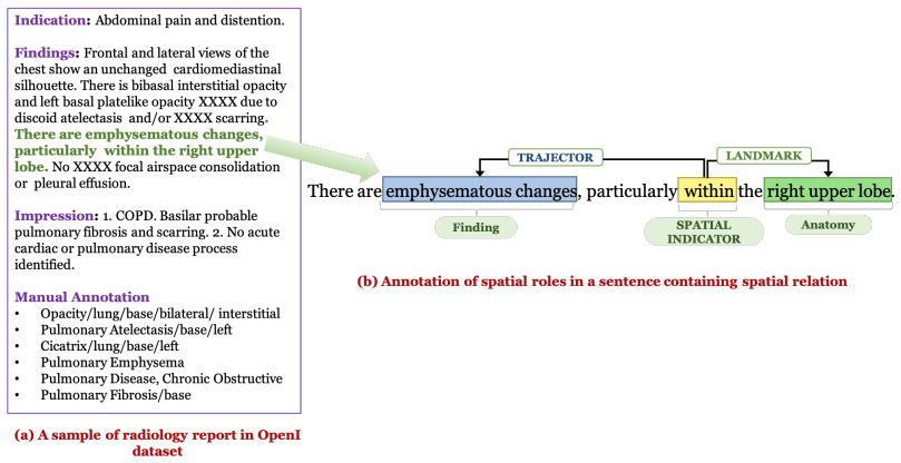
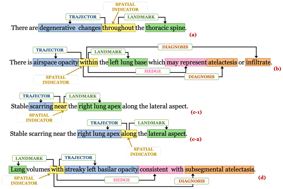
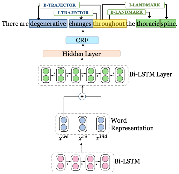
The load-bearing mechanism is Rad-SpRL, a representation framework that extends the standard spatial role labeling scheme by adding Diagnosis and Hedge as first-class roles tied to a Spatial Indicator. The extraction engine is a Bi-LSTM-CRF sequence tagger whose per-word input concatenates a pre-trained word embedding, a character-level embedding from a second Bi-LSTM, and a small indicator embedding that marks whether the word is the Spatial Indicator. The indicator embedding is what lets the model assign roles relative to one trigger, and the preprocessing step of creating a separate sentence instance for each Spatial Indicator resolves cases where the same phrase is a Trajector for one indicator and a Landmark or Diagnosis for another.
What would settle it
Re-annotate a random sample of about 200 of the 2000 reports with a fresh pair of annotators who use only the Rad-SpRL guidelines and do not see the original labels; if their agreement on Diagnosis and Trajector stays below roughly 0.6 $F_1$, then the reported model scores are substantially measuring consistency with one reconciliation process, and the claim that the corpus carries reliable spatial semantics is weakened.
Extended reading notes
Core claim
The central claim is that a single sequence-labeling model, given the location of the Spatial Indicator in a sentence, can recover all four Rad-SpRL roles, with strong performance on the two spatial roles and moderate performance on the interpretive roles. The authors show that the same phrase can legitimately play different roles in different spatial relations, so the framework does not impose a fixed finding-versus-diagnosis ontology; it records the role a phrase plays relative to a specific spatial trigger. The annotated corpus itself is part of the discovery: 2000 reports contain 1972 spatial relations, with 2293 Trajectors, 2167 Landmarks, 455 Diagnoses, and 388 Hedges, and only 304 relations include all four roles. The authors claim this shows the representation is usable and the baseline is a credible first step, while deferring joint extraction of Spatial Indicators and roles to future work.
Load-bearing premise
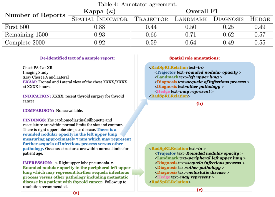
The load-bearing premise is that the reconciled annotations are a dependable reference for all four spatial roles; every reported $F_1$ is measured against these labels, yet raw inter-annotator agreement is low, with overall $F_1$ agreement of 0.59 for Trajector, 0.64 for Landmark, 0.49 for Diagnosis, and 0.55 for Hedge, so the model scores partly measure agreement with one reconciliation rather than stable spatial semantics.
Editorial extensions
If this is right
- Radiologists' free-text descriptions can be converted into role tuples of the form (Trajector, Spatial Indicator, Landmark) with optional (Diagnosis, Hedge), giving referring clinicians structured summaries of what was seen, where, and what it might be.
- Because the framework defines roles relative to a spatial trigger rather than as absolute categories, annotation effort stays close to the language actually used, and downstream systems can apply their own finding-versus-diagnosis rules.
- Diagnosis and Hedge extraction are the weak links at about 71 and 73 $F_1$, so a practical deployment should treat these roles as needing more data, longer context, or specialized modeling rather than as solved by the baseline.
- The framework is explicitly proposed to generalize beyond chest X-rays, since the same roles fit other modalities such as head CT reports where a lesion sits in a named anatomical structure and is consistent with a diagnosis.
- Spatial Indicator detection shows high recall but low precision (99.25 versus 78.86), so an end-to-end system would need to filter false prepositions before role extraction can run reliably on predicted indicators.
Reading between the lines
- If the corpus is released, the decisive follow-up experiment is to run the same model end-to-end with predicted Spatial Indicators; the paper's own precision gap predicts a measurable $F_1$ drop, and that gap is the practical bottleneck.
- A fresh annotation study using only the published guidelines, without the original reconciliation, could settle whether the low inter-annotator agreement comes from guideline ambiguity or from inherent ambiguity between findings and diagnoses.
- The framework's next natural extension is non-prepositional spatial triggers such as verbs like 'demonstrates' or 'shows', and medical devices as Trajectors; both are named in the paper and both would make the representation more useful for procedural reports.
- The role tuples could serve as weak supervision for image-text alignment, grounding each finding mention at its Landmark location on the X-ray; the paper does not propose this, but the structured roles make it a natural test.
Signed reviews
Editorial analysis
A structured set of objections, weighed in public.
Referee Report
Summary. The paper introduces Rad-SpRL, an extension of the SpRL framework for radiology reports, defining four spatial roles (Trajector, Landmark, Diagnosis, Hedge) anchored to a Spatial Indicator. The authors annotate 2000 chest X-ray reports from the OpenI dataset, report inter-annotator agreement, and train a Bi-LSTM-CRF sequence tagger with word, character, and indicator embeddings. Using 10-fold cross-validation and exact-match evaluation conditional on gold Spatial Indicators, they report average F1 of 90.28 for Trajector, 94.61 for Landmark, 71.47 for Diagnosis, and 73.27 for Hedge, as well as Spatial Indicator extraction precision/recall/F1 of 78.86/99.25/87.82. The paper also discusses error patterns and acknowledges several limitations, including the absence of inter-sentence relations.
Significance. If the corpus and evaluation are valid, Rad-SpRL is a useful resource: it is a radiology-specific spatial role framework that explicitly captures probable diagnoses and hedging terms, and the 2000-report annotation effort is substantial. The evaluation has notable strengths: exact-match scoring is strict, role extraction is evaluated conditional on gold Spatial Indicators, and the authors transparently report low annotator agreement and the ambiguous finding/diagnosis boundary. However, the corpus is not yet publicly available, and the quantitative claims currently lack per-fold variability and comparison to a baseline, so the reported numbers should be viewed as preliminary until these issues are addressed.
major comments (3)
- [Section 4.2-4.3] The evaluation may suffer from instance-level data leakage. Section 4.2 creates one training instance per Spatial Indicator, yielding 1867 instances derived from 1780 source sentences, with 221 source sentences containing more than one Spatial Indicator. Section 4.3 reports an 80/10/10 split (1495/186/186) without stating that all instances generated from the same source sentence are kept in the same fold. If the split is instance-level, the same source sentence can appear in both training and test sets in different forms, allowing the Bi-LSTM-CRF to memorize the shared lexical context and inflate the reported F1 scores. Please clarify the fold construction and, if instances are not grouped by source sentence, re-run the experiments with sentence-level grouping and report the resulting F1 scores.
- [Section 3.3.1 / Table 4] The gold standard underlying every reported F1 score has low inter-annotator agreement: Table 4 reports complete-corpus F1 agreement of 0.59 for Trajector, 0.64 for Landmark, 0.49 for Diagnosis, and 0.55 for Hedge, with Diagnosis agreement of only 0.25 in the first 500 reports. Because the system is trained and evaluated against the reconciled labels, the model may be fitting one reconciliation process rather than stable spatial semantics, especially given the authors' own observation that the same phrase can be annotated as Trajector or Diagnosis in different contexts. To support the validity of the corpus, please report system performance against each annotator's labels separately, provide a detailed analysis of disagreement patterns, and make the annotation guidelines and adjudication procedure available.
- [Section 5 / Table 6] Table 6 reports only average Precision, Recall, and F1 across 10 folds, with no standard deviations, confidence intervals, or significance tests. The reader cannot judge whether the differences between role F1 scores (e.g., 90.28 vs 94.61) are stable across folds. In addition, no baseline is evaluated, so the claim that the Bi-LSTM-CRF produces 'promising results' is not contextualized against simpler alternatives. Please report per-fold variance or confidence intervals and include at least one simple baseline, such as a CRF with lexical features or a rule-based system.
minor comments (4)
- [Section 1] In the radiology example, the sentence 'Mild streaky opacities are present in the left lung base' contains the preposition 'in', but the text says the spatial preposition is 'on'; please correct this typo.
- [Section 4.3] Please clarify the relationship between '10-fold cross-validation' and the single 80/10/10 split described as 1495/186/186 instances; in standard 10-fold cross-validation the role of the validation set should be explained more precisely.
- [Abstract] The abstract states that the corpus 'will soon be made available upon request'; in the final version, please provide a stable URL, DOI, or repository and a clear data-use license, since the reproducibility of the evaluation depends on access to the corpus.
- [Abstract and Section 5] The abstract does not state that the spatial role F1 scores are conditional on gold Spatial Indicators; this condition is explicit only in Section 5 and should appear in the abstract to avoid overstating end-to-end performance.
Circularity Check
No significant circularity; the paper's central results rest on a held-out supervised evaluation, not on an input-to-output reduction.
full rationale
This is an empirical annotation and supervised extraction paper rather than a derivation. The central claims are (1) the Rad-SpRL framework and corpus, and (2) the Bi-LSTM-CRF F1 scores. The F1 scores are computed by 10-fold cross-validation with exact-match scoring (Section 4.3, Table 6), so the reported numbers are not forced by construction from the training data. The model is trained on one subset and evaluated on held-out folds; no fitted parameter is later renamed as a prediction. Self-citations to Roberts et al. and Demner-Fushman et al. are used for background and data provenance, not as a load-bearing uniqueness theorem or as the source of the spatial role definitions, which the paper presents with its own annotation guidelines and examples. The low inter-annotator F1 agreement (Table 4) is a gold-standard reliability concern, and the one-instance-per-Spatial-Indicator preprocessing could create a sentence-level leakage risk in evaluation, but neither makes the derivation circular. No claim in the paper reduces to its inputs by definition, and no prediction is equivalent to a fitted quantity.
Assumptions & free parameters
free parameters (7)
- LSTM hidden size =
500
- Dropout rate =
0.5
- Learning rate =
0.01
- Learning rate decay =
0.99
- Character embedding dimension =
100
- Indicator embedding dimension =
5
- Maximum training epochs =
20
assumptions (5)
- domain assumption SpRL roles (Trajector, Landmark, Spatial Indicator) apply to radiology reports and capture clinically meaningful spatial relations.
- domain assumption Reconciled annotations are an acceptable gold standard despite low inter-annotator agreement.
- domain assumption Spatial role extraction performance is evaluated conditional on gold Spatial Indicators.
- domain assumption Pre-trained MIMIC-III word embeddings are appropriate for chest X-ray reports.
- standard math Bi-LSTM-CRF is a competent baseline sequence labeling architecture.
invented entities (1)
-
Rad-SpRL role set (Trajector, Landmark, Hedge, Diagnosis, Spatial Indicator)
Cite this review
Pith. "Pith review of Understanding Spatial Language in Radiology: Representation Framework, Annotation, and Spatial Relation Extraction from Chest X-ray Reports using Deep Learning." pith.science (2026). https://pith.science/paper/U4BDV34E
@misc{pith2026190804485,
  author       = {Pith},
  title        = {Pith review of: Understanding Spatial Language in Radiology: Representation Framework, Annotation, and Spatial Relation Extraction from Chest X-ray Reports using Deep Learning},
  year         = {2026},
  howpublished = {\url{https://pith.science/paper/U4BDV34E}},
  note         = {Machine review of arXiv:1908.04485}
}
read the original abstract
We define a representation framework for extracting spatial information from radiology reports (Rad-SpRL). We annotated a total of 2000 chest X-ray reports with 4 spatial roles corresponding to the common radiology entities. Our focus is on extracting detailed information of a radiologist's interpretation containing a radiographic finding, its anatomical location, corresponding probable diagnoses, as well as associated hedging terms. For this, we propose a deep learning-based natural language processing (NLP) method involving both word and character-level encodings. Specifically, we utilize a bidirectional long short-term memory (Bi-LSTM) conditional random field (CRF) model for extracting the spatial roles. The model achieved average F1 measures of 90.28 and 94.61 for extracting the Trajector and Landmark roles respectively whereas the performance was moderate for Diagnosis and Hedge roles with average F1 of 71.47 and 73.27 respectively. The corpus will soon be made available upon request.
Figures
Figures from the paper (1 more)
Reference graph
Works this paper leans on
-
[1]
D. Birchall, Spatial ability in radiologists: A necessary prerequisite?, British Journal of Radiology 88 (1049) (2015) 6–8.doi:10.1259/bjr.20140511
-
[2]
Corry, The future of recruitment and selection in radiology
C. Corry, The future of recruitment and selection in radiology. Is there a role for assessment of basic visuospatial skills?, Clinical Radiology 66 (5) (2011) 481–483. doi:10.1016/j.crad.2010.12.003
-
[3]
P. Kordjamshidi, M. V. Otterlo, M.-F. Moens, Spatial Role Labeling : Task Definition and Annotation Scheme, in: Proceedings of the Language Resources & Evaluation Con- ference, 2010, pp. 413–420. URL http://www.lrec-conf.org/proceedings/lrec2010/pdf/846{_}Paper.pdf 15
work page 2010
-
[4]
P. Kordjamshidi, T. Rahgooy, U. Manzoor, Spatial Language Understanding with Mul- timodal Graphs using Declarative Learning based Programming, in: Proceedings of the 2nd Workshop on Structured Prediction for Natural Language Processing, 2017, pp. 33–43.doi:10.18653/v1/w17-4306. URL https://www.aclweb.org/anthology/W17-4306
-
[5]
K. Roberts, L. Rodriguez, S. Shooshan, D. Demner-Fushman, Automatic Extraction and Post-coordination of Spatial Relations in Consumer Language, in: AMIA Annual Symposium Proceedings, Vol. 2015, 2015, pp. 1083–1092. URL https://www.ncbi.nlm.nih.gov/pubmed/26958247
-
[6]
D. Demner-Fushman, M. D. Kohli, M. B. Rosenman, S. E. Shooshan, L. Rodriguez, S. Antani, G. R. Thoma, C. J. McDonald, Preparing a collection of radiology exam- inations for distribution and retrieval, Journal of the American Medical Informatics Association 23 (2) (2016) 304–310.doi:10.1093/jamia/ocv080
-
[7]
X. Wang, Y. Peng, L. Lu, Z. Lu, M. Bagheri, R. M. Summers, ChestX-ray8: Hospital- scale chest X-ray database and benchmarks on weakly-supervised classification and localization of common thorax diseases, in: 2017 IEEE Conference on Computer Vision and Pattern Recognition (CVPR), 2017, pp. 3462–3471.arXiv:arXiv:1705.02315v5, doi:10.1109/CVPR.2017.369
arXiv 2017
-
[8]
J. Irvin, P. Rajpurkar, M. Ko, Y. Yu, S. Ciurea-Ilcus, C. Chute, H. Marklund, B. Hagh- goo, R. Ball, K. Shpanskaya, J. Seekins, D. A. Mong, S. S. Halabi, J. K. Sandberg, R. Jones, D. B. Larson, C. P. Langlotz, B. N. Patel, M. P. Lungren, A. Y. Ng, CheX- pert: A Large Chest Radiograph Dataset with Uncertainty Labels and Expert Compar- isonarXiv:1901.07031....
arXiv 1901
Show all 37 references
-
[9]
Y. Wang, L. Sun, Q. Jin, Enhanced Diagnosis of Pneumothorax with an Improved Real- time Augmentation for Imbalanced Chest X-rays Data Based on DCNN, IEEE/ACM Transactions on Computational Biology and Bioinformatics 14 (8) (2019) 1–1.doi: 10.1109/TCBB.2019.2911947. URL https://...
2019
-
[10]
Pesce, S
E. Pesce, S. J. Withey, P.-P. Ypsilantis, R. Bakewell, V. Goh, G. Montana, Learning to detect chest radiographs containing lung nodules using visual at- tention networks, Medical Image Analysis 53 (2019) 26–38. arXiv:1712.00996, doi:10.1016/j.media.2018.12.007. URL http://arxi...
2019 arXiv
-
[11]
Hassanpour, C
S. Hassanpour, C. P. Langlotz, Information extraction from multi-institutional radiology reports, Artificial Intelligence in Medicine 66 (2016) 29–39. doi: 10.1016/j.artmed.2015.09.007. 16 URL http://www.ncbi.nlm.nih.gov/pubmed/26481140{%}0Ahttp://www. pubmedcentral.nih.gov/art...
2016
-
[12]
Cornegruta, R
S. Cornegruta, R. Bakewell, S. Withey, G. Montana, Modelling Radiological Language with Bidirectional Long Short-Term Memory NetworksarXiv:1609.08409. URL http://arxiv.org/abs/1609.08409
-
[13]
Bustos, A
A. Bustos, A. Pertusa, J.-M. Salinas, M. de la Iglesia-Vayá, PadChest: A large chest x-ray image dataset with multi-label annotated reportsarXiv:1901.07441. URL http://arxiv.org/abs/1901.07441
1901 arXiv
-
[14]
Hassanpour, G
S. Hassanpour, G. Bay, C. P. Langlotz, Characterization of Change and Significance for Clinical Findings in Radiology Reports Through Natural Language Processing, Journal of Digital Imaging 30 (3) (2017) 314–322.doi:10.1007/s10278-016-9931-8
2017 doi
-
[15]
Annarumma, S
M. Annarumma, S. J. Withey, R. J. Bakewell, E. Pesce, V. Goh, G. Montana, Au- tomated Triaging of Adult Chest Radiographs with Deep Artificial Neural Networks, Radiology 291 (1) (2019) 196–202.doi:10.1148/radiol.2018180921
2019 doi
-
[16]
Sevenster, R
M. Sevenster, R. Van Ommering, Y. Qian, Automatically correlating clinical findings and body locations in radiology reports using MedLEE, Journal of Digital Imaging 25 (2) (2012) 240–249.doi:10.1007/s10278-011-9411-0. URL https://www.ncbi.nlm.nih.gov/pmc/articles/PMC3295967/pdf...
2012 doi
-
[17]
W.-W. Yim, T. Denman, S. W. Kwan, M. Yetisgen, Tumor information extraction in radiology reports for hepatocellular carcinoma patients., in: AMIA Joint Summits on Translational Science Proceedings, Vol. 2016, 2016, pp. 455–64. URL http://www.ncbi.nlm.nih.gov/pubmed/27570686{%}...
2016
-
[18]
B. Rink, K. Roberts, S. Harabagiu, R. H. Scheuermann, S. Toomay, T. Browning, T. Bosler, R. Peshock, Extracting actionable findings of appendicitis from radiology reports using natural language processing, in: AMIA Joint Summits on Translational Science Proceedings, Vol. 2013, ...
2013
-
[19]
Roberts, B
K. Roberts, B. Rink, S. M. Harabagiu, R. H. Scheuermann, S. Toomay, T. Browning, T. Bosler, R. Peshock, A machine learning approach for identifying anatomical locations of actionable findings in radiology reports, in: AMIA Annual Symposium Proceedings, Vol. 2012, 2012, pp. 779–...
2012
-
[20]
X. Wang, Y. Peng, L. Lu, Z. Lu, R. M. Summers, TieNet: Text-Image Embedding Network for Common Thorax Disease Classification and Reporting in Chest X-Rays, 17 in: The IEEE Conference on Computer Vision and Pattern Recognition (CVPR), 2018, pp. 9049–9058.doi:10.1109/CVPR.2018.00943
2018
-
[21]
Y. Peng, X. Wang, L. Lu, M. Bagheri, R. Summers, Z. Lu, NegBio: a high-performance tool for negation and uncertainty detection in radiology reports., in: AMIA Joint Summits on Translational Science Proceedings., Vol. 2018, 2018, pp. 188–196. URL http://www.ncbi.nlm.nih.gov/pub...
2018
-
[22]
URL http://zdaniels.com/papers/ISBI2019{_}camera.pdf
Z.A.Daniels, D.N.Metaxas, ExploitingVisualandReport-BasedInformationforChest X-Ray Analysis by Jointly Learning Visual Classifiers and Topic Models, in: IEEE 16th International Symposium on Biomedical Imaging (ISBI), 2019. URL http://zdaniels.com/papers/ISBI2019{_}camera.pdf
2019
-
[23]
J. R. Zech, M. A. Badgeley, M. Liu, A. B. Costa, J. J. Titano, E. K. Oermann, Variable generalization performance of a deep learning model to detect pneumonia in chest radiographs: A cross-sectional study, PLoS Medicine 15 (11) (2018) 1–17. doi:10.1371/journal.pmed.1002683. UR...
2018 doi
-
[24]
H.-C. Shin, K. Roberts, L. Lu, D. Demner-Fushman, J. Yao, R. M. Summers, Learning to Read Chest X-Rays : Recurrent Neural Cascade Model for Automated Image Annotation, in: The IEEE Conference on Computer Vision and Pattern Recognition (CVPR), 2016, pp. 2497–2506. URL http://op...
2016
-
[25]
Huang, Y
X. Huang, Y. Fang, M. Lu, Y. Yao, M. Li, An Annotation Model on End-to-End Chest Radiology Reports, in: IEEE Access, Vol. 7, IEEE, 2019.doi:10.1109/access.2019. 2917922. URL https://ieeexplore.ieee.org/stamp/stamp.jsp?tp={&}arnumber=8718588
2019 doi
-
[26]
Leaman, R
R. Leaman, R. Khare, Z. Lu, Challenges in Clinical Natural Language Processing for Automated Disorder Normalization, Journal of Biomedical Informatics 57 (2015) 28–37. doi:10.1016/j.jbi.2015.07.010. URL https://www.ncbi.nlm.nih.gov/pmc/articles/PMC4713367/
2015 doi
-
[27]
A. R. Aronson, F.-M. Lang, An overview of MetaMap: historical perspective and recent advances, Journal of the American Medical Informatics Association 17 (3) (2010) 229–
2010
-
[28]
Candemir, S
S. Candemir, S. Rajaraman, G. Thoma, S. Antani, Deep learning for grading car- diomegaly severity in chest x-rays: An investigation, in: IEEE Life Sciences Conference (LSC), IEEE, 2018, pp. 109–113.doi:10.1109/LSC.2018.8572113. URL https://ieeexplore.ieee.org/stamp/stamp.jsp?t...
2018
-
[29]
Lample, M
G. Lample, M. Ballesteros, S. Subramanian, K. Kawakami, C. Dyer, Neural Archi- tectures for Named Entity Recognition, in: Proceedings of NAACL-HLT, 2016, pp. 260–270.arXiv:1603.01360. URL https://www.aclweb.org/anthology/N16-1030
2016 arXiv
-
[30]
Abadi, A
M. Abadi, A. Agarwal, P. Barham, E. Brevdo, Z. Chen, C. Citro, G. S. Corrado, A. Davis, J. Dean, M. Devin, et al., Tensorflow: Large-scale machine learning on het- erogeneous distributed systems, arXiv preprint arXiv:1603.04467
-
[31]
F. Li, M. Zhang, G. Fu, D. Ji, A neural joint model for entity and rela- tion extraction from biomedical text, BMC Bioinformatics 18 (1) (2017) 198. doi:10.1186/s12859-017-1609-9. URL https://bmcbioinformatics.biomedcentral.com/track/pdf/10.1186/ s12859-017-1609-9
2017 doi
-
[32]
M. Miwa, M. Bansal, End-to-End Relation Extraction using LSTMs on Sequences and Tree Structures, in: Proceedings of the 54th Annual Meeting of the Association for Computational Linguistics, 2016, pp. 1105–1116.arXiv:arXiv:1601.00770v3, doi: 10.18653/v1/P16-1105. URL http://acl...
2016 arXiv
-
[33]
C. P. Langlotz, RadLex: a new method for indexing online educational materials, Ra- diographics 26 (6) (2006) 1595–1597.doi:10.1148/rg.266065168. URL https://pubs.rsna.org/doi/pdf/10.1148/rg.266065168
2006 doi
-
[34]
Devlin, M.-W
J. Devlin, M.-W. Chang, K. Lee, K. Toutanova, BERT: Pre-training of Deep Bidirec- tional Transformers for Language Understanding, in: Proceedings of the 2019 Confer- ence of the North American Chapter of the Association for Computational Linguistics: Human Language Technologie...
2019 doi
-
[35]
R. K. Srivastava, K. Greff, J. Schmidhuber, Highway NetWorks: Training Very Deep Networks, in: NIPS’15 Proceedings of the 28th International Conference on Neural Information Processing Systems, 2015, pp. 2377–2385.arXiv:1507.06228. URL http://arxiv.org/abs/1507.06228
2015 arXiv
-
[36]
S. He, Z. Li, H. Zhao, H. Bai, G. Liu, Syntax for Semantic Role Labeling, To Be, Or Not ToBe, in: Proceedingsofthe56thAnnualMeetingoftheAssociationforComputational Linguistics, 2018, pp. 2061–2071.doi:10.18653/v1/p18-1192. 19
2018 doi
-
[236]
doi:10.1136/jamia.2009.002733
2009 arXiv
Reviewed August 14, 2026 · model on record in the stance chip above.
Discussion (0). Continue with ORCID to comment.