REVIEW 5 major objections 4 minor 72 references
Multilabel Classification for Lung Disease Detection: Integrating Deep Learning and Natural Language Processing
T0 review · 5 major / 4 minor · reviewed 2026-08-11 · deepseek-v4-flash
Pith's one-line read Chest X-ray classifier with report parsing hits F1 0.69, AUROC 0.86
desk verdict The headline metrics are computed on a heavily filtered validation set and the NLP claim is unsupported, so the paper is not a credible research contribution despite a reasonable architecture comparison. read the letter →
The pith
A machine-rendered reading of the paper's core claim, the machinery that carries it, and where it could break.
The reading
What carries the argument
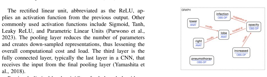
The mechanism is a two-stream representation: image features $X_{\text{image}}$ from three pretrained CNN backbones are combined with text-derived feature vectors $X_{\text{text}}$ obtained by parsing radiology reports into RadGraph entities and relations, yielding a joint representation $X=[X_{\text{text}}, X_{\text{image}}]$ that is mapped to per-disease probabilities by a sigmoid classifier with weighted binary cross-entropy loss. RadGraph is a graph-based NLP extractor that annotates each report with nodes (anatomy, observation, modifier, negation) and directed edges (located at, suggestive of, modification, negation); the paper uses this graph to consolidate condition labels as definitely present, definitely absent, or uncertain.
What would settle it
Re-evaluate the same trained model on the unmodified CheXpert validation split (with original class distribution) and compute AUROC and F1; if the numbers drop materially from 0.86 and 0.69, the clinical-potential claim is an artifact of the data filtering.
Extended reading notes
Core claim
The central claim is that an ensemble of transfer-learned CNN encoders—VGG16, ResNet50, and ConvNeXt Large—feeding a custom classifier, trained on frontal radiographs from the CheXpert dataset, can classify four chest findings at a level the paper calls clinically promising (F1 0.69, AUROC 0.86). The paper further claims that preprocessing radiology reports with RadGraph, an NLP parser that extracts typed entities (anatomy, observations, modifiers, negations) and their relations, improves the model's ability to turn uncertain reports into definite classifications, and that Grad-CAM visualizations verify the model attends to the correct structures. Per-condition AUROC values range from 0.85 for pneumothorax to 0.91 for no-finding.
Load-bearing premise
The reported AUROC and F1 are computed on a validation set from which 91% of No Finding and 30% of Pleural Effusion images were removed, so the metrics reflect that artificially balanced distribution rather than the real clinical prevalence of these conditions.
Editorial extensions
If this is right
- If the reported metrics hold on natural-prevalence data, the model could serve as a triage system that flags abnormal chest X-rays before radiologist review.
- The RadGraph-based preprocessing recipe could be ported to other chest imaging datasets to standardize report annotations for training.
- The ConvNeXt result suggests that modern vision backbones, not just residual networks, matter most for chest X-ray classification performance.
- Grad-CAM localization on pneumothorax and effusion cases provides a visual check that could help clinicians decide when to trust model output.
Reading between the lines
- The paper states, but does not experimentally verify, that comparing uncertain reports with certain ones lets the NLP-enhanced model resolve ambiguous labels; a clean ablation with and without RadGraph features would be needed to establish that claim.
- Since the reported metrics were computed after dropping 91% of No Finding and 30% of Pleural Effusion images, a reader should not compare them directly with published CheXpert benchmarks; a prevalence-corrected recalculation would be required.
- The architecture section describes a binary pneumonia output while the results and confusion matrices are for four classes; clarifying the actual output head is necessary for replication.
Editorial analysis
A structured set of objections, weighed in public.
Referee Report
Summary. The paper proposes a transfer-learning pipeline for multi-label classification of chest X-rays from the CheXpert dataset, combining image backbones (VGG16, ResNet50, ConvNeXt Large, etc.) with NLP-based preprocessing using spaCy and RadGraph. The authors report an F1 score of 0.69 and AUROC of 0.86 after applying a drastic class-imbalance mitigation step that removes 91% of No Finding and 30% of Pleural Effusion cases. The paper also includes Grad-CAM visualizations and claims that NLP-based comparison of uncertain reports improves classification, although no such experiment is presented.
Significance. If the reported metrics were trustworthy and the NLP claim were demonstrated, the paper could be of interest as a practical application of modern CNN backbones to a well-known benchmark. However, the central evaluation is compromised by the post-hoc rebalancing of the validation set, the F1 metric is prevalence-dependent and not corrected for the altered distribution, and the NLP enhancement is asserted without any supporting experiment. The paper also contains internal inconsistencies between the binary and multi-label descriptions. The study does provide a clear account of class-imbalance difficulties and uses Grad-CAM for interpretability, and it explicitly acknowledges the limitations of its data-dropping approach; these are strengths, but they do not compensate for the missing validation on the original distribution and the lack of reproducibility.
major comments (5)
- [Section IV, Table III, Abstract] The headline claims of F1=0.69 and AUROC=0.86 are computed on a validation set from which 91% of No Finding images and 30% of Pleural Effusion images were dropped. F1 is prevalence-dependent: for Pneumonia and Pneumothorax, the removal of almost all No Finding cases removes most negative examples, which can inflate precision and F1; for Pleural Effusion, dropping 30% of positives changes recall. The manuscript provides no prevalence correction, reweighting, or reporting on the original CheXpert distribution. Therefore the reported numbers do not estimate performance on the unaltered clinical population, and the conclusion about 'potential for clinical applications' is not supported by the evidence presented.
- [Section II, Table I, Abstract] The manuscript reports inconsistent dataset sizes. The abstract states 12,617 images; Section II states that the collection contains 12,716 images and that after removing uncertain scans 12,549 remain. Table I lists per-class present/absent counts that sum to 12,549 for every condition. Since the abstract's 12,617 is not used anywhere else, it is unclear which dataset size was actually analyzed, and this inconsistency prevents independent verification of the study population and the preprocessing steps.
- [Section V, Table II] There is a fundamental inconsistency in the task formulation. Section V repeatedly describes the model as performing binary pneumonia classification: 'predict the presence of pneumonia', 'a binary output that predicts the presence (y=1) or absence (y=0) of pneumonia', and 'weighted binary cross-entropy loss'. Table II, titled 'BINARY ENCODING OF MEDICAL CONDITIONS', shows mutually exclusive one-hot rows for No Finding, Pneumonia, Pneumothorax, and Effusion. In contrast, the abstract and Section VI claim a multi-label classification of multiple lung diseases. This internal contradiction affects the central claim because the model output, loss function, and evaluation metrics are not consistently defined.
- [Abstract, Section III.B, Section VI] The abstract and conclusion state that 'By comparing uncertain reports with more certain cases, the NLP-enhanced model improves its ability to conclusively classify conditions.' However, no experiment, table, or figure compares a model with NLP/RadGraph integration against an image-only baseline. The NLP preprocessing is described in detail in Sections III.A and III.B, but the reported results in Section VI and Table III come from image-only models. The claimed NLP-driven improvement is therefore unsupported and cannot be evaluated.
- [Section VI, Table V, Reproducibility] The evaluation is not reproducible. The manuscript provides no code, checkpoints, train/validation split definition, hyperparameters, or random seeds. Table III reports single-point precision, recall, F1, and AUROC values for five architectures without confidence intervals or statistical significance tests. The confusion-matrix counts in Table V sum to 614 samples per class, but the paper does not explain how this validation set relates to the 12,549-image dataset after the described 91% and 30% drops, or what fraction of the rebalanced data was held out for validation. Without these details, the reported metrics cannot be verified or compared against prior work.
minor comments (4)
- [Section VIII] The first sentence of the Discussion ends mid-phrase: 'With a validation accuracy of 92' is not a complete sentence and the reported accuracy value is never given.
- [Figures 10 and 11] The figure captions appear to be swapped relative to the text: Figure 10 is described in the text as plotting validation loss and training loss, while Figure 11 is described as plotting validation metrics, but the captions are assigned the opposite content.
- [Figures 7 and 8] Figures 7 and 8 have the identical caption 'Class distribution before dataset reduction.' If one of them depicts the distribution after reduction, the captions should be corrected to reflect the difference.
- [Section II, Reference [8]] The text states that the CheXpert dataset 'consist[s] of 220,763 MIMIC-CXR reports,' which conflates CheXpert with the MIMIC-CXR dataset. CheXpert is a collection of chest radiographs with reports; the reported number and dataset identity should be corrected.
Circularity Check
No significant circularity: the headline F1/AUROC are empirical measurements on a filtered validation set, not predictions derived by construction from their inputs; the unsupported NLP-improvement claim is missing evidence, not circularity.
full rationale
The central result — F1=0.69, AUROC=0.86 — comes from training a ConvNeXt-based classifier on a CheXpert subset and evaluating on a validation slice; it is a measurement of the pipeline actually run, not a parameter fitted to the test set and then relabeled as a prediction. Section IV's decision to drop 91% of No Finding and 30% of Pleural Effusion changes the validation distribution and likely inflates prevalence-dependent F1, so the abstract's 'clinical potential' inference is not valid for the original CheXpert population; that is a serious external-validity/reporting flaw, but no equation in the paper makes the reported metric equal to an input or makes the conclusion a restatement of the data-processing choice. The abstract's claim that 'the NLP-enhanced model improves its ability to conclusively classify conditions' is never tested: Section V's architecture is image-only, and Section VIII lists NLP as future work, so the claim is unsupported rather than circularly derived. There are no load-bearing self-citations, no imported uniqueness theorems, and no ansatz smuggled in by citation; RadGraph, CheXpert, and the pretrained backbones are external resources. The No Finding label is derived from the absence of the other labels (Section III.C), which makes its AUROC partly non-independent, but this is a label-construction detail and not the paper's main derivation. Accordingly, no circular step meets the required bar of Eq.-equals-Eq. or fitted parameter renamed as prediction.
Assumptions & free parameters
free parameters (4)
- No Finding drop rate =
91%
- Pleural Effusion drop rate =
30%
- Early stopping epoch =
6
- Class weights w_i =
not reported
assumptions (3)
- domain assumption CheXpert labels are reliable ground truth after removing uncertain scans
- domain assumption ImageNet-pretrained features transfer to chest X-rays
- domain assumption Validation set performance after early stopping generalizes to unseen data
Cite this review
Pith. "Pith review of Multilabel Classification for Lung Disease Detection: Integrating Deep Learning and Natural Language Processing." pith.science (2026). https://pith.science/paper/U7WGEHHZ
@misc{pith2026241211452,
  author       = {Pith},
  title        = {Pith review of: Multilabel Classification for Lung Disease Detection: Integrating Deep Learning and Natural Language Processing},
  year         = {2026},
  howpublished = {\url{https://pith.science/paper/U7WGEHHZ}},
  note         = {Machine review of arXiv:2412.11452}
}
read the original abstract
Classifying chest radiographs is a time-consuming and challenging task, even for experienced radiologists. This provides an area for improvement due to the difficulty in precisely distinguishing between conditions such as pleural effusion, pneumothorax, and pneumonia. We propose a novel transfer learning model for multi-label lung disease classification, utilizing the CheXpert dataset with over 12,617 images of frontal radiographs being analyzed. By integrating RadGraph parsing for efficient annotation extraction, we enhance the model's ability to accurately classify multiple lung diseases from complex medical images. The proposed model achieved an F1 score of 0.69 and an AUROC of 0.86, demonstrating its potential for clinical applications. Also explored was the use of Natural Language Processing (NLP) to parse report metadata and address uncertainties in disease classification. By comparing uncertain reports with more certain cases, the NLP-enhanced model improves its ability to conclusively classify conditions. This research highlights the connection between deep learning and NLP, underscoring their potential to enhance radiological diagnostics and aid in the efficient analysis of chest radiographs.
Figures


Figures from the paper (11 more)



Reference graph
Works this paper leans on
-
[1]
A. Ramalingam, A. Karunamurthy, T. A. Victoire, and B. Pavithra, “Impact of artificial intelligence on healthcare: A review of current applications and future possibilities,” Quing International Journal of Innovative Research in Science and Engineering , vol. 2, no. 2, pp. 37– 49, 2023
work page 2023
-
[2]
Artificial intelligence and machine learning: What you always wanted to know but were afraid to ask,
P. Rattan, D. D. Penrice, and D. A. Simonetto, “Artificial intelligence and machine learning: What you always wanted to know but were afraid to ask,” Gastro Hep Advances , vol. 1, no. 1, pp. 70–78, 2022
work page 2022
-
[3]
Addressing burnout in radiologists,
A. L. Chetlen, T. L. Chan, D. H. Ballard, L. A. Frigini, A. Hildebrand, S. Kim, J. M. Brian, E. A. Krupinski, and D. Ganeshan, “Addressing burnout in radiologists,” Academic Radiology, vol. 26, no. 4, pp. 526– 533, 2018
work page 2018
-
[4]
Physician burnout statistics 2024: The latest changes and trends in physician burnout by specialty,
A. M. Association, “Physician burnout statistics 2024: The latest changes and trends in physician burnout by specialty,” https://www.ama-assn.org/practice-management/physician-health/ physician-burnout-statistics-2024-latest-changes-and-trends, 2024, accessed: 2024-08-19
work page 2024
-
[5]
Machine learning augmented interpretation of chest x-rays: A systematic review,
H. K. Ahmad, M. R. Milne, Q. D. Buchlak, N. Ektas, G. Sanderson, H. Chamtie, S. Karunasena, J. Chiang, X. Holt, C. H. M. Tang, J. C. Y . Seah, G. Bottrell, N. Esmaili, P. Brotchie, and C. Jones, “Machine learning augmented interpretation of chest x-rays: A systematic review,” Diagnostics, vol. 13, no. 4, p. 743, 2023
work page 2023
-
[6]
X-ray: Imaging test quickly helps find diagnosis,
M. Clinic, “X-ray: Imaging test quickly helps find diagnosis,” https: //www.mayoclinic.org/tests-procedures/x-ray/about/pac-20395303, n.d., accessed: 2024-08-19
work page 2024
-
[7]
Radgraph dataset and chexpert results,
S. A. Center, “Radgraph dataset and chexpert results,” https://aimi. stanford.edu/datasets/radgraph-chexpert-results, n.d., accessed: 2024-12- 08
work page 2024
-
[8]
Chexpert: A large chest radiograph dataset with uncertainty labels and expert comparison,
J. Irvin, P. Rajpurkar, M. Ko, Y . Yu, S. Ciurea-Ilcus, C. Chute, H. Mark- lund, B. Haghgoo, R. L. Ball, K. Shpanskaya, J. Seekins, D. A. Mong, S. S. Halabi, J. K. Sandberg, R. Jones, D. B. Larson, C. P. Langlotz, B. N. Patel, M. P. Lungren, and A. Y . Ng, “Chexpert: A large chest radiograph dataset with uncertainty labels and expert comparison,” Proceedi...
work page 2019
Show all 72 references
-
[9]
medspacy: A library for clinical natural language processing with spacy,
H. Eyre, P. Renuka, N. Chen, and W. W. Chapman, “medspacy: A library for clinical natural language processing with spacy,” Journal of the American Medical Informatics Association , vol. 28, no. 10, pp. 2245–2246, 2021
2021
-
[10]
spacy: Industrial-strength natural language processing in python,
M. Honnibal, I. Montani, S. Van Landeghem, and A. Boyd, “spacy: Industrial-strength natural language processing in python,” https://spacy. io/, 2020, accessed: 2024-12-08
2020
-
[12]
Extracting relations in radiology reports using dependency parsing and nlp techniques,
I. Dima, L. Wang, and W. W. Chapman, “Extracting relations in radiology reports using dependency parsing and nlp techniques,” Journal of Biomedical Informatics , vol. 130, p. 103121, 2022
2022
-
[13]
Neural architectures for named entity recognition,
G. Lample, M. Ballesteros, S. Subramanian, K. Kawakami, and C. Dyer, “Neural architectures for named entity recognition,” Proceedings of the 2016 Conference of the North American Chapter of the Association for Computational Linguistics: Human Language Technologies , pp. 260– 270, 2016
2016
-
[14]
Multimodal deep learning,
J. Ngiam, A. Khosla, M. Kim, J. Nam, H. Lee, and A. Y . Ng, “Multimodal deep learning,” Proceedings of the 28th International Conference on Machine Learning (ICML-11) , pp. 689–696, 2011, http: //proceedings.mlr.press/v15/ngiam11a/ngiam11a.pdf
2011
-
[15]
Chexpert: A large chest x-ray dataset with uncertainty labels and expert comparison,
G. Shih and et al., “Chexpert: A large chest x-ray dataset with uncertainty labels and expert comparison,” Proceedings of the 36th International Conference on Machine Learning , 2020. [Online]. Available: https://arxiv.org/abs/1901.07031
2020 arXiv
-
[17]
Imagenet large scale visual recognition challenge,
O. Russakovsky and et al., “Imagenet large scale visual recognition challenge,” International Journal of Computer Vision , vol. 115, no. 3, pp. 211–252, 2015. [Online]. Available: https://arxiv.org/abs/1409.0575
2015 arXiv
-
[19]
Learning deep features for discriminative localization,
B. Zhou and et al., “Learning deep features for discriminative localization,” arXiv preprint arXiv:1603.03610 , 2016. [Online]. Available: https://arxiv.org/abs/1603.03610
2016 arXiv
-
[20]
Learning from imbalanced data,
H. He and E. A. Garcia, “Learning from imbalanced data,” IEEE Transactions on Knowledge and Data Engineering , vol. 29, no. 6, pp. 1253–1271, 2017. [Online]. Available: https://ieeexplore.ieee.org/ document/8106846
2017
-
[21]
Very deep convolutional networks for large-scale image recognition,
K. Simonyan and A. Zisserman, “Very deep convolutional networks for large-scale image recognition,” arXiv preprint arXiv:1409.1556 , 2014. [Online]. Available: https://arxiv.org/abs/1409.1556
2014 arXiv
-
[22]
Going deeper with convolutions,
C. Szegedy, W. Liu, Y . Jia, P. Sermanet, S. Reed, D. Anguelov, D. Erhan, V . Vanhoucke, and A. Rabinovich, “Going deeper with convolutions,” CoRR, vol. abs/1409.4842, 2015
2015 arXiv
-
[24]
Deep learning,
I. Goodfellow, Y . Bengio, and A. Courville, “Deep learning,” 2016. [Online]. Available: https://www.deeplearningbook.org/
2016
-
[25]
A comprehensive survey on transfer learning,
F. Zhuang, Z. Qi, K. Duan, D. Xi, Y . Zhu, H. Zhu, H. Xiong, and Q. He, “A comprehensive survey on transfer learning,” arXiv preprint arXiv:1911.02685 , Dec 2019. [Online]. Available: https: //arxiv.org/abs/1911.02685
1911 arXiv
-
[26]
Imagenet: A large-scale hierarchical image database,
J. Deng, W. Dong, R. Socher, L.-J. Li, K. Li, and L. Fei-Fei, “Imagenet: A large-scale hierarchical image database,” in 2009 IEEE Conference on Computer Vision and Pattern Recognition , Jun 2009
2009
-
[27]
Pytorch: An imperative style, high- performance deep learning library,
A. Paszke, S. Gross, F. Massa, A. Lerer, J. Bradbury, G. Chanan, T. Killeen, Z. Lin, N. Gimelshein, L. Antiga, A. Desmaison, A. Kopf, E. Yang, Z. DeVito, M. Raison, A. Tejani, S. Chilamkurthy, B. Steiner, L. Fang, J. Bai, and S. Chintala, “Pytorch: An imperative style, high- p...
1912 arXiv
-
[28]
Torchvision the machine-vision package of torch,
S. Marcel and Y . Rodriguez, “Torchvision the machine-vision package of torch,” Proceedings of the ACM International Conference on Multi- media, Oct 2010
2010
-
[29]
A comprehensive study on torchvision pre-trained models for fine-grained inter-species classification,
F. Albardi, H. M. D. Kabir, M. M. I. Bhuiyan, P. M. Kebria, A. Khosravi, and S. Nahavandi, “A comprehensive study on torchvision pre-trained models for fine-grained inter-species classification,” arXiv preprint arXiv:2110.07097, Oct 2021, accessed Jan. 13, 2024. [Online]. Avai...
-
[30]
An introduction to roc analysis,
T. Fawcett, “An introduction to roc analysis,” Pattern Recognition Letters, vol. 27, no. 8, pp. 861–874, 2006. [Online]. Available: https://doi.org/10.1016/j.patrec.2005.10.010
2006 doi
-
[31]
A comparison of confusion matrix- based evaluation metrics for multi-class classification problems,
R. Bhatnagar and et al., “A comparison of confusion matrix- based evaluation metrics for multi-class classification problems,” International Journal of Computer Applications , vol. 97, no. 8, pp. 38–44, 2014. [Online]. Available: https://www.ijcaonline.org/archives/ volume97/n...
2014
-
[32]
Grad-cam++: Generalized gradient-based visual explanations for deep convolutional networks,
A. Chattopadhay and et al., “Grad-cam++: Generalized gradient-based visual explanations for deep convolutional networks,” Proceedings of the IEEE/CVF Conference on Computer Vision and Pattern Recognition (CVPR) , pp. 2868–2877, 2018. [Online]. Available: https://arxiv.org/abs/...
2018 arXiv
-
[33]
Grad-cam: Visual explanations from deep networks via gradient-based localization,
R. R. Selvaraju, M. Cogswell, and et al., “Grad-cam: Visual explanations from deep networks via gradient-based localization,” International Conference on Computer Vision (ICCV) , pp. 618–626,
-
[34]
Learning important features through propagating activation differences,
A. Shrikumar, P. Greenside, and A. Kundaje, “Learning important features through propagating activation differences,” Proceedings of the 34th International Conference on Machine Learning , pp. 3145–3153,
-
[35]
Network in network,
M. Lin, Q. Chen, and S. Yan, “Network in network,” arXiv preprint arXiv:1312.4400, 2013. [Online]. Available: https://arxiv.org/abs/1312. 4400
2013 arXiv
-
[36]
Available: https://arxiv.org/abs/1704.02685
[Online]. Available: https://arxiv.org/abs/1704.02685
-
[37]
Grad-cam: Visual explanations from deep networks via gradient-based localization,
R. R. Selvaraju, M. Cogswell, A. Das, R. Vedantam, D. Parikh, and D. Batra, “Grad-cam: Visual explanations from deep networks via gradient-based localization,” International Journal of Computer Vision , vol. 128, no. 2, pp. 336–359, 2019
2019
-
[38]
Convnext v2: Co-designing and scaling convnets with masked autoencoders,
S. Woo et al. , “Convnext v2: Co-designing and scaling convnets with masked autoencoders,” arXiv.org, 2023, accessed: 2024-12-09. [Online]. Available: https://arxiv.org/abs/2301.00808
2023 arXiv
-
[39]
Swin transformer: Hierarchical vision transformer using shifted windows,
Z. Liu et al., “Swin transformer: Hierarchical vision transformer using shifted windows,” arXiv.org, 2021, accessed: 2024-12-09. [Online]. Available: https://arxiv.org/abs/2103.14030
2021 arXiv
-
[40]
An image is worth 16x16 words: Transformers for image recognition at scale,
A. Dosovitskiy et al. , “An image is worth 16x16 words: Transformers for image recognition at scale,” arXiv.org, 2020, accessed: 2024-12-09. [Online]. Available: https://arxiv.org/abs/2010.11929
2020 arXiv
-
[41]
Clinicalbert: Modeling clinical notes and predicting hospital readmission,
K. Huang, J. Altosaar, and R. Ranganath, “Clinicalbert: Modeling clinical notes and predicting hospital readmission,” arXiv.org, 2019, accessed: 2024-12-09. [Online]. Available: https://arxiv.org/abs/1904. 05342
2019
-
[42]
Biobert: a pre-trained biomedical language representation model for biomedical text mining,
J. Lee et al., “Biobert: a pre-trained biomedical language representation model for biomedical text mining,” Bioinformatics, vol. 36, no. 4, pp. 1234–1240, 2019
2019
-
[43]
Class balancing gan with a classifier in the loop,
H. Rangwani, M. K. Reddy, and B. R. Venkatesh, “Class balancing gan with a classifier in the loop,” arXiv.org, 2021, accessed: 2024-12-09. [Online]. Available: https://arxiv.org/abs/2106.09402
2021 arXiv
-
[44]
Smote: Synthetic minority over-sampling technique,
N. V . Chawla, K. W. Bowyer, L. O. Hall, and W. P. Kegelmeyer, “Smote: Synthetic minority over-sampling technique,” Journal of Artificial Intel- ligence Research, vol. 16, pp. 321–357, 2002
2002
-
[45]
Radgraph: Extracting clinical entities and relations from radiology reports,
S. Jain, S. Liao, Y . Gao, D. Van Nguyen, and W. W. Chapman, “Radgraph: Extracting clinical entities and relations from radiology reports,” Proceedings of the Conference on Empirical Methods in Natural Language Processing (EMNLP) , pp. 1237–1247, 2021
2021
-
[46]
Zhang and Z.-H
M.-L. Zhang and Z.-H. Zhou, A Review on Multi-Label Learning Algorithms. IEEE Transactions on Knowledge and Data Engineering, 2014, vol. 26, no. 8
2014
-
[47]
A comprehensive survey on imbalanced data handling in machine learning,
V . L ´opez, S. Garc ´ıa, and et al., “A comprehensive survey on imbalanced data handling in machine learning,” IEEE Access , vol. 8, pp. 109 463–109 492, 2020. [Online]. Available: https: //ieeexplore.ieee.org/document/9170381
2020
-
[48]
Modeling the shape of the scene: A holistic representation of the spatial envelope,
A. Oliva and A. Torralba, “Modeling the shape of the scene: A holistic representation of the spatial envelope,” International Journal of Computer Vision , vol. 42, no. 3, pp. 145–175, 2001. [Online]. Available: https://www.springer.com/gp/book/9780387202972
2001
-
[49]
Adam: A method for stochastic optimization,
D. Kingma and J. Ba, “Adam: A method for stochastic optimization,” arXiv preprint arXiv:1412.6980 , 2014. [Online]. Available: https: //arxiv.org/abs/1412.6980
2014 arXiv
-
[50]
Early stopping—but when?
L. Prechelt, “Early stopping—but when?” Neural Networks: Tricks of the Trade , pp. 55–69, 1998. [Online]. Available: https://link.springer. com/chapter/10.1007/3-540-49430-8 6
1998 doi
-
[51]
C. D. Manning, P. Raghavan, and H. Sch ¨utze, Introduction to Information Retrieval . Cambridge University Press, 2008. [Online]. Available: https://nlp.stanford.edu/IR-book/pdf/irbook.pdf
2008
-
[52]
The meaning and use of the area under a receiver operating characteristic (roc) curve,
J. A. Hanley and B. J. McNeil, “The meaning and use of the area under a receiver operating characteristic (roc) curve,” Radiology, vol. 143, no. 1, pp. 29–36, 1982. [Online]. Available: https://doi.org/10.1148/radiology.143.1.7063747
1982 doi
-
[53]
Imagenet classification with deep convolutional neural networks,
A. Krizhevsky, I. Sutskever, and G. E. Hinton, “Imagenet classification with deep convolutional neural networks,” in Proceedings of Neural Information Processing Systems (NIPS) , 2012
2012
-
[54]
Delving deep into rectifiers: Surpassing human-level performance on imagenet classification,
K. He, X. Zhang, S. Ren, and J. Sun, “Delving deep into rectifiers: Surpassing human-level performance on imagenet classification,” CoRR, vol. abs/1502.01852, 2015
2015 arXiv
-
[55]
Human-centric vision: A robust approach for multi-scale object detection,
S. Bai, G. Cheng, L. Guo, and J. Xu, “Human-centric vision: A robust approach for multi-scale object detection,” in Proceedings of the IEEE Conference on Computer Vision and Pattern Recognition (CVPR), 2019
2019
-
[56]
Xception: Deep learning with depthwise separable convo- lutions,
F. Chollet, “Xception: Deep learning with depthwise separable convo- lutions,” CoRR, vol. abs/1610.02357, 2017
2017 arXiv
-
[57]
Focal loss for dense object detection,
T.-Y . Lin, P. Goyal, R. Girshick, K. He, and P. Dollar, “Focal loss for dense object detection,” CoRR, vol. abs/1708.02002, 2017
2017 arXiv
-
[58]
Fully convolutional networks for semantic segmentation,
J. Long, E. Shelhamer, and T. Darrell, “Fully convolutional networks for semantic segmentation,” CoRR, vol. abs/1411.4038, 2015
2015 arXiv
-
[59]
Automatic radiol- ogy report generation with deep learning,
D. Neumann, J. Reinhardt, B. Sarikaya, and M. Sam, “Automatic radiol- ogy report generation with deep learning,” CoRR, vol. abs/1901.01994, 2019
1901 arXiv
-
[60]
Rethinking the inception architecture for computer vision,
C. Szegedy, V . Vanhoucke, S. Ioffe, J. Shlens, and Z. Wojna, “Rethinking the inception architecture for computer vision,” in Proceedings of the IEEE Conference on Computer Vision and Pattern Recognition (CVPR) , 2016
2016
-
[61]
Dermatologist-level classification of skin cancer with deep neural networks,
A. Esteva, B. Kuprel, R. A. Novoa, J. Ko, S. M. Swetter, H. M. Blau, and S. Thrun, “Dermatologist-level classification of skin cancer with deep neural networks,” Nature, vol. 542, pp. 115–118, 2017
2017
-
[62]
Intelligent systems in medicine,
R. Caruana, P. Geurts, C. Mues et al., “Intelligent systems in medicine,” AI Magazine, 2015
2015
-
[63]
Natural lan- guage processing: from bedside to everywhere,
E. Aramaki, S. Wakamiya, S. Yada, and Y . Nakamura, “Natural lan- guage processing: from bedside to everywhere,” Yearbook of Medical Informatics, vol. 31, no. 01, pp. 243–253, 2022
2022
-
[64]
An ai eye on chest x-rays,
S. I. of Technology, “An ai eye on chest x-rays,” September 2024. [Online]. Available: https://www.stevens.edu/news/ an-ai-eye-on-chest-x-rays
2024
-
[65]
A survey of convolutional neural networks: Analysis, applications, and prospects,
Z. Li, W. Yang, S. Peng, and F. Liu, “A survey of convolutional neural networks: Analysis, applications, and prospects,” 2020. [Online]. Available: https://arxiv.org/abs/2004.02806
2020 arXiv
-
[66]
Artificial intelligence methods in natural language processing: A comprehensive review,
Y . Chen, H. Wang, K. Yu, and R. Zhou, “Artificial intelligence methods in natural language processing: A comprehensive review,” Highlights in Science Engineering and Technology , vol. 85, pp. 545–550, 2024
2024
-
[67]
Understanding of convolutional neural network (cnn): A review,
P. Purwono, A. Ma’arif, W. Rahmaniar, H. I. K. Fathurrahman, A. Z. K. Frisky, and Q. M. U. Haq, “Understanding of convolutional neural network (cnn): A review,”International Journal of Robotics and Control Systems, vol. 2, no. 4, pp. 739–748, 2023
2023
-
[68]
An introduction to convolutional neural networks,
K. O’Shea and R. Nash, “An introduction to convolutional neural networks,” 2015. [Online]. Available: https://arxiv.org/abs/1511.08458
2015 arXiv
-
[69]
’why should i trust you?’: Explaining the predictions of any classifier,
M. T. Ribeiro, S. Singh, and C. Guestrin, “’why should i trust you?’: Explaining the predictions of any classifier,” arXiv.org, 2016, accessed: 2024-12-09. [Online]. Available: https://arxiv.org/abs/1602.04938
2016 arXiv
-
[70]
Con- volutional neural networks: an overview and application in radiology,
R. Yamashita, M. Nishio, R. K. Gian, R. Do, and K. Togashi, “Con- volutional neural networks: an overview and application in radiology,” Insights Into Imaging , vol. 9, no. 4, pp. 611–629, 2018
2018
-
[71]
Densely connected convolu- tional networks,
G. Huang, Z. Liu, and K. Q. Weinberger, “Densely connected convolu- tional networks,” arXiv preprint arXiv:1608.06993 , Aug 2016
2016 arXiv
-
[72]
Deep residual learning for image recognition,
K. He, X. Zhang, S. Ren, and J. Sun, “Deep residual learning for image recognition,” arXiv preprint arXiv:1512.03385 , Dec 2015
2015 arXiv
-
[73]
Searching for mobilenetv3,
A. Howard, R. Pang, H. Adam, Q. V . Le, M. Sandler, B. Chen, W. Wang, M. Zhu, A. Zhmoginov, and L.-C. Chen, “Searching for mobilenetv3,” arXiv preprint arXiv:1905.02244 , Nov 2019. [Online]. Available: https://arxiv.org/abs/1905.02244v5
1905 arXiv
-
[74]
Mobilenetv2: Inverted residuals and linear bottlenecks,
M. Sandler, A. W. Howard, M. Zhu, A. Zhmoginov, and L.-C. Chen, “Mobilenetv2: Inverted residuals and linear bottlenecks,” arXiv preprint arXiv:1801.04381, Jan 2018
2018 arXiv
-
[76]
A convnet for the 2020s,
Z. Liu, H. Mao, C.-Y . Wu, C. Feichtenhofer, T. Darrell, and S. Xie, “A convnet for the 2020s,” arXiv preprint arXiv:2201.03545 , Jan 2022
2022 arXiv
-
[2017]
Available: https://arxiv.org/abs/1610.02391
[Online]. Available: https://arxiv.org/abs/1610.02391
Reviewed August 11, 2026 · model on record in the stance chip above.
Discussion (0). Continue with ORCID to comment.