REVIEW 3 major objections 6 minor 25 references
Dual-Domain Cross-Modal Decoding for Clinical Text-Guided Medical Image Segmentation
T0 review · 3 major / 6 minor · reviewed 2026-08-15 · deepseek-v4-flash
Pith's one-line read Clinical text guides segmentation in both space and frequency, lifting Dice to 91.46%.
desk verdict A plausible dual-domain decoder with a solid ablation, but the headline SOTA claim is undercut by a missing closest baseline and per-metric cherry-picking of the comparator. read the letter →
The pith
A machine-rendered reading of the paper's core claim, the machinery that carries it, and where it could break.
The reading
What carries the argument
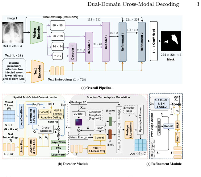
The load-bearing mechanism is the dual-domain decoder built from two named modules. TGSA (Text-Guided Spatial Cross-Attention) uses locally mixed visual tokens as queries and clinical text embeddings as keys and values, scaling the attention output by a learned image-text agreement gate. STAM (Spectral-Text Adaptive Modulation) applies a channel-wise 2D discrete cosine transform, computes learnably gated per-channel band energy, concatenates it with the pooled text vector, and predicts FiLM parameters $(\gamma,\beta)$ that are applied with a tanh bound to recalibrate channels. The coarse-to-fine schedule keeps cross-modal attention at low resolutions, while the two-stage refinement module restores full-resolution boundaries through shallow-feature fusion and FiLM conditioning from the text.
What would settle it
Run the frequency-domain multimodal fusion method cited as [22] on the same QaTa-COV19 and MosMedData+ train/val/test splits with the same augmentation and preprocessing; if its Dice/mIoU reaches or exceeds 91.46/84.26 and 81.95/69.42 on those exact splits, the paper's central claim of beating the strongest prior baselines would be contradicted.
Extended reading notes
Core claim
At its core, the paper claims that clinical text should steer segmentation in two complementary channels during decoding: spatially, by aligning visual features with text semantics, and spectrally, by recalibrating feature channels according to text-conditioned frequency-band statistics. It embodies this in DD-CMD, whose decoder interleaves TGSA and STAM at three progressively finer resolutions from 7x7 to 56x56, followed by a lightweight refinement module that restores 224x224 masks with FiLM conditioning and shallow skip features. On QaTa-COV19 the method reaches 91.46% Dice and 84.26% mIoU, and on MosMedData+ it reaches 81.95% Dice and 69.42% mIoU, yielding average gains of +1.96 Dice and +2.67 mIoU over the strongest prior baselines in the comparison table. The paper interprets these results as evidence that spatial alignment and frequency calibration are complementary forms of language guidance.
Load-bearing premise
That DD-CMD beats the strongest prior baselines assumes the methods in Table 1 are the strongest relevant ones under the same splits; in particular, the cited frequency-domain fusion method [22] is never compared, so it could tie or beat the reported numbers under identical conditions.
Editorial extensions
If this is right
- DD-CMD reports the highest Dice and mIoU on both benchmarks among all compared text-free and text-guided methods.
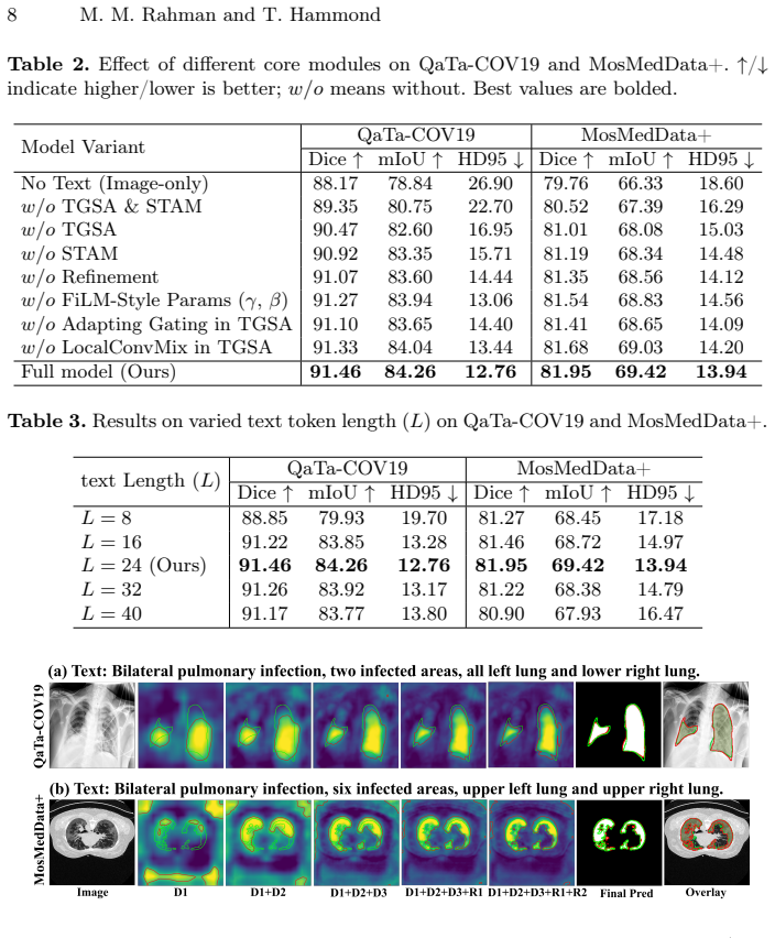
- Ablations show that adding STAM to TGSA improves overlap and lowers HD95 on both datasets, so frequency-aware channel recalibration contributes beyond spatial alignment.
- Ablations show that removing either TGSA or STAM degrades performance, with TGSA providing the larger individual jump.
- The high-resolution refinement module mainly sharpens boundaries and closes small gaps rather than changing coarse localization, consistent with the coarse-to-fine design.
- Text length near L=24 is needed for the reported performance; shorter prompts truncate lesion descriptors and longer prompts add noise.
Reading between the lines
- Because STAM's DCT and FiLM conditioning do not depend on pulmonary texture specifically, the same dual-domain decoder could transfer to other lesion types or imaging modalities where boundary fidelity is frequency-sensitive; the paper does not test this.
- The learned frequency gate inside STAM could be visualized to show which DCT bands clinical text emphasizes, offering a direct check on whether the gains come from semantically meaningful frequency channels; the paper does not report such an analysis.
- The frozen text encoder and L=24 truncation suggest that the reported gains rely on clinical descriptors appearing within the first 24 tokens, a dependency the paper's own text-length ablation supports.
Signed reviews
Editorial analysis
A structured set of objections, weighed in public.
Referee Report
Summary. The paper proposes DD-CMD, a text-guided medical image segmentation model that combines spatial cross-attention (TGSA) with frequency-domain channel calibration (STAM) in a coarse-to-fine decoder, followed by a high-resolution refinement module. The method is evaluated on QaTa-COV19 and MosMedData+, reporting 91.46% Dice / 84.26% mIoU and 81.95% Dice / 69.42% mIoU respectively, with claimed average gains of +1.96 Dice and +2.67 mIoU over the strongest prior baselines. The architecture is specified with equations, ablations support the contribution of each component, and code is released. The main weaknesses concern the comparison protocol: the cited frequency-domain baseline [22] is absent from Table 1, the headline gains are computed against different baselines per dataset and metric, and no error bars are reported.
Significance. If the empirical claims hold after a corrected comparison, DD-CMD would be a useful contribution to text-guided medical image segmentation, particularly in demonstrating that frequency-domain conditioning and spatial attention are complementary. The paper is transparent about its architecture and ablations, and the release of code is a practical strength. However, the central claim of outperforming the strongest prior baselines is currently supported only by a comparison set that omits the most relevant frequency-domain competitor [22] and by a per-metric best-baseline averaging protocol. The significance is therefore conditional on strengthening the empirical evidence rather than on the architectural novelty, which is incremental.
major comments (3)
- [§3, Table 1] The comparison in Table 1 omits reference [22], a frequency-domain multimodal fusion method for language-guided medical image segmentation, even though §3 states that the authors follow [13,9,3,22] for identical train/val/test splits. This omission is load-bearing because the paper's motivating narrative argues that prior work overlooks frequency content, yet [22] operates in exactly that niche. The authors should add [22] to Table 1 (using its reported numbers under the same splits, or by re-running it) or explicitly justify its exclusion. Without this, the claim that DD-CMD outperforms the strongest prior baselines is not fully supported.
- [§3, Comparison with SOTA Methods] The reported average gains of +1.96 Dice and +2.67 mIoU are computed by selecting a different strongest baseline per dataset and metric: MMI-UNet for QaTa-COV19, MAdapter for MosMedData+ Dice, and RecLMIS for MosMedData+ mIoU. This means no single baseline serves as the 'strongest prior baseline' across all four cells. The comparison protocol should be stated explicitly and consistently, for example by reporting gains against the best baseline per dataset or against a single overall strongest baseline, so the reader can verify the claimed margins.
- [§3, Comparison with SOTA Methods] No error bars or standard deviations are reported for any method. The smallest headline gain, +0.58 Dice on QaTa-COV19 over MMI-UNet, is within typical seed-to-seed variance for segmentation models at this scale. The authors should report mean and standard deviation over at least three random seeds for the proposed method and, where feasible, for the closest baselines, and indicate whether the observed differences are statistically significant.
minor comments (6)
- [§2.2] The sentence 'An overview of DD-CMD is shown in 1' is missing the figure number; it should read 'shown in Fig. 1.'
- [§2.2, Eq. (2)] The notation V(s) is used both for the input visual tokens and for the attended output in Eq. (2); please use separate symbols, e.g., V_in and V_att, to avoid ambiguity.
- [§3, Table 2] The 'w/o FiLM-Style Params' row is ambiguous about whether the high-resolution refinement FiLM conditioning is also removed; please clarify what remains in this variant.
- [§3, Table 1] Several baselines report N/R for parameters and FLOPs, which makes the efficiency comparison incomplete; consider citing the original sources or recomputing these values for a fair comparison.
- [§3, Fig. 2] The qualitative comparison in Fig. 2 does not identify which baseline methods are shown; please label the columns or add a descriptive caption.
- [References] References [17] and [18] appear to be self-citations with only marginal relevance to the DCT and lightweight-conditioning discussions; please verify that they are necessary and, if possible, cite the original sources of these ideas.
Circularity Check
No circularity: the claims are empirical against public benchmarks, and the self-citations are not load-bearing.
full rationale
The paper's central claim is an empirical performance comparison on two public benchmarks, QaTa-COV19 and MosMedData+. The methodology defines an architecture (TGSA, STAM, coarse-to-fine decoder, refinement module) whose parameters are learned from training data and evaluated on held-out test splits. No equation in the paper constructs a reported metric from a fitted constant, and no fitted parameter is renamed as a prediction. The only self-citations are [17], a DWT/DCT/SVD watermarking paper cited alongside the standard 2D DCT definition, and [18], the authors' prior skin-lesion segmentation paper cited illustratively for 'lightweight conditioning.' Neither citation is load-bearing: the DCT is a standard external transform, and the lightweight-conditioning claim is independently supported by the paper's own ablations. There is no uniqueness theorem imported from the authors' prior work, no ansatz smuggled in via a self-citation, and no known empirical result merely renamed in new coordinates. The concern that frequency-domain baseline [22] is cited for the split protocol but absent from Table 1 is a comparison-completeness issue, not a circularity issue; it weakens the strength of the SOTA claim without making the derivation reduce to its inputs. Consequently, no circular step can be exhibited, and the appropriate score is 0.
Assumptions & free parameters
free parameters (2)
- Text length L =
24 tokens
- Training schedule =
160 epochs, AdamW, initial LR 5e-5, eta_min 1e-6, batch size 8
assumptions (4)
- standard math 2D DCT-II coefficients and their squared energies are informative for lesion boundary segmentation
- domain assumption Frozen PubMedBERT embeddings carry the clinical semantics needed for segmentation guidance
- ad hoc to paper Medical anomalies exhibit distinct frequency signatures
- domain assumption The adopted train/val/test splits from [13,9,3,22] are correctly reproduced
Cite this review
Pith. "Pith review of Dual-Domain Cross-Modal Decoding for Clinical Text-Guided Medical Image Segmentation." pith.science (2026). https://pith.science/paper/U7XXXKYO
@misc{pith2026260811335,
  author       = {Pith},
  title        = {Pith review of: Dual-Domain Cross-Modal Decoding for Clinical Text-Guided Medical Image Segmentation},
  year         = {2026},
  howpublished = {\url{https://pith.science/paper/U7XXXKYO}},
  note         = {Machine review of arXiv:2608.11335}
}
read the original abstract
Clinical text can narrow down what to segment, but recent text-guided designs emphasize spatial alignment while overlooking frequency content that governs texture and boundaries. We propose Dual-Domain Cross-Modal Decoding (DD-CMD) for clinical text-guided pulmonary infection segmentation, integrating two complementary forms of language guidance during decoding. In the spatial domain, Text-Guided Spatial Cross-Attention (TGSA) aligns multi-scale visual tokens with text semantics and updates features through gated residual fusion. In the frequency domain, Spectral-Text Adaptive Modulation (STAM) applies a 2D DCT to compute learnable band-energy statistics and predicts text-conditioned FiLM parameters to recalibrate decoder channels for frequency-aware decoding. DD-CMD embeds TGSA and STAM into a coarse-to-fine decoder (7x7 to 56x56) and restores full-resolution masks using a lightweight two-stage refinement module. Experiments on QaTa-COV19 and MosMedData+ show that DD-CMD achieves 91.46% Dice / 84.26% mIoU and 81.95% Dice / 69.42% mIoU, respectively, with average gains of +1.96 Dice and +2.67 mIoU over the strongest prior baselines. Code: https://github.com/maklachur/DD-CMD.
Figures

Reference graph
Works this paper leans on
-
[22]
In: International Conference on Medical Image Computing and Computer-Assisted Intervention
Yu, B., Yang, J., Du, Z., Huang, Y., Li, C., Wang, L.: Frequency-domain multi- modal fusion for language-guided medical image segmentation. In: International Conference on Medical Image Computing and Computer-Assisted Intervention. pp. 278–288. Springer (2025)
work page 2025
-
[17]
arXiv preprint arXiv:1307.3294 (2013)
Rahman, M.M.: A dwt, dct and svd based watermarking technique to protect the image piracy. arXiv preprint arXiv:1307.3294 (2013)
arXiv 2013
-
[18]
In: Proceedings of the IEEE/CVF Conference on Computer Vision and Pattern Recognition
Rahman, M.M., Jung, S.K., Hammond, T.: Mambaliteunet: Cross-gated adap- tive feature fusion for robust skin lesion segmentation. In: Proceedings of the IEEE/CVF Conference on Computer Vision and Pattern Recognition. pp. 8556– 8565 (2026)
work page 2026
-
[1]
Ahmed,N.,Natarajan,T.,Rao,K.R.:Discretecosinetransform.IEEEtransactions on Computers100(1), 90–93 (1974)
work page 1974
-
[2]
arXiv preprint arXiv:2312.05391 (2023)
Azad, R., Heidary, M., Yilmaz, K., Hüttemann, M., Karimijafarbigloo, S., Wu, Y., Schmeink, A., Merhof, D.: Loss functions in the era of semantic segmentation: A survey and outlook. arXiv preprint arXiv:2312.05391 (2023)
arXiv 2023
-
[3]
In: International Conference on Medical Image Computing and Computer-Assisted Intervention
Bhardwaj, R., Tambe, U.Y., Neog, D.R.: Vitexnet: Vision-text guided dynamic convolution network for medical image segmentation. In: International Conference on Medical Image Computing and Computer-Assisted Intervention. pp. 690–699. Springer (2025)
work page 2025
-
[4]
In: International Conference on Medical Image Computing and Computer-Assisted Intervention
Bui, P.N., Le, D.T., Choo, H.: Visual-textual matching attention for lesion segmen- tation in chest images. In: International Conference on Medical Image Computing and Computer-Assisted Intervention. pp. 702–711. Springer (2024)
work page 2024
-
[5]
In: European conference on computer vision
Cao, H., Wang, Y., Chen, J., Jiang, D., Zhang, X., Tian, Q., Wang, M.: Swin- unet: Unet-like pure transformer for medical image segmentation. In: European conference on computer vision. pp. 205–218. Springer (2022)
2022
Show all 25 references
-
[6]
In: 2022 IEEE International Conference on Image Processing (ICIP)
Degerli, A., Kiranyaz, S., Chowdhury, M.E., Gabbouj, M.: Osegnet: Operational segmentation network for covid-19 detection using chest x-ray images. In: 2022 IEEE International Conference on Image Processing (ICIP). pp. 2306–2310. IEEE (2022)
2022
-
[7]
In: International Conference on Medical Image Computing and Computer-Assisted Intervention
Ding,S.,Li,M.,Wang,C.:Mg-unet:Amemory-guidedunetforlesionsegmentation in chest images. In: International Conference on Medical Image Computing and Computer-Assisted Intervention. pp. 355–365. Springer (2025)
2025
-
[8]
ACM Transactions on Computing for Healthcare3(1), 2:1– 2:23 (Jan 2022)
Gu, Y., Tinn, R., Cheng, H., Lucas, M., Usuyama, N., Liu, X., Naumann, T., Gao, J., Poon, H.: Domain-specific language model pretraining for biomedical natural language processing. ACM Transactions on Computing for Healthcare3(1), 2:1– 2:23 (Jan 2022). https://doi.org/10.1145/3458754
2022 doi
-
[9]
In: International Conference on Medical Image Computing and Computer-Assisted Intervention
Guo, Y., Zeng, X., Zeng, P., Fei, Y., Wen, L., Zhou, J., Wang, Y.: Common vision- language attention for text-guided medical image segmentation of pneumonia. In: International Conference on Medical Image Computing and Computer-Assisted Intervention. pp. 192–201. Springer (2024)
2024
-
[10]
In: International Conference on Medical Image Computing and Computer-Assisted Intervention
Hu, J., Li, Y., Sun, H., Song, Y., Zhang, C., Lin, L., Chen, Y.W.: Lga: A lan- guage guide adapter for advancing the sam model’s capabilities in medical im- age segmentation. In: International Conference on Medical Image Computing and Computer-Assisted Intervention. pp. 610–62...
2024
-
[11]
IEEE Transactions on Medical Imaging44(4), 1821–1835 (2024)
Huang, X., Li, H., Cao, M., Chen, L., You, C., An, D.: Cross-modal conditioned re- construction for language-guided medical image segmentation. IEEE Transactions on Medical Imaging44(4), 1821–1835 (2024)
2024
-
[12]
Nature methods18(2), 203–211 (2021)
Isensee, F., Jaeger, P.F., Kohl, S.A., Petersen, J., Maier-Hein, K.H.: nnu-net: a self-configuring method for deep learning-based biomedical image segmentation. Nature methods18(2), 203–211 (2021)
2021
-
[13]
IEEE transactions on medical imaging43(1), 96–107 (2023) 10 M
Li, Z., Li, Y., Li, Q., Wang, P., Guo, D., Lu, L., Jin, D., Zhang, Y., Hong, Q.: Lvit: language meets vision transformer in medical image segmentation. IEEE transactions on medical imaging43(1), 96–107 (2023) 10 M. M. Rahman and T. Hammond
2023
-
[14]
In: Proceedings of the IEEE/CVF conference on computer vision and pattern recognition
Liu, Z., Mao, H., Wu, C.Y., Feichtenhofer, C., Darrell, T., Xie, S.: A convnet for the 2020s. In: Proceedings of the IEEE/CVF conference on computer vision and pattern recognition. pp. 11976–11986 (2022)
2022
-
[15]
arXiv preprint arXiv:2005.06465 (2020)
Morozov, S.P., Andreychenko, A.E., Pavlov, N.A., Vladzymyrskyy, A., Ledikhova, N.V., Gombolevskiy, V.A., Blokhin, I.A., Gelezhe, P.B., Gonchar, A., Chernina, V.Y.: Mosmeddata: Chest ct scans with covid-19 related findings dataset. arXiv preprint arXiv:2005.06465 (2020)
2020 arXiv
-
[16]
In: Proceedings of the AAAI conference on artificial intelligence
Perez, E., Strub, F., De Vries, H., Dumoulin, V., Courville, A.: Film: Visual rea- soning with a general conditioning layer. In: Proceedings of the AAAI conference on artificial intelligence. vol. 32 (2018)
2018
-
[19]
In: International Conference on Medical image computing and computer-assisted intervention
Ronneberger, O., Fischer, P., Brox, T.: U-net: Convolutional networks for biomedi- cal image segmentation. In: International Conference on Medical image computing and computer-assisted intervention. pp. 234–241. Springer (2015)
2015
-
[20]
In: International Conference on Medical Image Computing and Computer-Assisted Intervention
Wang, Q., Lin, X., Yan, Z.: Towards robust medical image referring segmentation with incomplete textual prompts. In: International Conference on Medical Image Computing and Computer-Assisted Intervention. pp. 636–646. Springer (2025)
2025
-
[21]
In: Proceedings of the IEEE/CVF conference on computer vision and pattern recognition
Yang, Z., Wang, J., Tang, Y., Chen, K., Zhao, H., Torr, P.H.: Lavt: Language- aware vision transformer for referring image segmentation. In: Proceedings of the IEEE/CVF conference on computer vision and pattern recognition. pp. 18155– 18165 (2022)
2022
-
[23]
In: International Conference on Medical Image Computing and Computer- Assisted Intervention
Zeng, Q., Luo, H., Ma, X., Lu, Z., Hu, Y., Xia, Y.: Exploring text-enhanced mixture-of-expertsforsemi-supervisedmedicalimagesegmentationwithcomposite data. In: International Conference on Medical Image Computing and Computer- Assisted Intervention. pp. 226–236. Springer (2025)
2025
-
[24]
In: International Conference on Medical Image Computing and Computer-Assisted Intervention
Zhang, X., Ni, B., Yang, Y., Zhang, L.: Madapter: A better interaction between image and language for medical image segmentation. In: International Conference on Medical Image Computing and Computer-Assisted Intervention. pp. 425–434. Springer (2024)
2024
-
[25]
In: Deep learning in medical image analysis and multimodal learning for clinical decision support, pp
Zhou, Z., Rahman Siddiquee, M.M., Tajbakhsh, N., Liang, J.: Unet++: A nested u-net architecture for medical image segmentation. In: Deep learning in medical image analysis and multimodal learning for clinical decision support, pp. 3–11. Springer (2018)
2018
Reviewed August 15, 2026 · model on record in the stance chip above.
Discussion (0). Continue with ORCID to comment.