REVIEW 4 major objections 5 minor 25 references
A combined Machine Learning and Finite Element Modelling tool for the surgical planning of craniosynostosis correction
T0 review · 4 major / 5 minor · reviewed 2026-08-07 · deepseek-v4-flash
Pith's one-line read A multi-output support vector regressor trained on finite-element simulations predicts post-operative skull shape from 3D photographs, with $R^2 = 0.95$ on synthetic test data.
desk verdict A genuinely tested surrogate model whose abstract oversells it: the synthetic R2=0.95 is real, the real-patient validation (R2=-3.58) is honestly reported but fatal to the clinical claim. read the letter →
The pith
A machine-rendered reading of the paper's core claim, the machinery that carries it, and where it could break.
The reading
What carries the argument
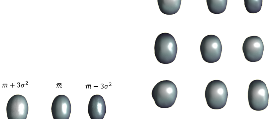
The engine is a statistical shape model (SSM) paired with a multi-output support vector regressor. SSM applies principal component analysis to skull meshes, representing each skull as the population mean plus a weighted combination of eigenmodes; the same compression is applied to the 2,356 finite-element output meshes. The regressor learns the mapping from input mode weights plus age, osteotomy parameters, and spring stiffness and free length to output mode weights, so a prediction is a full skull surface. This is what reduces hours of finite-element computation to a function evaluation.
What would settle it
Take a prospective cohort of at least 30 sagittal craniosynostosis patients, feed each preoperative 3D photograph and the actual surgical parameters into the trained model, and compare predicted surfaces with 3-week follow-up scans using the same $R^2$ and per-vertex error metrics as the paper's validation; if $R^2$ stays near or below zero rather than approaching the synthetic 0.95, the synthetic FEM training data do not transfer to real surgery.
Extended reading notes
Core claim
The paper's central claim is that post-operative skull shape after spring-assisted cranioplasty can be predicted as a function of patient age, surgical parameters (osteotomy position and width), spring characteristics, and a compressed description of the pre-operative head shape, using a machine-learning surrogate of finite-element simulations. The surrogate is a multi-output support vector regressor trained on 2,356 synthetic outcomes generated by finite-element models built from population-averaged skull and skin thickness and average suture positions; on the synthetic test set it achieves $R^2 = 0.95$, MSE = 0.042, and MAE = 0.122. The authors further claim this enables outcome prediction from 3D photographs in real time, eliminating CT and the engineering expertise otherwise needed for finite-element planning. They report an average surface error of $2.70 \pm 0.58$ mm on 13 real patients, alongside a validation $R^2$ of -3.58, and identify growth and population-averaged modelling assumptions as the main sources of the discrepancy.
Load-bearing premise
The load-bearing premise is that finite-element simulations built from population-average skull and skin thickness, average suture positions, and uniform material properties faithfully reproduce what actually happens when a real baby's skull is operated on.
Editorial extensions
If this is right
- Preoperative planning can be done from a 3D photograph and planned surgical parameters, with no CT scan and no radiation exposure.
- Surgeons could simulate many spring placements and choices in seconds, rather than committing to one configuration based on experience and age.
- Because the surrogate inherits the finite-element model's behaviour, the machine-learning step introduces little additional error on synthetic data; the practical limit of the tool is how faithfully the FEM represents real surgery.
- The same model could be inverted to search for surgical parameters that maximise the cranial index, a direction the paper explicitly identifies for future work.
Reading between the lines
- My reading: the negative real-patient $R^2$ is evidence that the bottleneck is the finite-element model's fidelity, not the regressor, since the regressor fits the synthetic dataset almost perfectly; improvements should target growth modelling and personalised thickness and suture inputs before model architecture.
- A testable extension would be to retrain the surrogate on FEM cases that include per-patient skull thickness and suture geometry inferred from photographs, and check whether real-patient $R^2$ rises above the current -3.58; the paper cites skull-inference from photos as future work.
- If validated, the same photo-to-surrogate pipeline could transfer to other cranial vault procedures or to longitudinal follow-up, where the surrogate is trained once on a population and evaluated on surface scans.
- The clinically meaningful quantity is the $2.70 \pm 0.58$ mm surface error on real patients, not the synthetic $R^2$; whether that error is acceptable depends on a clinical threshold the paper has not defined.
Editorial analysis
A structured set of objections, weighed in public.
Referee Report
Summary. The paper proposes a pipeline for real-time prediction of postoperative skull shape after spring-assisted cranioplasty (SAC) for sagittal craniosynostosis. The method uses population-average skull and soft-tissue thickness to convert 3D photographs into synthetic skull models, simulates surgical outcomes with finite element modelling (FEM) across a design of experiments (2356 simulations), and trains a multi-output support vector regressor to predict FEM output shape modes from patient age, surgical parameters, spring properties, and preoperative shape modes. The abstract reports a test-set R² of 0.95 with MSE and MAE below 0.13. The paper also reports an independent validation on 13 real patients in Section 3.4, where the model achieves R² = -3.58, MSE = 1.41, and MAE = 0.88.
Significance. If the reported accuracy held on real patients, the tool would be a valuable non-ionising alternative to CT-based FEM planning for SAC. The pipeline is methodologically coherent, uses an independent external cohort for validation, and the authors are transparent about the poor validation performance in Section 3.4. However, the central claim of the paper—that the surrogate model predicts real surgical outcomes from 3D photographs—is contradicted by that validation. The negative R² means the model is worse than predicting the mean postoperative shape, so the proposed tool currently has no demonstrated clinical predictive value. The abstract and discussion substantially overstate the findings by foregrounding the synthetic test metrics and the mean surface error without a null-model baseline.
major comments (4)
- [Abstract and Section 3.3-3.4] The abstract's central claim ('the resulting multi-output support vector regressor model achieves a R2 metric of 0.95 and MSE and MAE below 0.13') is based solely on the held-out split of FEM simulations in Section 3.3. The independent validation on 13 real patients in Section 3.4 reports R² = -3.58, MSE = 1.41, and MAE = 0.88. A negative R² means the model performs worse than a trivial predictor returning the mean postoperative shape. Presenting the synthetic R² without the validation result is misleading and does not support the stated goal of predicting surgical outcomes for new patients.
- [Section 4 (Discussion)] The Discussion claims clinical usefulness from a mean surface error of 2.70 ± 0.58 mm and compares this to Borghi et al. (2020). This comparison is not benchmarked against a null model. A constant prediction equal to the average follow-up shape could plausibly achieve a mean surface error at or below 2.70 mm on these 13 patients, in which case the model adds no predictive value. The paper does not report the baseline surface error for such a null predictor, so the error-rate claim does not establish that the model generalises to real outcomes.
- [Sections 2.2-2.4 and 3.4] The load-bearing assumption that a surrogate trained on FEM outputs can generalise to real surgical outcomes is empirically rejected by the validation in Section 3.4. The FEM models are built from population-averaged skull/skin thickness, uniform material properties, and artificial suture locations (Sections 2.2-2.3), and the trained model reproduces those simulations well. However, the validation shows no predictive signal on real postoperative 3D photographs (R² = -3.58). The Discussion lists possible causes such as growth and population-averaged parameters, but the magnitude of the negative R² indicates that the model is not merely noisy; it fails to capture the actual surgical response. This invalidates the clinical prediction claim unless the model is retrained or fundamentally revised.
- [Section 2.5.1] The validation preprocessing is under-specified: the postoperative 3D photographs are 'scaled down to omit the growth factor' that was not in the FEM, but the scaling method (e.g., global uniform scaling, landmark-based alignment, or volume normalisation) is not described. Because the validation metric is a surface-distance error, the scaling choice directly affects the reported 2.70 mm error and the R² value. Without details and a sensitivity analysis, the validation result cannot be fully interpreted or reproduced.
minor comments (5)
- [Section 2.4.1] Equations (2) and (3) use inconsistent notation: the preoperative vector is indexed to N_preop while the postoperative vector is indexed to N_postop, but the text later states the mean shapes are both in ℝ^{N×3}; the relationship between N_preop and N_postop and the template's node count should be clarified.
- [Section 2.1] The text states the CT population has 30 patients and the validation population has 13 patients, but the age ranges are given as '5.8 ± 1.15 months' and '5.1 ± 1.0' respectively; the second entry is missing the unit 'months'.
- [Section 3.2] The SSM retains 11 modes for both input and output shapes, but the output modes account for only 90% of variance while the input modes account for 94%. The potential impact of discarding 10% of output variance on the prediction metrics is not discussed.
- [Figure 12] The predicted-versus-actual plots for the first four modes would benefit from showing the R² and error metrics for each individual mode, since the aggregate R² of 0.95 may conceal modes with poor prediction that nonetheless contribute to the final shape error.
- [Section 2.3] The paper states that bone and suture are assigned viscoelastic material properties via a Prony series, but the actual parameter values are only cited to earlier work; for reproducibility, the key material constants (or a reference table) should be included.
Circularity Check
The clinical prediction claim rests on a self-citation chain: the reported R2=0.95 is accuracy on FEM-generated outputs, and the FEM-to-clinical bridge is asserted via the same group's prior papers, while the paper's own real-patient validation contradicts that bridge.
-
self citation load bearing
[Section 4 (Discussion and Conclusions)]
"The feasibility of FEM for predicting surgical outcomes in SAC has been established, validated, and optimized by the research group (Borghi et al., 2017, 2020, 2018; Garate Andikoetxea et al., 2023)."
The ML surrogate's training labels are FEM outputs (Sections 2.3-2.5). The only support offered for equating those FEM outputs with actual post-SAC skull shapes is this sentence, which cites exclusively papers with overlapping authorship. No external validation of that FEM is provided in this paper; instead, Section 3.4 reports R2=-3.58 on 13 real patients, directly contradicting the asserted feasibility. The clinical prediction claim therefore depends on a load-bearing self-citation rather than on an independently verified result.
-
fitted input called prediction
[Abstract; Section 3.3 vs Section 3.4]
"The resulting multi-output support vector regressor model achieves a R2 metric of 0.95 and MSE and MAE below 0.13."
The R2=0.95 is Section 3.3's test-set performance on a held-out split of 2356 FEM-generated outputs; it is not measured on any real patient outcome. Section 3.4 states that 'performance declined when compared to the actual surgical output' and reports R2=-3.58. Presenting the synthetic FEM-reproduction accuracy as 'the resulting model' performance for surgical planning substitutes the surrogate's internal fit for clinical prediction: the quantity actually predicted in the headline metric is the FEM simulator's output, not the surgical outcome.
1 more flagged steps
-
ansatz smuggled in via citation
[Section 2.2.2 and Section 2.3 (Image Processing and Finite Element Modelling)]
"Sutures were artificially created on the model following the method by (Garate Andikoetxea et al., 2023), assuming a width of 2 mm ... The material properties assigned to each part were defined based on (Borghi et al., 2020)."
The synthetic training population is constructed from population-average skull and skin thickness, average suture positions, and material/spring parameters imported from the same group's earlier works. The ML model then learns this synthetic input-output mapping. Because the real surgical outcome is never directly observed in training, every ansatz in those prior papers is inherited by construction. The paper does not re-derive or independently validate these assumptions; the Discussion instead calls the numerical models 'already validated' by self-citations. This is how the earlier FEM ansatz is carried into the ML prediction without new evidence.
full rationale
The surrogate itself is a legitimate technical object: R2=0.95 on held-out FEM outputs shows that the SVM learned the FEM mapping from inputs to output shape modes. That part is not circular. The circularity enters at the level of the clinical claim. The abstract and Discussion present this surrogate accuracy as predictive of surgical outcomes, but the only bridge from FEM outputs to real post-SAC shapes is a set of same-group citations (Borghi et al. 2017, 2018, 2020; Garate Andikoetxea et al. 2023). Moreover, the paper's own independent validation on 13 real patients gives R2=-3.58, MSE=1.41 and MAE=0.88, meaning the bridge is not merely unverified but empirically contradicted. The 2.70 +/- 0.58 mm mean error cited in the Discussion is not benchmarked against a mean-shape null model, so it does not restore the link. These observations fit patterns 3 and 2: a load-bearing self-citation chain, and a synthetic reproduction accuracy being called a prediction. The score is 4 rather than higher because the ML surrogate does have independent internal content, and the paper at least reports the negative real-patient validation openly. The central clinical claim, however, is not supported by an independent derivation; it reduces to the same group's prior FEM assertions.
Assumptions & free parameters
free parameters (4)
- Population mean skull thickness (t_skull) =
2.02 ± 0.33 mm (population average)
- Population mean soft tissue thickness (t_skin) =
3.42 ± 0.51 mm (population average)
- Number of retained SSM modes =
11 (94% input variance, 90% output variance)
- Design of experiments ranges (A, AP, LAT) =
A: 18-30%, AP: 47-63%, LAT: 10-25%
assumptions (5)
- domain assumption Population-average skull thickness, soft tissue thickness, and suture width/location are representative of each individual patient.
- ad hoc to paper Uniform inward offset of a 3D photograph by t_skin reconstructs an accurate skull geometry.
- domain assumption The FEM setup (isotropic viscoelastic bone, linear springs, no suture tearing) reproduces spring-assisted cranioplasty biomechanics.
- domain assumption PCA modes capturing 94% input and 90% output variance preserve enough information for clinically relevant outcome prediction.
- ad hoc to paper A surrogate model trained on FEM outputs can generalize to real surgical outcomes.
Cite this review
Pith. "Pith review of A combined Machine Learning and Finite Element Modelling tool for the surgical planning of craniosynostosis correction." pith.science (2026). https://pith.science/paper/V5JRFMWY
@misc{pith2026250603202,
  author       = {Pith},
  title        = {Pith review of: A combined Machine Learning and Finite Element Modelling tool for the surgical planning of craniosynostosis correction},
  year         = {2026},
  howpublished = {\url{https://pith.science/paper/V5JRFMWY}},
  note         = {Machine review of arXiv:2506.03202}
}
read the original abstract
Craniosynostosis is a medical condition that affects the growth of babies' heads, caused by an early fusion of cranial sutures. In recent decades, surgical treatments for craniosynostosis have significantly improved, leading to reduced invasiveness, faster recovery, and less blood loss. At Great Ormond Street Hospital (GOSH), the main surgical treatment for patients diagnosed with sagittal craniosynostosis (SC) is spring assisted cranioplasty (SAC). This procedure involves a 15x15 mm2 osteotomy, where two springs are inserted to induce distraction. Despite the numerous advantages of this surgical technique for patients, the outcome remains unpredictable due to the lack of efficient preoperative planning tools. The surgeon's experience and the baby's age are currently relied upon to determine the osteotomy location and spring selection. Previous tools for predicting the surgical outcome of SC relied on finite element modeling (FEM), which involved computed tomography (CT) imaging and required engineering expertise and lengthy calculations. The main goal of this research is to develop a real-time prediction tool for the surgical outcome of patients, eliminating the need for CT scans to minimise radiation exposure during preoperative planning. The proposed methodology involves creating personalised synthetic skulls based on three-dimensional (3D) photographs, incorporating population average values of suture location, skull thickness, and soft tissue properties. A machine learning (ML) surrogate model is employed to achieve the desired surgical outcome. The resulting multi-output support vector regressor model achieves a R2 metric of 0.95 and MSE and MAE below 0.13. Furthermore, in the future, this model could not only simulate various surgical scenarios but also provide optimal parameters for achieving a maximum cranial index (CI).
Figures


Figures from the paper (10 more)




Reference graph
Works this paper leans on
-
[1]
author Beaumont, C.A.A. , author Knoops, P.G.M. , author Borghi, A. , author Owase Jeelani, N.U. , author Koudstaal, M.J. , author Schievano, S. , author Dunaway, D.J. , author Rodriguez-Florez, N. , year 2017 . title Three-dimensional surface scanners compared with standard anthropometric measurements for head shape . journal Journal of Cranio-Maxillo-Fa...
-
[2]
author Bellman, R. , year 1957 . title DYNAMIC PROGRAMMING . edition 6 ed
work page 1957
-
[3]
author Borghi, A. , author Rodriguez-Florez, N. , author Rodgers, W. , author James, G. , author Hayward, R. , author Dunaway, D. , author Jeelani, O. , author Schievano, S. , year 2018 . title Spring assisted cranioplasty: A patient specific computational model . journal Medical Engineering & Physics volume 53 , pages 58--65 . :10.1016/J.MEDENGPHY.2018.01.001
-
[4]
author Borghi, A. , author Rodriguez Florez, N. , author Ruggiero, F. , author James, G. , author O'hara, J. , author Ong, J. , author Jeelani, O. , author Dunaway, D. , author Schievano, S. , year 2020 . title A population-specific material model for sagittal craniosynostosis to predict surgical shape outcomes . journal Biomechanics and Modeling in Mecha...
-
[5]
author Borghi, A. , author Schievano, S. , author Florez, N.R. , author McNicholas, R. , author Rodgers, W. , author Ponniah, A. , author James, G. , author Hayward, R. , author Dunaway, D. , author Owase Jeelani, N.U. , year 2017 . title Assessment of spring cranioplasty biomechanics in sagittal craniosynostosis patients . journal Journal of Neurosurgery...
-
[6]
author Erasmie, U. , author Ringertz, H. , year 1976 . title Normal width of cranial sutures in the neonate and infant. An objective method of assessment . journal Acta Radiologica - Series Diagnosis volume 17 , pages 565--572 . https://journals.sagepub.com/doi/10.1177/028418517601705A03, 10.1177/028418517601705A03/ASSET/028418517601705A03.FP.PNG \_ V03
-
[7]
author Fearon, J.A. , year 2014 . title Evidence-Based Medicine . journal Plastic and Reconstructive Surgery volume 133 , pages 1261--1275 . https://journals.lww.com/plasreconsurg/fulltext/2014/05000/evidence_based_medicine__craniosynostosis.36.aspx, :10.1097/PRS.0000000000000093
-
[8]
author Garate Andikoetxea, B. , author Ajami, S. , author Rodriguez-Florez, N. , author Jeelani, N.U.O. , author Dunaway, D. , author Schievano, S. , author Borghi, A. , author Owase Jeelani, N.U. , year 2023 . title Towards a radiation free numerical modelling framework to predict spring assisted correction of scaphocephaly . journal Computer Methods in ...
Show all 25 references
-
[9]
, author Knoops, P
author Heutinck, P. , author Knoops, P. , author Florez, N.R. , author Biffi, B. , author Breakey, W. , author James, G. , author Koudstaal, M. , author Schievano, S. , author Dunaway, D. , author Jeelani, O. , author Borghi, A. , year 2021 . title Statistical shape modelling ...
2021 doi
-
[10]
, author Morris, A
author Iyer, K. , author Morris, A. , author Zenger, B. , author Karanth, K. , author Khan, N. , author Orkild, B.A. , author Korshak, O. , author Elhabian, S. , year 2023 . title Statistical shape modeling of multi-organ anatomies with shared boundaries . journal Frontiers in...
2023
-
[11]
, author Borghi, A
author Jeelani, N.u.O. , author Borghi, A. , author Florez, N.R. , author Bozkurt, S. , author Dunaway, D. , author Schievano, S. , year 2020 . title The science behind the springs: Using biomechanics and finite element modeling to predict outcomes in spring-assisted sagittal ...
2020
-
[12]
, author M Wilkie, A.O
author Johnson, D. , author M Wilkie, A.O. , year 2011 . title PRACTICAL GENETICS . journal European Journal of Human Genetics www.nature.com/ejhg, :10.1038/ejhg.2010.235
2011 doi
-
[13]
, author Preisig, H.A
author Karolius, S. , author Preisig, H.A. , author Rusche, H. , year 2016 . title Multi-scale modelling software framework facilitating simulation of interconnected scales using surrogate-models . journal Computer Aided Chemical Engineering volume 38 , pages 463--468 . :10.10...
2016 doi
-
[14]
, author Bakhaty, A
author Madani, A. , author Bakhaty, A. , author Kim, J. , author Mubarak, Y. , author Mofrad, M.R.K. , year 2019 . title Bridging Finite Element and Machine Learning Modeling: Stress Prediction of Arterial Walls in Atherosclerosis . journal Journal of biomechanical engineering...
2019 doi
-
[15]
, author Figl, M
author Mei, L. , author Figl, M. , author Rueckert, D. , author Darzi, A. , author Edwards, P. , year 2008 . title Statistical shape modelling: How many modes should be retained? journal 2008 IEEE Computer Society Conference on Computer Vision and Pattern Recognition Workshops...
2008
-
[16]
, author Peter, D
author Milojevic, A. , author Peter, D. , author Huber, N.B. , author Azevedo, L. , author Latyshev, A. , author Sailer, I. , author Gross, M. , author Thomaszewski, B. , author Solenthaler, B. , author G \" o zc \" u , B. , year 2024 . title AutoSkull: Learning-based Skull Es...
2024 doi
-
[17]
, year 2016
author Morris, L.M. , year 2016 . title Nonsyndromic Craniosynostosis and Deformational Head Shape Disorders . journal Facial Plastic Surgery Clinics of North America volume 24 , pages 517--530 . :10.1016/J.FSC.2016.06.007
2016 doi
-
[18]
, year 2008
author Pascoletti, G. , year 2008 . title O R I G I N A L P A P E R Statistical shape modelling of the human mandible: 3D shape predictions based on external morphometric features . journal International Journal on Interactive Design and Manufacturing (IJIDeM) volume 16 , page...
2008 doi
-
[19]
, author Salotti, J.A
author Pearce, M.S. , author Salotti, J.A. , author Howe, N.L. , author Little, M.P. , author Lee, C. , author Ronckers, C.M. , author Rajaraman, P. , author Korea, S. , author Pearce, M.S. , author Salotti, J.A. , author Little, M.P. , author McHugh, K. , author Lee, C. , aut...
2012
-
[20]
, author Glass, G.E
author Rodgers, W. , author Glass, G.E. , author Schievano, S. , author Borghi, A. , author Rodriguez-Florez, N. , author Tahim, A. , author Angullia, F. , author Breakey, W. , author Knoops, P. , author Tenhagen, M. , author O'Hara, J. , author Ponniah, A. , author James, G. ...
2017
-
[21]
, author Bruse, J.L
author Rodriguez-Florez, N. , author Bruse, J.L. , author Borghi, A. , author Vercruysse, H. , author Ong, J. , author James, G. , author Pennec, X. , author Dunaway, D.J. , author Owase Jeelani, N.U. , author Schievano, S. , year 2017 a. title Statistical shape modelling to a...
2017
-
[22]
, author G \" o ktekin, O.K
author Rodriguez-Florez, N. , author G \" o ktekin, O.K. , author Bruse, J.L. , author Borghi, A. , author Angullia, F. , author Knoops, P.G.M. , author Tenhagen, M. , author O Hara, J.L. , author Koudstaal, M.J. , author Schievano, S. , author Owase Jeelani, N.U. , author Jam...
2017
-
[23]
o lby, L. , author Maltese, G. , author S \
author Tarnow, P. , author K \" o lby, L. , author Maltese, G. , author S \" o fteland, M.B. , author Lew \' e n, A. , author Nilsson, P. , author Enblad, P. , author Nowinski, D. , year 2022 . title Incidence of Non-Syndromic and Syndromic Craniosynostosis in Sweden . journal...
2022 doi
-
[24]
, author Bruse, J.L
author Tenhagen, M. , author Bruse, J.L. , author Rodriguez-Florez, N. , author Angullia, F. , author Borghi, A. , author Koudstaal, M.J. , author Schievano, S. , author Jeelani, O. , author Dunaway, D. , year 2016 . title Three-Dimensional Handheld Scanning to Quantify Head-S...
2016
-
[25]
write newline
" write newline "" before.all 'output.state := FUNCTION n.dashify 't := "" t empty not t #1 #1 substring "-" = t #1 #2 substring "--" = not "--" * t #2 global.max substring 't := t #1 #1 substring "-" = "-" * t #2 global.max substring 't := while if t #1 #1 substring * t #2 gl...
Reviewed August 7, 2026 · model on record in the stance chip above.
Discussion (0). Sign in to comment.