REVIEW 4 major objections 6 minor 90 references
Rapid MRI-Based Synthetic CT Simulations for Precise tFUS Targeting
T0 review · 4 major / 6 minor · reviewed 2026-08-06 · deepseek-v4-flash
Pith's one-line read MRI-only brain ultrasound planning matches CT accuracy.
desk verdict A useful six-pipeline comparison undermined by a load-bearing sign error in the HU-to-acoustic conversion that makes the reported absolute accuracy figures invalid as written. read the letter →
The pith
A machine-rendered reading of the paper's core claim, the machinery that carries it, and where it could break.
The reading
What carries the argument
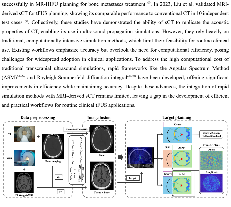
The argument rests on two pieces of machinery. The first is a synthetic-CT generator: an encoder-decoder U-Net with four strided 3D convolutions, two vision-transformer blocks, and subpixel upsampling, trained with a loss that blends global, bone-masked, and brain-masked mean absolute error with global and brain SSIM terms. Converting the generated Hounsfield units into density, sound speed, and attenuation via the Marsac bone model turns the sCT into an acoustic medium for simulation. The second is the hybrid propagation scheme: both accelerated pipelines first compute the transducer's field at a plane near the skull (one using a full-wave solver, the other using a Rayleigh–Sommerfeld diffraction integral), then propagate that plane wavefield through the skull with the angular spectrum method, which decomposes the field into plane waves and applies a phase factor in the spatial-frequency domain. This split avoids time-stepping through the skull, which is why the runtime drops by roughly 90–94% while keeping focal geometry comparable to full-wave simulation.
What would settle it
Take the trained synthetic-CT network and apply it to T1-weighted images acquired on a different scanner vendor, field strength, or patient age group, comparing the output skull Hounsfield units against paired real CT; if the skull-region mean absolute error rises substantially above the reported ~178 HU or if full-wave simulations through the sCT shift the focus by more than 1 mm, the central claim fails. A physical test would be to measure the pressure field with a calibrated hydrophone through an ex vivo human skull using phase corrections computed from the sCT, and check that the measured focal position and width match the sub-millimeter and ~3.3–3.8 mm values claimed.
Extended reading notes
Core claim
The central discovery is that a 3D U-Net with transformer blocks, trained on 17 paired T1-weighted MRI and CT volumes, can generate a synthetic CT whose Hounsfield-unit values—especially in the skull—are accurate enough to drive acoustic simulations for transcranial focused ultrasound. The paper validates this by comparing six pipelines: full-wave and hybrid angular-spectrum solvers, each run on real CT and on synthetic CT. The sCT-based full-wave fields reproduce the CT-based fields with normalized pressure differences below 0.2 outside the focal zone, and the hybrid solvers (k-Wave plus angular spectrum, and Rayleigh–Sommerfeld plus angular spectrum) keep sub-millimeter lateral targeting error while cutting runtime to 187 s and 34 s, respectively, from about 3320 s for the gold-standard solver. The authors conclude that MRI-derived sCT combined with rapid solvers enables fast, accurate, and radiation-free tFUS planning.
Load-bearing premise
The whole pipeline assumes that T1-weighted MRI carries enough information, after skull stripping, for the trained network to reconstruct skull geometry and bone density in a new patient, even though cortical bone is nearly silent on T1-weighted MRI and the network was trained on only 17 subjects.
Editorial extensions
If this is right
- tFUS treatment planning can be performed with MRI alone, eliminating CT scanning and its ionizing radiation for patients who need repeated or longitudinal neuromodulation sessions.
- Simulation time falls from about 55 minutes to roughly 3 minutes with the k-Wave–angular-spectrum hybrid and to about 34 seconds with the Rayleigh–Sommerfeld hybrid, making per-patient planning practical in a clinical workflow.
- Deep targets such as the thalamus and shallower 4-cm targets are both simulated with sub-millimeter lateral targeting error and focal widths of 3.3–3.8 mm, consistent with CT-based gold standard.
- Because sCT-based and CT-based pipelines agree in focal geometry, the sCT pipeline can serve as a drop-in replacement for CT in existing tFUS planning systems.
Reading between the lines
- Beyond the paper, the same sCT-plus-rapid-solver stack could be tested on MR-guided blood–brain barrier opening or histotripsy sessions, where repeated planning per session makes the radiation-free workflow most valuable.
- Beyond the paper, the ~34-second RS-ASM runtime suggests that interactive planning—where a clinician moves the transducer and sees the focus update in near real time—is now within reach, a use case the paper does not demonstrate.
- Beyond the paper, the claim would be strengthened by a physical check: measuring the focal field through an ex vivo skull with a hydrophone using sCT-derived phase corrections, to confirm the simulated sub-millimeter targeting survives real propagation.
Signed reviews
Editorial analysis
A structured set of objections, weighed in public.
Referee Report
Summary. The paper proposes a fully MRI-based pipeline for transcranial focused ultrasound (tFUS) planning: a 3D U-Net with transformer blocks generates synthetic CT (sCT) from T1-weighted MRI, and the resulting skull acoustic property maps are used in three simulation pipelines (full-wave k-Wave, hybrid k-Wave/Angular Spectrum Method, and Rayleigh–Sommerfeld/Angular Spectrum Method). The authors compare sCT-based simulations against real-CT-based k-Wave 'gold standard' simulations on five test skulls, reporting sub-millimeter targeting deviation, consistent focal widths, normalized pressure errors below 0.2, and roughly 90% reduction in simulation time for the hybrid methods. The manuscript claims this enables fast, accurate, radiation-free tFUS planning.
Significance. If correct, the proposed framework would be a practical step toward CT-free, rapid tFUS treatment planning, leveraging open-source tools (k-Wave, MONAI, ANTs) and offering a clear time advantage over full-wave simulation. The paper includes a quantitative comparison of six pipelines, which is a useful design. However, the central claims are currently undermined by a likely sign error in the density/sound-speed conversion equations, by an abstract that overstates the targeting accuracy and misreports the RS-ASM runtime, and by evaluation on only five subjects with no external validation. These issues are fixable in principle, but they must be addressed before the results can be considered physically meaningful.
major comments (4)
- [Section 2.2, Eqs. (7)-(8)] The Hounsfield-unit-to-acoustic-property conversion is written with inverted signs. Eq. (7) gives rho = rho_min + (rho_min - rho_max)*(HU-HU_min)/(HU_max-HU_min), so at HU = HU_max the density is 2*rho_min - rho_max = 2000 - 3200 = -1200 kg/m^3. Eq. (8) similarly gives c = 2*c_min - c_max = 2960 - 4050 = -1090 m/s at the upper end. These are unphysical values, and because the same conversion is applied to both rCT and sCT, the reported rCT-versus-sCT agreement cannot reveal the error. Every simulated pressure field, focal metric, and timing result is therefore computed on nonphysical skull maps, and the k-Wave 'gold standard' is itself invalid. If this is a typographical error and the code uses the standard forms (rho_max - rho_min) and (c_max - c_min), the authors must correct the equations and provide the corrected code or the corrected numerical results; otherwise the central claim of accurate tFUS planning is unsupported as written.
- [Abstract and Figure 8] The abstract's claim of 'sub-millimeter targeting deviation' is contradicted by the reported quantitative results. Figure 8B shows longitudinal (axial) deviations of 1.5-2.6 mm for all pipelines; only transverse errors (Figure 8D) are below 0.7 mm. The abstract should either state 'sub-millimeter transverse deviation' or also report the axial errors. Additionally, the abstract states that RS-ASM reduced simulation time to 34 s, but Figure 8E reports 345 +/- 85 s for RS-ASM; 34 s would correspond to ~99% time savings, not the claimed ~90%. The abstract's runtime number is inconsistent with the results by an order of magnitude.
- [Section 3.1, Table 1] Table 1 reports MAE values for N=5 test skulls without standard deviations, confidence intervals, or any statistical test comparing the proposed method with the pseudo-CT baseline. With only five samples, the improvements in head, brain, and skull MAE may not be statistically meaningful. Furthermore, the table header says 'Skull MAE (0<=HU<=2000)' but the text in Section 3.1 refers to 'skull region (HU > 2000)', and the training loss uses a bone mask of HU [100,1500]. These inconsistent HU thresholds make the skull MAE result difficult to interpret and should be reconciled.
- [Section 3.2.3] The 'targeting deviation' results are relative to k-Wave simulations using real CT, not to an absolute ground truth such as hydrophone measurements, ex vivo skull experiments, or clinical outcomes. The paper therefore demonstrates consistency between simulation pipelines, not absolute tFUS targeting accuracy. The authors should explicitly state this distinction and temper the conclusion that the framework provides 'precise tFUS targeting' in an absolute sense. Additionally, the evaluation uses only five test skulls from the same dataset used for training, with no external cohort, different field strengths, or different MRI protocols, so generalizability to clinical populations remains unestablished.
minor comments (6)
- [Section numbering] Section numbering is inconsistent: Section 1.1.3 is followed by Section 2.2 ('MRI-Derived Acoustic Parameter Estimation') and Section 2.3; these should be renumbered to maintain a coherent hierarchy (e.g., 1.2, 1.3).
- [Eq. (5)] In the brain SSIM loss, the mask is multiplied only with the SSIM fraction, so outside the mask the loss equals 1 - 0 = 1 (or 1 - mask*SSIM), rather than ignoring non-brain voxels. Consider using masked averaging over the brain region or applying the mask to both the numerator and the denominator consistently.
- [Abstract] The phrase 'fully CT free simulation framework' is inaccurate because the sCT model is trained on CT and the gold standard uses real CT; the framework is CT-free only at inference. Please clarify this to avoid overstatement.
- [Highlights] The third highlight contains a typo: 'approachs' should be 'approaches'.
- [Section 1.1.1] The sentence 'We developed a deep learning pipeline to sCT images from paired T1-weighted MRI data' is missing a verb; it should be 'to generate sCT images'.
- [Table 1] The notation 'Skull MAE (0<=HU<=2000)' is inconsistent with the text's 'HU > 2000' and with the bone mask range used in training ([100,1500]); please define the HU bins precisely and use the same definitions throughout.
Circularity Check
No circularity: sCT is trained on held-out rCT pairs and compared against an independent k-Wave rCT reference; the acoustic conversion is an external Marsac/Aubry model.
full rationale
The paper's derivation chain is empirical and externally referenced rather than self-referential. The sCT network (Section 1.1) is trained on 17 T1/rCT pairs and evaluated on 5 held-out skulls, so the sCT-vs-rCT MAE and R² comparisons are genuine test-set performance, not a fitted-input recollection. Acoustic parameter maps are obtained from rCT or sCT by the Marsac et al. conversion (Eqs. 7-8) and the attenuation model of Aubry/Constans (Eq. 9), both cited external models; the same conversion is applied to both arms, which makes the sCT/rCT comparison a controlled surrogate test rather than a circular reduction. The k-Wave, kW-ASM, and RS-ASM solvers are standard, independently developed methods (Treeby & Cox; Leung et al.; Rayleigh-Sommerfeld), and the hybrid pipelines are benchmarked against kWave-rCT as reference. No parameter is fitted to the reported targeting/FWHM/pressure-error outcomes. The self-citations in the reference list are background or methodological citations and do not carry the central claim. The printed sign of Eqs. 7-8 appears physically nonphysical at high HU if taken literally, and the Discussion's stated limitations (skull-base HU error, planar-propagation approximation) are acknowledged, but these are correctness/generalizability concerns, not circularity. Therefore no circular step is identified.
Assumptions & free parameters
free parameters (3)
- sCT loss weights lambda_MAE, lambda_SSIM, lambda_bone =
20, 5, 0.5
- bone mask HU threshold =
[100, 1500]
- training/test split =
17 train / 5 test
assumptions (4)
- domain assumption T1-weighted MRI contains sufficient information to reconstruct skull Hounsfield units
- domain assumption Linear HU-to-density and HU-to-sound-speed mappings of Marsac are valid for both rCT and sCT
- domain assumption Angular spectrum propagation through the heterogeneous skull is an acceptable approximation
- standard math k-Wave pseudospectral simulation is an adequate reference gold standard
Cite this review
Pith. "Pith review of Rapid MRI-Based Synthetic CT Simulations for Precise tFUS Targeting." pith.science (2026). https://pith.science/paper/VASMV4NN
@misc{pith2026250708688,
  author       = {Pith},
  title        = {Pith review of: Rapid MRI-Based Synthetic CT Simulations for Precise tFUS Targeting},
  year         = {2026},
  howpublished = {\url{https://pith.science/paper/VASMV4NN}},
  note         = {Machine review of arXiv:2507.08688}
}
read the original abstract
Accurate targeting is critical for the effectiveness of transcranial focused ultrasound (tFUS) neuromodulation. While CT provides accurate skull acoustic properties, its ionizing radiation and poor soft tissue contrast limit clinical applicability. In contrast, MRI offers superior neuroanatomical visualization without radiation exposure but lacks skull property mapping. This study proposes a novel, fully CT free simulation framework that integrates MRI-derived synthetic CT (sCT) with efficient modeling techniques for rapid and precise tFUS targeting. We trained a deep-learning model to generate sCT from T1-weighted MRI and integrated it with both full-wave (k-Wave) and accelerated simulation methods, hybrid angular spectrum (kWASM) and Rayleigh-Sommerfeld ASM (RSASM). Across five skull models, both full-wave and hybrid pipelines using sCT demonstrated sub-millimeter targeting deviation, focal shape consistency (FWHM ~3.3-3.8 mm), and <0.2 normalized pressure error compared to CT-based gold standard. Notably, the kW-ASM and RS-ASM pipelines reduced simulation time from ~3320 s to 187 s and 34 s respectively, achieving ~94% and ~90% time savings. These results confirm that MRI-derived sCT combined with innovative rapid simulation techniques enables fast, accurate, and radiation-free tFUS planning, supporting its feasibility for scalable clinical applications.
Figures

Reference graph
Works this paper leans on
-
[1]
While CT provides accurate skull acoustic properties, its ionizing radiation and poor soft tissue contrast limit clinical applicability
Abstract: Accurate targeting is critical for the effectiveness of transcranial focused ultrasound (tFUS ) neuromodulation. While CT provides accurate skull acoustic properties, its ionizing radiation and poor soft tissue contrast limit clinical applicability. In contrast, MRI offers superior neuroanatomical visualization without radiation exposure but lac...
2019
-
[2]
The assessment included direct structural comparison, voxel-wise intensity correlation, and quantitative error analysis
Results 3.1 Validation of sCT Accuracy Against Real CT for tFUS Planning To establish the feasibility of MRI-derived sCT as a substitute for rCT in tFUS planning, we conducted a comprehensive evaluation of its accuracy across five skull samples (Skull I–V). The assessment included direct structural comparison, voxel-wise intensity correlation, and quantit...
2000
-
[3]
The pipeline includes data preprocessing, a U-Net-based network architecture, and training objectives
Method 1.1 Generation of Synthesis CT from MRI We developed a deep learning pipeline to sCT images from paired T1 -weighted (T1w) MRI data, achieving high consistency in bone Hounsfield Unit (HU) values compared to reference CT images. The pipeline includes data preprocessing, a U-Net-based network architecture, and training objectives. 1.1.1 Data Preproc...
-
[4]
Krishna, V. et al. Trial of Globus Pallidus Focused Ultrasound Abla�on in Parkinson’s Disease. N. Engl. J. Med. 388, 683–693 (2023)
2023
-
[5]
Lipsman, N. et al. MR-guided focused ultrasound thalamotomy for essen�al tremor: A proof -of- concept study. Lancet Neurol. 12, 462–468 (2013)
2013
-
[6]
Elias, W. J. et al. A pilot study of focused ultrasound thalamotomy for essen�al tremor. N. Engl. J. Med. 369, 640–648 (2013)
2013
-
[7]
Follet, K. A. et al. Pallidal versus Subthalamic Deep-Brain S�mula�on for Parkinson’s Disease. N. Engl. J. Med. 362, 2077–2091 (2010)
2010
-
[8]
Kolabas, Z. I. et al. Dis�nct molecular profiles of skull bone marrow in health and neurological disorders. Cell 186, 3706-3725.e29 (2023)
work page 2023
Show all 90 references
-
[9]
Mar�nez-Fernández, R. et al. Randomized Trial of Focused Ultrasound Subthalamotomy for Parkinson’s Disease. N. Engl. J. Med. 383, 2501–2513 (2020)
2020
-
[10]
B., Huang, H., Walker, H
Montgomery, E. B., Huang, H., Walker, H. C., Guthrie, B. L. & Wats, R. L. High-frequency deep brain s�mula�on of the putamen improves bradykinesia in Parkinson’s disease. Mov. Disord. 26, 2232– 2238 (2011)
2011
-
[11]
Hu, Z. et al. Airy-beam holographic sonogene�cs for advancing neuromodula�on precision and flexibility. Proc. Natl. Acad. Sci. 121, 2017 (2024)
2024
-
[12]
Lipsman, N. et al. Blood–brain barrier opening in Alzheimer’s disease using MR -guided focused ultrasound. Nat. Commun. 9, 2336 (2018)
2018
-
[13]
Bae, S. et al. Transcranial blood –brain barrier opening in Alzheimer’s disease pa�ents using a portable focused ultrasound system with real-�me 2-D cavita�on mapping. Theranostics 14, 4519– 4535 (2024)
2024
-
[14]
& Ying, M
Shen, Y., Hua, L., Yeh, C., Shen, L. & Ying, M. Ultrasound with Microbubbles Improves Memory , Ameliorates Pathology and Modulates Hippocampal Proteomic Changes in a Triple Transgenic Mouse Model of Alzheimer ’ s Disease. Theranostics 10, 1–37 (2020)
2020
-
[15]
Beisteiner, R. et al. Transcranial Pulse S�mula�on with Ultrasound in Alzheimer’s Disease—A New Navigated Focal Brain Therapy. Adv. Sci. 7, (2020)
2020
-
[16]
Y., Lee, W
Lee, K., Park, T. Y., Lee, W. & Kim, H. A review of func�onal neuromodula�on in humans using low- intensity transcranial focused ultrasound. Biomed. Eng. Lett. (2024) doi:10.1007/s13534- 024- 00369-0. 22
2024 doi
-
[17]
Rezai, A. R. et al. Noninvasive hippocampal blood−brain barrier opening in Alzheimer’s disease with focused ultrasound. Proc. Natl. Acad. Sci. U. S. A. 117, 9180–9182 (2020)
2020
-
[18]
Niu, X., Yu, K. & He, B. Transcranial focused ultrasound induces sustained synap�c plas�city in rat hippocampus. Brain Stimul. 15, 352–359 (2022)
2022
-
[19]
Tsai, S. J. Transcranial focused ultrasound as a possible treatment for major depression. Med. Hypotheses 84, 381–383 (2015)
2015
-
[20]
Schoen, S. et al. Towards controlled drug delivery in brain tumors with microbubble -enhanced focused ultrasound. Adv. Drug Deliv. Rev. 180, 114043 (2022)
2022
-
[21]
Ye, D. et al. Mechanically manipula�ng glympha�c transport by ultrasound combined with microbubbles. Proc. Natl. Acad. Sci. 120, 2017 (2023)
2023
-
[22]
& Mitragotri, S
Sun, T., Dasgupta, A., Zhao, Z., Nurunnabi, M. & Mitragotri, S. Physical triggering strategies for drug delivery. Adv. Drug Deliv. Rev. (2020) doi:10.1016/j.addr.2020.06.010
2020 doi
-
[23]
Chen, K.-T. et al. Neuronaviga�on-guided focused ultrasound for transcranial blood -brain barrier opening and immunos�mula�on in brain tumors. Sci. Adv. 7, (2021)
2021
-
[24]
Perolina, E. et al. Transla�ng ultrasound-mediated drug delivery technologies for CNS applica�ons. Adv. Drug Deliv. Rev. 208, 115274 (2024)
2024
-
[25]
& Chen, H
Hu, Z., Chen, S., Yang, Y., Gong, Y. & Chen, H. An Affordable and Easy -to-Use Focused Ultrasound Device for Noninvasive and High Precision Drug Delivery to the Mouse Brain. IEEE Trans. Biomed. Eng. 69, 2723–2732 (2022)
2022
-
[26]
& Chen, H
Ye, D., Chukwu, C., Yang, Y., Hu, Z. & Chen, H. Adeno-associated virus vector delivery to the brain: Technology advancements and clinical applica�ons. Adv. Drug Deliv. Rev. 211, 115363 (2024)
2024
-
[27]
Beccaria, K. et al. Ultrasound-induced blood-brain barrier disrup�on for the treatment of gliomas and other primary CNS tumors. Cancer Lett. 479, 13–22 (2020)
2020
-
[28]
Han, H. et al. Imaging-guided bioresorbable acous�c hydrogel microrobots. Sci. Robot. 9, 1 –13 (2024)
2024
-
[29]
Bez, M. et al. Nonviral ultrasound-mediated gene delivery in small and large animal models. Nat. Protoc. 14, 1015–1026 (2019)
2019
-
[30]
E., Blesa, J
Karakatsani, M. E., Blesa, J. & Konofagou, E. E. Blood –brain barrier opening with focused ultrasound in experimental models of Parkinson’s disease. Mov. Disord. 34, 1252–1261 (2019)
2019
-
[31]
Han, M. et al. Janus micropar�cles-based targeted and spa�ally -controlled piezoelectric neural s�mula�on via low-intensity focused ultrasound. Nat. Commun. 15, 1–17 (2024)
2024
-
[32]
Zhang, Y. et al. Defining the Op�mal Age for Focal Lesioning in a Rat Model of Transcranial HIFU. Ultrasound Med. Biol. 41, 449–455 (2015)
2015
-
[33]
Ye, D. et al. Incisionless targeted adeno -associated viral vector delivery to the brain by focused ultrasound-mediated intranasal administra�on. eBioMedicine 84, 104277 (2022)
2022
-
[34]
Hu, Z. et al. Targeted delivery of therapeu�c agents to the mouse brain using a stereotac�c-guided focused ultrasound device. STAR Protoc. 4, 102132 (2023)
2023
-
[35]
B., Khokhlova, T
Rosnitskiy, P. B., Khokhlova, T. D., Schade, G. R., Sapozhnikov, O. A. & Khokhlova, V. A. Treatment Planning and Aberra�on Correc�on Algorithm for HIFU Abla�on of Renal Tumors. IEEE Trans. Ultrason. Ferroelectr. Freq. Control 71, 341–353 (2024)
2024
-
[36]
Pacia, C. P. et al. Focused Ultrasound –mediated Liquid Biopsy in a Tauopathy Mouse Model. Radiology 307, (2023)
2023
-
[37]
D., Cho, C
Xu, Z., Khokhlova, T. D., Cho, C. S. & Khokhlova, V. A. Histotripsy: A Method for Mechanical Tissue Abla�on with Ultrasound. Annu. Rev. Biomed. Eng. 26, 141–167 (2024). 23
2024
-
[38]
Bawiec, C. R. et al. A Prototype Therapy System for Boiling Histotripsy in Abdominal Targets Based on a 256 -Element Spiral Array. IEEE Trans. Ultrason. Ferroelectr. Freq. Control 68, 1496– 1510 (2021)
2021
-
[39]
Meng, Y. et al. MR-guided focused ultrasound liquid biopsy enriches circula�ng biomarkers in pa�ents with brain tumors. Neuro. Oncol. 23, 1789–1797 (2021)
2021
-
[40]
Pinton, G. et al. Atenua�on, scatering, and absorp�on of ultrasound in the skull bone. Med. Phys. 39, 299–307 (2012)
2012
-
[41]
Xu, L. et al. Characteriza�on of the Targe�ng Accuracy of a Neuronaviga�on -Guided Transcranial FUS System In Vitro, In Vivo, and In Silico. IEEE Trans. Biomed. Eng. 70, 1528–1538 (2023)
2023
-
[42]
S., Hall, M
Gordon, M. S., Hall, M. D., Gaston, J., Foots, A. & Suwangbutra, J. Individual differences in the acous�c proper�es of human skulls. J. Acoust. Soc. Am. 146, EL191–EL197 (2019)
2019
-
[43]
& Chen, H
Hu, Z., Yang, Y., Xu, L., Hao, Y. & Chen, H. Binary acous�c metasurfaces for dynamic focusing of transcranial ultrasound. Front. Neurosci. 16, 1–9 (2022)
2022
-
[44]
Pichardo, S., Sin, V. W. & Hynynen, K. Mul�-frequency characteriza�on of the speed of sound and atenua�on coefficient for longitudinal transmission of freshly excised human skulls. Phys. Med. Biol. 56, 219–250 (2011)
2011
-
[45]
Marsac, L. et al. Ex vivo op�misa�on of a heterogeneous speed of sound model of the human skull for non-invasive transcranial focused ultrasound at 1 MHz. Int. J. Hyperth. 33, 635–645 (2017)
2017
-
[46]
Jiménez-Gambín, S., Jiménez, N., Benlloch, J. M. & Camarena, F. Holograms to Focus Arbitrary Ultrasonic Fields through the Skull. Phys. Rev. Appl. 12, 014016 (2019)
2019
-
[47]
Jiang, C. et al. Numerical Evalua�on of the Influence of Skull Heterogeneity on Transcranial Ultrasonic Focusing. Front. Neurosci. 14, 1–12 (2020)
2020
-
[48]
Hu, Z. et al. Transcranial cavita�on localiza�on by �me difference of arrival algorithm using four sensors. J. Acoust. Soc. Am. 148, 2560–2560 (2020)
2020
-
[49]
& Chen, H
Hu, Z., Yang, Y., Xu, L., Jing, Y. & Chen, H. Airy beam -enabled binary acous�c metasurfaces (AB- BAM). J. Acoust. Soc. Am. 153, A140–A140 (2023)
2023
-
[50]
& Chen, H
Hu, Z., Yang, Y., Xu, L., Hao, Y. & Chen, H. Binary acous�c metasurfaces for transcranial focused ultrasound. J. Acoust. Soc. Am. 153, A140–A140 (2023)
2023
-
[51]
& Chen, H
Hu, Z., Chen, S., Yang, Y., Gong, Y. & Chen, H. Affordable stereotac�c -guided focused ultrasound device for tunable drug delivery to the mouse brain. J. Acoust. Soc. Am. 150, A129–A129 (2021)
2021
-
[52]
Liu, Y. et al. CT synthesis from MRI using mul� -cycle GAN for head -and-neck radia�on therapy. Comput. Med. Imaging Graph. 91, 101953 (2021)
2021
-
[53]
Zuo, W., An, Z., Zhang, B. & Hu, Z. Solu�on of nonlinear Lamb waves in plates with discon�nuous thickness. J. Acoust. Soc. Am. 155, 2171–2180 (2024)
2024
-
[54]
& Hus�nx, R
Salmon, E., Bernard Ir, C. & Hus�nx, R. Pi�alls and Limita�ons of PET/CT in Brain Imaging. Semin. Nucl. Med. 45, 541–551 (2015)
2015
-
[55]
utilized UTE sequences, employing a 2D CNN- based model that achieved less than 2°C error in acoustic and biothermal simulations 56
Similarly, Su et al. utilized UTE sequences, employing a 2D CNN- based model that achieved less than 2°C error in acoustic and biothermal simulations 56. More recently, Leung et al. evaluated the use of MRI -derived sCT for correcting skull -induced phase aberrations during tF...
2023
-
[56]
Lei, Y. et al. MRI-only based synthe�c CT genera�on using dense cycle consistent genera�ve adversarial networks. Med. Phys. 46, 3565–3581 (2019). 24
2019
-
[57]
Y., Chung, Y
Koh, H., Park, T. Y., Chung, Y. A., Lee, J. -H. & Kim, H. Acous�c Simula�on for Transcranial Focused Ultrasound Using GAN-Based Synthe�c CT. IEEE J. Biomed. Heal. Informatics 26, 161–171 (2022)
2022
-
[58]
Yaakub, S. N. et al. Pseudo-CTs from T1 -weighted MRI for planning of low -intensity transcranial focused ultrasound neuromodula�on: An open-source tool. Brain Stimul. 16, 75–78 (2023)
2023
-
[59]
Guo, S. et al. Feasibility of ultrashort echo �me images using full- wave acous�c and thermal modeling for transcranial MRI -guided focused ultrasound (tcMRgFUS) planning. Phys. Med. Biol. 64, (2019)
2019
-
[60]
Su, P. et al. Transcranial MR imaging⇓guided focused ultrasound interven�ons using deep learning synthesized CT. Am. J. Neuroradiol. 41, 1841–1848 (2020)
2020
-
[61]
Leung, S. A. et al. Comparison between MR and CT imaging used to correct for skull-induced phase aberra�ons during transcranial focused ultrasound. Sci. Rep. 12, 13407 (2022)
2022
-
[62]
A., Stagg, C
Miscouridou, M., Pineda-Pardo, J. A., Stagg, C. J., Treeby, B. E. & Stanziola, A. Classical and Learned MR to Pseudo-CT Mappings for Accurate Transcranial Ultrasound Simula�on. IEEE Trans. Ultrason. Ferroelectr. Freq. Control 69, 2896–2905 (2022)
2022
-
[63]
Lena, B. et al. Synthe�c CT for the planning of MR -HIFU treatment of bone metastases in pelvic and femoral bones: a feasibility study. Eur. Radiol. 32, 4537–4546 (2022)
2022
-
[64]
Liu, H. et al. Evalua�on of synthe�cally generated computed tomography for use in transcranial focused ultrasound procedures. J. Med. Imaging 10, 1–16 (2023)
2023
-
[65]
& Jing, Y
Gu, J. & Jing, Y. mSOUND: An Open Source Toolbox for Modeling Acous�c Wave Propaga�on in Heterogeneous Media. IEEE Trans. Ultrason. Ferroelectr. Freq. Control 68, 1476–1486 (2021)
2021
-
[66]
Aubry, J.-F. et al. Benchmark problems for transcranial ultrasound simula�on: Intercomparison of compressional wave models. J. Acoust. Soc. Am. 152, 1003–1019 (2022)
2022
-
[67]
A., Webb, T
Leung, S. A., Webb, T. D., Biton, R. R., Ghanouni, P. & Buts Pauly, K. A rapid beam simula�on framework for transcranial focused ultrasound. Sci. Rep. 9, 1–11 (2019)
2019
-
[68]
& Shen, G
Xu, P., Wu, N. & Shen, G. A rapid element pressure field simula�on method for transcranial phase correc�on in focused ultrasound therapy. Phys. Med. Biol. 68, (2023)
2023
-
[69]
Leung, S. A. et al. Transcranial focused ultrasound phase correc�on using the hybrid angular spectrum method. Sci. Rep. 11, 1–13 (2021)
2021
-
[70]
Top, C. B. A Generalized Split -Step Angular Spectrum Method for Efficient Simula�on of Wave Propaga�on in Heterogeneous Media. IEEE Trans. Ultrason. Ferroelectr. Freq. Control 68, 2687– 2696 (2021)
2021
-
[71]
Angla, C., Larrat, B., Gennisson, J. L. & Cha�llon, S. Transcranial ultrasound simula�ons: A review. Med. Phys. 50, 1051–1072 (2023)
2023
-
[72]
Jones, R. M. & Hynynen, K. Comparison of analy�cal and numerical approaches for CT -based aberra�on correc�on in transcranial passive acous�c imaging. Phys. Med. Biol. 61, 23–36 (2015). 25
2015
-
[73]
& Arridge, S
van ’t Wout, E., Gélat, P., Betcke, T. & Arridge, S. A fast boundary element method for the scatering analysis of high-intensity focused ultrasound. J. Acoust. Soc. Am. 138, 2726–2737 (2015)
2015
-
[74]
Angla, C. et al. New semi-analy�cal method for fast transcranial ultrasonic field simula�on. Phys. Med. Biol. 69, 095017 (2024)
2024
-
[75]
Tus�son, N. J. et al. N4ITK: Improved N3 bias correc�on. IEEE Trans. Med. Imaging 29, 1310–1320 (2010)
2010
-
[76]
S., Dalca, A
Hoopes, A., Mora, J. S., Dalca, A. V., Fischl, B. & Hoffmann, M. SynthStrip: skull -stripping for any brain image. Neuroimage 260, 119474 (2022)
2022
-
[77]
& Johnson, H
Avants, B., Tus�son, N. & Johnson, H. Advanced Normaliza�on Tools (ANTS). Insight J. 1–35 (2009)
2009
-
[78]
Taniguchi, H. et al. Improving Convenience and Reliability of 5 -ALA-Induced Fluorescent Imaging for Brain Tumor Surgery. Lecture Notes in Computer Science (including subseries Lecture Notes in Artificial Intelligence and Lecture Notes in Bioinformatics) vol. 9351 (2015)
2015
-
[79]
Dosovitskiy, A. et al. AN IMAGE IS WORTH 16X16 WORDS: TRANSFORMERS FOR IMAGE RECOGNITION AT SCALE. in ICLR 2021 - 9th International Conference on Learning Representations (2021)
2021
-
[80]
T r e e b y , B . E . & C o x , B . T . k-Wave: MATLAB toolbox for the simula�on and reconstruc�on of photoacous�c wave fields. J. Biomed. Opt. 15, 021314 (2010)
2010
-
[81]
Y., Pahk, K
Park, T. Y., Pahk, K. J. & Kim, H. Method to op�mize the placement of a single-element transducer for transcranial focused ultrasound. Comput. Methods Programs Biomed. 179, 104982 (2019)
2019
-
[82]
Wu, N. et al. An efficient and accurate parallel hybrid acous�c signal correc�on method for transcranial ultrasound. Phys. Med. Biol. 65, 215019 (2020)
2020
-
[83]
Hu, Z. et al. 3-D Transcranial Microbubble Cavita�on Localiza�on by Four Sensors. IEEE Trans. Ultrason. Ferroelectr. Freq. Control 68, 3336–3346 (2021)
2021
-
[84]
& Aubry, J
Constans, C., Deffieux, T., Pouget, P., Tanter, M. & Aubry, J. F. A 200 -1380-kHz quadrifrequency focused ultrasound transducer for neuros�mula�on in rodents and rrimates: Transcranial in vitro calibra�on and numerical study of the influence of skull cavity. IEEE Trans. Ultrason....
2017
-
[85]
-F., Tanter, M., Pernot, M., Thomas, J
Aubry, J. -F., Tanter, M., Pernot, M., Thomas, J. -L. & Fink, M. Experimental demonstra�on of noninvasive transskull adap�ve focusing based on prior computed tomography scans. J. Acoust. Soc. Am. 113, 84–93 (2003)
2003
-
[86]
& Jie Mao
Hu, Z., Cui, H., An, Z. & Jie Mao. Measurements of backward wave propaga�on using the dynamic photoelas�c technique. in 2016 IEEE International Ultrasonics Symposium (IUS) vols 2016-Novem 1–4 (IEEE, 2016)
2016
-
[87]
& Wang, X.-M
Hu, Z.-T., An, Z.-W., Lian, G.-X. & Wang, X.-M. Propaga�on Proper�es of Backward Lamb Waves in Plate Inves�gated by Dynamic Photoelas�c Technique *. Chinese Phys. Lett. 34, 114301 (2017)
2017
-
[88]
& Wang, X
Hu, Z., An, Z., Kong, Y., Lian, G. & Wang, X. The nonlinear S0 Lamb mode in a plate with a linearly - varying thickness. Ultrasonics 94, 102–108 (2019). 26
2019
-
[89]
, H u , Z
Z u o , W . , H u , Z . , A n , Z . & K o n g , Y . L D V-based measurement of 2D dynamic stress fields in transparent solids. J. Sound Vib. 476, 115288 (2020)
2020
-
[90]
Daneshzand, M. et al. Model-based naviga�on of transcranial focused ultrasound neuromodula�on in humans: Applica�on to targe�ng the amygdala and thalamus. Brain Stimul. 17, 958–969 (2024)
2024
Reviewed August 6, 2026 · model on record in the stance chip above.
Discussion (0). Continue with ORCID to comment.