REVIEW 5 major objections 6 minor 13 references
The paper claims that a fully automated MRI pipeline, from raw pre- and post-contrast scans to a survival score, predicts post-surgical survival in colorectal liver metastasis better than existing clinical and genomic biomarkers (C-index 0.
Reviewed by Pith at T0; open to challenge. T0 means a machine referee read the full paper against a public rubric. the ladder, T0–T4 →
A fully automated pre/post-contrast MRI framework, combining prompt-based segmentation with autoencoder multiple-instance survival analysis, improves CRLM post-surgery survival prediction over clinical and genomic biomarkers in 227 patients.
T0 review reviewed 2026-08-04 challenge →
load-bearing objection Two real algorithmic contributions (SAMONAI, SurvAMINN) and a sensible internal evaluation, but the headline survival statistics are built on a confidence interval that cannot be right for 82 events, and the outcome test set size is never reported. the 5 major comments →
Live(r) Die: Predicting Survival in Colorectal Liver Metastasis
The pith
A machine-rendered reading of the paper's core claim, the machinery that carries it, and where it could break.
The reading
Core claim
The central claim is that the full chain—automatic segmentation of liver, tumors, and spleen on both pre- and post-contrast MRI, extraction of hand-crafted radiomic features per tumor, and a neural network that jointly learns dimensionality reduction and proportional-hazards survival prediction—can outperform established prognostic markers for CRLM. The decisive design choices are: (1) incomplete liver and spleen labels are completed by a zero-shot 3D prompt-propagation method (SAMONAI), which lets the segmentation model become fully automated without dense manual annotation; (2) SurvAMINN treats each tumor as an instance in a patient-level bag and computes the patient hazard as the log-sum-
What carries the argument
The two load-bearing mechanisms are SAMONAI and SurvAMINN. SAMONAI is a zero-shot 3D prompt-propagation algorithm: from a single positive point in one slice of an MRI volume, it segments that slice with a general-purpose promptable segmentation model, propagates candidate prompts along perpendicular sagittal and coronal views, then segments slice-by-slice in all three views, fusing the probability maps and binarizing with an adaptive threshold (mean plus two standard deviations of logits) to suppress false positives. It exists to create reliable liver and spleen labels from only a small annotated sample, which then trains a few-shot model that autocompletes labels for the whole cohort. SurvA
Load-bearing premise
The load-bearing premise is that the liver and spleen pseudo-labels generated by SAMONAI on just 20 cases, without quality control, are accurate enough for the few-shot model that then autocompletes labels for the whole cohort; if those pseudo-labels contain systematic segmentation errors, the tumor masks, radiomic features, and survival scores inherit the errors and the reported improvement over biomarkers may be an artifact.
What would settle it
Manually re-segment liver and spleen on the 20 annotated cases, correct all SAMONAI errors, and rerun the stage-1 training, autocompletion, radiomics, and SurvAMINN on a held-out cohort from a different institution. If the corrected-pipeline C-index falls to roughly 0.5 or no longer beats the clinical biomarkers, or if the same pipeline on a similar external MRI set fails to reproduce the hazard ratio of 1.93, the central claim is falsified.
If this is right
- If the framework holds up, pre-surgical MRI alone can assign CRLM patients to high- and low-risk groups after resection, without expert tumor outlining.
- The smallest drop from ground-truth to predicted segmentations (about 0.01 in C-index when both contrast phases are used) means automated segmentation does not destroy the survival signal.
- Combining pre- and post-contrast phases yields a statistically significant improvement, so multi-phase acquisition is worth retaining in radiomics-based prognostication.
- Jointly learning dimensionality reduction with the survival objective outperforms independent feature selection plus a classifier, implying that feature selection should be tuned to the downstream outcome rather than done separately.
- The most aggressive tumor, not the average or the largest, appears to drive post-surgical survival, so patient risk should be computed with worst-tumor pooling rather than aggregate tumor statistics.
Where Pith is reading between the lines
- Editorial inference: log-sum-exp 'worst-tumor' pooling is a general principle that could transfer to other multifocal cancers (e.g., lung, renal, or ovarian metastases) where per-lesion imaging features are available; the paper only tests it in CRLM.
- Because the stage-1 pseudo-labels come from 20 cases with no quality control, an external test of this framework should measure how sensitive the final C-index is to random versus corrected liver/spleen labels; the paper acknowledges this as an open issue.
- Editorial inference: the reported comparison is against weak conventional biomarkers in a single institution; a practical extension would be an independent multi-center cohort with the same MRI protocol to test whether the 10% C-index improvement generalizes or partly reflects site-specific radiomics.
- A practical improvement the paper leaves open is to model 'no predicted tumor' as a separate risk stratum rather than excluding those patients, since the current exclusion biases the performance estimate.
Editorial analysis
A structured set of objections, weighed in public.
Referee Report
Summary. The paper proposes a fully automated pipeline for predicting overall survival after resection of colorectal liver metastases (CRLM) from pre- and post-contrast T1-weighted MRI. The segmentation component uses SAMONAI, a zero-shot 3D prompt-propagation method based on SAM, to pseudo-label liver and spleen on 20 cases, fine-tunes a 3DINO-initialized UNETR on these labels, and then auto-completes labels for the full cohort. The outcome component extracts PyRadiomics features from each predicted tumor and trains SurvAMINN, an autoencoder-based multiple-instance network with log-sum-exp pooling, on right-censored survival data. On an internal 227-patient cohort the authors report a C-index of 0.691, an HR of 1.93 (95% CI 1.89–1.98, p<0.001) against clinical/genomic biomarkers, and liver/spleen Dice scores up to 0.96.
Significance. If the statistical claims held, this would be a practically useful contribution: it targets a real clinical decision problem, combines pre- and post-contrast MRI, addresses multifocality through worst-tumor pooling, and reduces annotation burden via foundation-model pseudo-labeling. The paper deserves credit for its internal controls (repeated 3-fold cross-validation, a randomization test over shuffled labels, Kaplan–Meier/log-rank analysis, and a radiologist reassessment of detection errors) and for comparing with several standard baselines. However, the headline hazard-ratio confidence interval is not a valid patient-level interval, and the label-completion chain rests on unvalidated pseudo-labels. The claimed improvement over biomarkers is therefore not yet established. With corrected patient-level inference and pseudo-label validation, the pipeline's promise could plausibly be confirmed.
major comments (5)
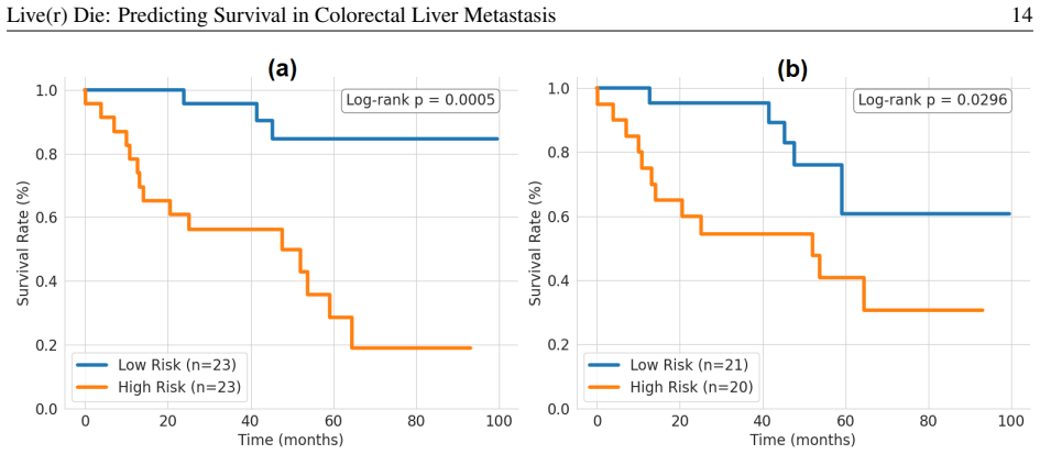
- [§3.4.3, Table 3, Figure 10] The headline HR CI is not a patient-level confidence interval. The cohort has only 82 uncensored events; a patient-level Cox model would give SE(log HR) ≈ 0.11 and a 95% CI of roughly 1.55–2.40. The reported CI of 1.89–1.98 implies SE(log HR) ≈ 0.012, consistent with bootstrapping over the 15 repeated 3-fold CV runs—where the same patients appear in multiple folds—rather than over patients. Repeated CV splits are not independent, so the reported interval and Wald p-value do not establish patient-level uncertainty. The randomization test only assesses the null of no association. Please report the outcome test-set size and event count, compute patient-level bootstrapped CIs for the HR and C-index, and use an appropriate test for comparing against biomarkers.
- [§3.1, Figure 2, §4.2] The test set for the survival experiments appears to be the genomic-data subset excluded from training, but its size, censoring distribution, and follow-up time are never reported. If this subset is small, the C-index of 0.691 has a wide confidence interval and the multivariate HRs in Table 5 are unstable. Please report the number of patients, number of events, and median follow-up for every held-out test subset, and give confidence intervals for the reported C-indices.
- [§3.2.1, §5] The entire segmentation training chain depends on SAMONAI pseudo-labels for liver and spleen generated on 20 cases with no quality control. The authors explicitly state that a QC step is future work and that SAMONAI can fail when the initial axial/sagittal/coronal slice is missegmented. Because the stage-2 model is trained on these labels and stage 3 auto-completes the full cohort, any systematic label error propagates into tumor segmentations, radiomic features, and survival predictions. Please validate pseudo-label quality against manual labels on held-out cases, or perform a sensitivity analysis by re-running the pipeline with alternative pseudo-labeling strategies. The current reliance on unvalidated pseudo-labels is load-bearing for the central claim.
- [§4.1, Table 2, Figure 9] Segmentation evaluation is limited to 20 post-contrast cases, and pre-contrast liver/spleen labels are obtained by registration from post-contrast labels rather than independent manual annotation. The tumor detection F1 is only 0.79 (post-contrast) and 0.71 (pre-contrast) before the expert reassessment, and the post-hoc reassessment in §5 changes the error counts substantially. In addition, five patients with no predicted tumors are excluded from the survival analysis in Figure 9(b); this acknowledged selection bias should be quantified and addressed, e.g., by reporting results with and without those patients and by modeling empty-prediction cases as a separate category.
- [§4.2, Table 5] The comparison of SurvAMINN with existing biomarkers lacks a formal statistical test for differences in C-index. Table 5 reports univariate and multivariate Cox HRs for each biomarker, but the 'exceeding 10% improvement' claim is not accompanied by a confidence interval for the C-index difference or a significance test. Please add a paired comparison (e.g., DeLong test or bootstrap over patients) between SurvAMINN and each biomarker baseline, and clarify whether the 10% improvement is absolute or relative.
minor comments (6)
- [§2.3] Typo: 'autencoder' should be 'autoencoder' in the contributions bullet list.
- [§3.3.1, Eq. (4)] The notation for min-max normalization in the prompt-selection criteria is ambiguous. State explicitly that the standardization is computed over the candidate set P for each criterion separately.
- [§3.4.3] The formatting '1 × 10³' should be '1 × 10^3' (superscript) for the number of bootstrap and randomization iterations.
- [Table 4, Table 5] Several reported HR 95% CIs, especially for RSF and biomarkers, are implausibly narrow given the event count. Please check the standard-error calculations and report the number of events used in each Cox model.
- [§4.2] Figure 8 shows box plots of repeated CV runs, but the number of runs and the exact definition of the 'both' fusion are not fully described in the caption. Please clarify how the late-fusion hazards are combined and how many independent runs are summarized.
- [§5] The radiologist reassessment that reclassifies many detection errors as 'not real' is important, but it is presented only qualitatively. Please provide the exact corrected precision/recall/F1 numbers and a clear protocol for how the reassessment was completed.
Circularity Check
No significant circularity: survival predictions are tested on held-out folds and benchmarked against external clinical/genomic biomarkers.
full rationale
The derivation chain is self-contained. The segmentation model is trained on manual tumor annotations plus foundation-model pseudo-labels for liver/spleen, with the manually annotated liver/spleen cases held out for evaluation (Sections 3.1, 3.4.2, 4.1). SurvAMINN optimizes a Cox partial likelihood (Eq. 7) and is scored with the concordance index on 15 repeated 3-fold CV splits; the headline C-index is therefore an out-of-sample prediction, not a fitted quantity. Self-citations to AMINN and 3DINO supply architectural/pre-training choices, but the central comparison against biomarkers (Table 5) and standard baselines (Table 4) is anchored to held-out performance and external metrics. The paper's own Section 5 notes unresolved pseudo-label quality control and patient-exclusion bias; these are correctness/robustness limitations, not circular reductions. The very narrow reported HR CI suggests bootstrap over CV runs rather than patients, but that is a statistical inference concern, not a circularity of the derivation.
Axiom & Free-Parameter Ledger
free parameters (8)
- SAMONAI prompt criterion weights =
alpha=1, beta=1, gamma=2
- SAMONAI logit binarization threshold T_L =
mu_L + 2*sigma_L
- Negative-point intensity threshold T_N =
min(I) + 0.1*(max(I)-min(I))
- Small predicted tumor diameter cutoff =
1st percentile of ground-truth longest diameters in training set
- Small predicted tumor volume cutoff =
100 mm^3
- SAMONAI slice sampling rate =
1/3 with linear interpolation
- Radiomics preprocessing hyperparameters =
2 mm resampling, mu +/- 3*sigma outlier clip, bin width 5, z-score and scale by 100
- Loss schedule alpha =
current_epoch / total_epochs
axioms (8)
- domain assumption SAM and 3DINO foundation models provide useful zero-shot or few-shot representations for abdominal MRI.
- domain assumption Manual tumor segmentations by a single radiologist are reliable ground truth for tumors of at least 10 mm, and small tumors or benign lesions are intentionally absent.
- domain assumption Pre-contrast liver and spleen labels, obtained by registering post-contrast labels, are accurate enough for evaluation.
- standard math Cox proportional hazards and independent censoring hold for the cohort.
- domain assumption LogSumExp pooling over tumor hazards approximates the biology that the most aggressive tumors drive survival.
- ad hoc to paper The random 20-case sample used for SAMONAI pseudo-labeling is representative of the full cohort.
- domain assumption PyRadiomics features extracted from predicted segmentations contain prognostic signal beyond known biomarkers.
- domain assumption The reported data splits prevent patient overlap between segmentation training and outcome testing.
Cite this review
Pith. "Pith review of Live(r) Die: Predicting Survival in Colorectal Liver Metastasis." pith.science (2026). https://pith.science/paper/VTIE2EGC
@misc{pith2026250908935,
  author       = {Pith},
  title        = {Pith review of: Live(r) Die: Predicting Survival in Colorectal Liver Metastasis},
  year         = {2026},
  howpublished = {\url{https://pith.science/paper/VTIE2EGC}},
  note         = {Machine review of arXiv:2509.08935}
}
read the original abstract
Colorectal cancer frequently metastasizes to the liver, significantly reducing long-term survival. While surgical resection is the only potentially curative treatment for colorectal liver metastasis (CRLM), patient outcomes vary widely depending on tumor characteristics along with clinical and genomic factors. Current prognostic models, often based on limited clinical or molecular features, lack sufficient predictive power, especially in multifocal CRLM cases. We present a fully automated framework for surgical outcome prediction from pre- and post-contrast MRI acquired before surgery. Our framework consists of a segmentation pipeline and a radiomics pipeline. The segmentation pipeline learns to segment the liver, tumors, and spleen from partially annotated data by leveraging promptable foundation models to complete missing labels. Also, we propose SAMONAI, a novel zero-shot 3D prompt propagation algorithm that leverages the Segment Anything Model to segment 3D regions of interest from a single point prompt, significantly improving our segmentation pipeline's accuracy and efficiency. The predicted pre- and post-contrast segmentations are then fed into our radiomics pipeline, which extracts features from each tumor and predicts survival using SurvAMINN, a novel autoencoder-based multiple instance neural network for survival analysis. SurvAMINN jointly learns dimensionality reduction and hazard prediction from right-censored survival data, focusing on the most aggressive tumors. Extensive evaluation on an institutional dataset comprising 227 patients demonstrates that our framework surpasses existing clinical and genomic biomarkers, delivering a C-index improvement exceeding 10%. Our results demonstrate the potential of integrating automated segmentation algorithms and radiomics-based survival analysis to deliver accurate, annotation-efficient, and interpretable outcome prediction in CRLM.
Figures



Reference graph
Works this paper leans on
-
[8]
Foundation models for generalist medi- cal artificial intelligence. Nature 616, 259–265. doi:10.1038/s41586-023-05881-4. Nakai, Y ., Gonoi, W., Kurokawa, R., Nishioka, Y ., Abe, H., Arita, J., Ushiku, T., Hasegawa, K., Abe, O.,
-
[11]
BMC Cancer 24, 426
Prognostic factors associated with early re- currence following liver resection for colorectal liver metastases: a systematic review and meta-analysis. BMC Cancer 24, 426. doi:10.1186 /s12885-024- 12162-4. Ulrich, C., Isensee, F., Wald, T., Zenk, M., Baumgart- ner, M., Maier-Hein, K.H., 2023. Multitalent: A multi-dataset approach to medical image segmenta...
2023
-
[70]
Zhu, J., Hamdi, A., Qi, Y ., Jin, Y ., Wu, J., 2024
doi:10.1038 /s41392-022-00922-2. Zhu, J., Hamdi, A., Qi, Y ., Jin, Y ., Wu, J., 2024. Medical sam 2: Segment medical images as video via segment anything model 2. arXiv preprint arXiv:2408.00874
Pith/arXiv arXiv 2024
-
[2008]
The Annals of Ap- plied Statistics 2
Random survival forests. The Annals of Ap- plied Statistics 2. doi:10.1214 /08-AOAS169. Kirienko, M., Sollini, M., Corbetta, M., V oulaz, E., Gozzi, N., Interlenghi, M., Gallivanone, F., Cas- tiglioni, I., Asselta, R., Duga, S., Soldà, G., Chiti, A., 2021. Radiomics and gene expression profile to characterise the disease and predict outcome in pa- tients w...
2021
-
[2017]
Attention is all you need 30. Wang, Y ., Zhong, Xinyangand He, X., Hu, Z., Huang, H., Chen, J., Chen, K., Zhao, S., Wei, P ., Li, D., 2023. Liver metastasis from colorectal cancer: pathogenetic development, immune landscape of the tumour mi- croenvironment and therapeutic approaches. Journal of Experimental & Clinical Cancer Research 42, 177. doi:10.1186/...
Pith/arXiv arXiv 2023
-
[2018]
Liver imaging reporting and data system (li- rads) version 2018: Imaging of hepatocellular carci- noma in at-risk patients. Radiology 289, 816–830. doi:10.1148/radiol.2018181494. Cheung, H.M.C., Karanicolas, P .J., Coburn, N., Seth, V ., Law, C., Milot, L., 2019. Delayed tumour en- hancement on gadoxetate-enhanced mri is associated with overall survival i...
arXiv 2018
-
[2019]
Chen, T., Kornblith, S., Norouzi, M., Hinton, G., 2020
Unsupervised clustering of quantitative imag- ing phenotypes using autoencoder and gaussian mix- ture model , 575–582. Chen, T., Kornblith, S., Norouzi, M., Hinton, G., 2020. A simple framework for contrastive learning of visual representations . Chen, Z., Duan, Y ., Wang, W., He, J., Lu, T., Dai, J., Qiao, Y ., 2022. Vision transformer adapter for dense ...
Pith/arXiv arXiv 2020
-
[2020]
Mri findings of liver parenchyma peripheral to colorectal liver metastasis: A potential predictor of long-term prognosis. Radiology 297, 584–594. doi:10.1148/radiol.2020202367. Peng, H., Long, F., Ding, C., 2005. Fea- ture selection based on mutual information crite- ria of max-dependency, max-relevance, and min- redundancy. IEEE Transactions on Pattern An...
Pith/arXiv arXiv 2005
-
[2021]
Chen, J., Milot, L., Cheung, H.M.C., Martel, A.L.,
Aminn: Autoencoder-based multiple instance neural network improves outcome prediction in mul- tifocal liver metastases , 752–761. Chen, J., Milot, L., Cheung, H.M.C., Martel, A.L.,
-
[2022]
Tian, Y ., Wang, Y ., Wen, N., Wang, S., Li, B., Liu, G.,
Self-supervised pre-training of swin trans- formers for 3d medical image analysis , 20698– 20708doi:10.1109/CVPR52688.2022.02007. Tian, Y ., Wang, Y ., Wen, N., Wang, S., Li, B., Liu, G.,
arXiv 2022
-
[2023]
Deep learning framework for liver segmen- tation from t1-weighted mri images. Sensors 23. doi:10.3390/s23218890. Isensee, F., Jaeger, P .F., Kohl, S.A.A., Petersen, J., Maier-Hein, K.H., 2021. nnu-net: a self-configuring method for deep learning-based biomedical im- age segmentation. Nature Methods 18, 203–211. doi:10.1038/s41592-020-01008-z. Isensee, F., ...
Pith/arXiv arXiv 2021
-
[2024]
Segment anything in medical images. Nature Communications 15, 654. doi:10.1038 /s41467-024- 44824-z. Ma, J., Y ang, Z., Kim, S., Chen, B., Baharoon, M., Fal- lahpour, A., Asakereh, R., Lyu, H., Wang, B., 2025. Medsam2: Segment anything in 3d medical images and videos. arXiv preprint arXiv:2504.03600 . Mann, H.B., Whitney, D.R., 1947. On a Test of Whether ...
Pith/arXiv arXiv 2025
-
[3655]
doi:10.1007 /s00259-021-05371-7. Kirillov, A., Mintun, E., Ravi, N., Mao, H., Rolland, C., Gustafson, L., Xiao, T., Whitehead, S., Berg, A.C., Lo, W.Y ., Dollár, P ., Girshick, R., 2023. Segment anything. arXiv preprint arXiv:2304.02643 . Klein, S., Staring, M., Murphy, K., Viergever, M.A., Pluim, J.P .W., 2010. elastix: A toolbox for intensity-based medi...
Pith/arXiv arXiv 2023
This paper was first reviewed by deepseek-v4-flash on August 4, 2026.
discussion (0)
Sign in with ORCID, Apple, or X to comment. Anyone can read and Pith papers without signing in.