REVIEW 3 major objections 4 minor 41 references
The study of intrinsic defect state of FeSe with scanning tunneling microscopy
T0 review · 3 major / 4 minor · reviewed 2026-08-14 · deepseek-v4-flash
Pith's one-line read STM identifies FeSe's dumbbell defects as an iron vacancy and a selenium antisite, with wide-bias spectroscopy and electron microscopy as support.
desk verdict A careful STM/STS catalogue of four FeSe defect types with a plausible but unproven assignment of the two dumbbell features to Fe vacancy and Se_Fe antisite; the 'determined' language overshoots the evidence. read the letter →
The pith
A machine-rendered reading of the paper's core claim, the machinery that carries it, and where it could break.
The reading
What carries the argument
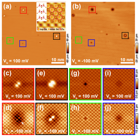
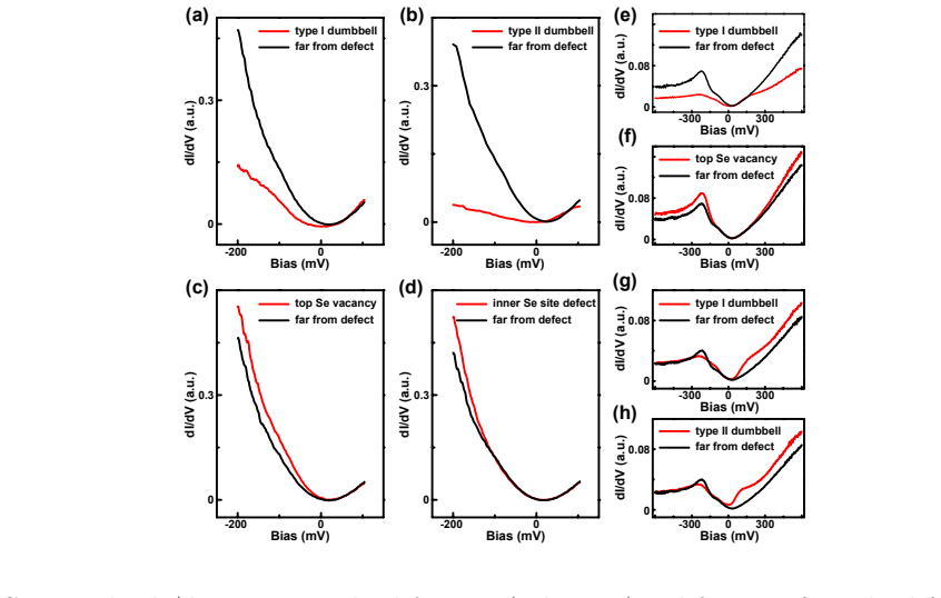
The load-bearing tool is scanning tunneling spectroscopy over a wide bias range (roughly $-600$ to $+600$ mV), which reveals defect-induced changes in the local density of states. The interpretive rule is a valence-change ordering: the more the substituted ion's valence drops relative to the host ion, the more the occupied-state DOS is depressed. Under that rule, a Fe vacancy is a milder perturbation than a Se$_{\mathrm{Fe}}$ antisite, and both depress the negative-bias DOS while selenium vacancies enhance it. The dumbbell shape itself is tied to the two protruding selenium orbitals neighboring the iron site, following a density functional theory image for Fe vacancies on monolayer FeSe/SrTiO$_3$.
What would settle it
A first-principles or model calculation of the local density of states around an isolated Fe vacancy and an isolated Se$_{\mathrm{Fe}}$ antisite in bulk FeSe would settle the ordering: if the computed occupied-state depression is reversed, the type I and type II labels swap. An experiment that identifies the same individual defect by STM and by atomically resolved electron microscopy on the same flake could also test the assignment directly.
Extended reading notes
Core claim
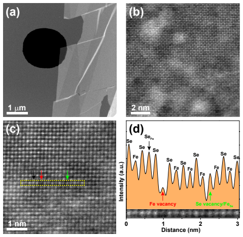
The central claim is that the type I dumbbell is a Fe vacancy and the type II dumbbell is a Se$_{\mathrm{Fe}}$ antisite, with the top-layer Se vacancy and inner-layer Se-site vacancy as the other two defect species. The identification rests on the observed relation between valence change at the defect site and the depression or enhancement of the local density of states at negative bias: replacing Fe$^{2+}$ by a Fe vacancy ($V_{\mathrm{Fe}}^{0+}$) depresses the occupied-state DOS less than replacing it by Se$^{2-}$ (Se$_{\mathrm{Fe}}$), while removing Se$^{2-}$ enhances it. The paper reports that ADF-STEM on three-layer exfoliated FeSe observes both Fe vacancies and Se$_{\mathrm{Fe}}$ antisites, consistent with the STM assignments, though the two measurements are made on different samples.
Load-bearing premise
The assignment assumes that defect-induced changes in the local density of states at negative bias are ordered monotonically by the valence change of the substitution, so the Fe vacancy must depress the occupied-state DOS less than the Se$_{\mathrm{Fe}}$ antisite.
Editorial extensions
If this is right
- If the assignments are correct, every prior STM work that treated dumbbells as a single defect species must now distinguish Fe vacancies from Se$_{\mathrm{Fe}}$ antisites when interpreting scattering data.
- The wide-range spectra provide a reference dataset against which future density functional calculations of correlated FeSe can be tested.
- The observation that type I dumbbell density decreases with cooling cycles or annealing supports the picture that Fe vacancies can be annealed out, offering a handle on defect concentration.
- Identifying the inner-layer Se-site defect as a second-layer Se vacancy implies that subsurface defects leave measurable STM signatures, not just surface-layer ones.
Reading between the lines
- This goes beyond the paper: if the valence-change ordering is general, the same spectroscopy-based reasoning could be applied to antisite and vacancy defects in other iron-based superconductors, and the predicted depression ordering could be checked by controlled doping studies.
- The paper's assignment implies that the low-energy in-gap states previously attributed to 'the dumbbell' may actually come from two different impurities, which could explain sample-dependent variations in earlier low-bias STM experiments.
- As a testable extension, one could combine STM with atomically resolved electron energy-loss spectroscopy on the same exfoliated flake to correlate a specific defect's chemical identity with its STS spectrum, removing the sample-mismatch ambiguity.
Signed reviews
Editorial analysis
A structured set of objections, weighed in public.
Referee Report
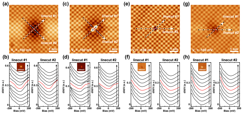
Summary. The paper reports a scanning tunneling microscopy/spectroscopy study of intrinsic defects in bulk FeSe at 77 K. Four defect types are identified in top-layer Se lattice images: two types of dumbbell defects centered at Fe sites, a top-layer Se vacancy, and an inner-layer Se-site defect. From the relative depression or enhancement of the dI/dV spectra over a wide bias range, the authors assign the type I dumbbell to an Fe vacancy and the type II dumbbell to a Se_Fe antisite defect; the two Se-site defects are assigned to a top-layer Se vacancy and a possible inner-layer Se vacancy. ADF-STEM on an exfoliated few-layer FeSe sample is presented as supporting evidence for the existence of Fe vacancies and Se_Fe antisites. The paper also provides spatial mapping of the defect-induced DOS and notes the need for future detailed theoretical calculations.
Significance. If the assignments are correct, the paper provides a systematic catalog of intrinsic defect states in bulk FeSe and distinguishes two Fe-site dumbbell defects that previous studies treated together. The measurements are reproducible across samples and tips, the large-energy-range spectroscopy is a useful dataset, and the ADF-STEM comparison independently demonstrates that Fe vacancies and Se_Fe antisites occur in this material. However, the central type I/type II assignment rests on an asserted, rather than derived or computed, monotonic relation between formal valence change and occupied-state DOS depression. The ADF-STEM data confirm species existence but do not atomically correlate them with the specific STM signatures. These caveats mean the paper's main conclusion is plausible but not yet quantitatively established.
major comments (3)
- [Section III, paragraph beginning 'The asgrown bulk FeSe is a bad metal'] The assignment of the type I and type II dumbbells to the Fe vacancy and Se_Fe antisite rests entirely on the asserted ordering that a larger valence change (Fe2+ to Se2-) depresses the occupied-state DOS more than a smaller change (Fe2+ to V_Fe^0+). The paper itself states that a simple defect-DOS argument is not available for a bad metal and that general DFT is inadequate due to strong correlations, but no model, calculation, or independent calibration of this monotonicity is provided. In addition, treating a vacancy as a substitutional positive ion V_Fe^0+ is not a physically well-defined substitution; removing an Fe atom changes the local electronic structure through rehybridization and lattice relaxation. If the ordering of the DOS depression were reversed or non-monotonic, the type I/type II assignment would flip. Please either supply a quantitative calculation or a chemically and physically justified argument for the ordering, or explicitly recast the assignment as a tentative inference that is not yet determined.
- [Section III, ADF-STEM paragraph and Fig. 4] The ADF-STEM measurement is performed on a different exfoliated three-layer sample, and the paper states that the intermediate FeSe is protected by terminal layers; the STM and STEM samples are therefore not the same. The STEM images show Fe vacancies and Se_Fe antisites exist in FeSe, but they do not identify which of the two dumbbell classes seen in STM corresponds to which defect. The abstract and summary state the determination is 'largely confirmed' by STEM, which overstates the correlation. Please soften the claim to 'consistent with the existence of the proposed defect species' unless a correlated STM-STEM measurement on the same sample or a defect-species-specific signature is provided.
- [Section III, discussion of inner-layer Se-site defect] The assignment of the inner-layer Se-site defect to an inner-layer Se vacancy is presented as 'most possibly' and 'possible', which is appropriately cautious. However, the reasoning that the subtle spectral difference from the top-layer Se vacancy is simply due to the larger tip-defect distance is not supported by any calculation or distance-dependent measurement. This is a secondary claim and can be addressed by explicitly labeling it as speculative or by providing additional evidence, such as a comparison of apparent defect size in conductance maps with the expected depth.
minor comments (4)
- [Throughout] There are several typographical errors, including 'paring symmetry' in the Introduction (should be 'pairing symmetry') and inconsistent notation such as 'Se Fe' instead of 'Se_Fe' in the abstract and Fig. 4 labels. Please proofread the text.
- [Fig. 2] The dI/dV curves in Fig. 2 are normalized in arbitrary units, but the normalization procedure is not described. Please state explicitly how the spectra were normalized, since the central argument compares the relative depression/enhancement across defect types.
- [Section III, paragraph on large-range spectra] The sentence 'a protruding kink below 200 mV is the main dumbbell-induced change of DOS at positive energy' is ambiguous: the kink is visible in the negative-bias region of Figs. 2(g,h) as well. Please clarify whether the kink is at positive or negative bias, and define the energy range precisely.
- [References] Reference [41] (Huang et al., Nano Lett. 16, 4224 (2016)) is cited for the DFT result that two protruding Se orbitals around an Fe vacancy lead to bright dumbbell lobes; this is helpful, but the paper should also cite related STM studies of defects in FeSe that report similar dumbbell features, such as Refs. [25, 35], when discussing the variation of defect-site spectroscopy.
Circularity Check
No significant circularity: the defect assignment is an interpretive comparison of spectra with independent STEM evidence, not a fit or self-referential reduction.
full rationale
The paper's central claim—that the type I dumbbell is an Fe vacancy and the type II dumbbell is a Se_Fe antisite—does not reduce by construction to its inputs. The STS spectra are measured experimental data; the assignment is made by a qualitative valence-change ordering argument in Section III, in which a larger valence reduction (Fe2+ to Se2-) is associated with stronger depression of occupied-state DOS relative to a smaller valence reduction (Fe2+ to V_Fe^0+). This argument is an interpretive assumption that could be wrong or non-monotonic, but it is not circular: the defect labels are not defined as 'the dumbbell with the larger negative-bias DOS depression,' and no parameter is fitted to the spectra and then renamed as a prediction. The ADF-STEM measurements independently demonstrate that Fe vacancies and Se_Fe antisites exist in exfoliated FeSe, even though those images do not atomically correlate the species with the specific STM dumbbell classes; this is independent supporting evidence for species existence, not a self-referential or construction-based reduction. There is no load-bearing self-citation, no imported uniqueness theorem, no ansatz smuggled in via citation, and no renaming of a known pattern as unification. The paper also explicitly concedes the limits of its own analysis by noting that the defect-induced DOS change cannot be simply explained or calculated by general DFT, and it calls for future complex calculations. Any weakness in the monotonic valence-change assumption is a soundness or evidentiary concern, not a circularity concern. The derivation chain is therefore self-contained with respect to circularity, and the appropriate score is 0.
Assumptions & free parameters
assumptions (4)
- domain assumption The top-layer termination after cleavage is a Se layer, and bright spots in STM topographies are Se atoms.
- standard math The dI/dV signal is proportional to the local density of states for the relevant bias range.
- ad hoc to paper The relative depression of negative-bias DOS is a monotonic function of the valence change of the substitution at the Fe site.
- domain assumption Defects observed in the as-grown crystal are intrinsic, not introduced by cleaving or imaging.
Cite this review
Pith. "Pith review of The study of intrinsic defect state of FeSe with scanning tunneling microscopy." pith.science (2026). https://pith.science/paper/WVZXR7AJ
@misc{pith2026190803427,
  author       = {Pith},
  title        = {Pith review of: The study of intrinsic defect state of FeSe with scanning tunneling microscopy},
  year         = {2026},
  howpublished = {\url{https://pith.science/paper/WVZXR7AJ}},
  note         = {Machine review of arXiv:1908.03427}
}
abstract
We apply high resolution scanning tunneling microscopy to study intrinsic defect states of bulk FeSe. Four types of intrinsic defects including the type I dumbbell, type II dumbbell, top-layer Se vacancy and inner-layer Se-site defect are extensively analyzed by scanning tunneling spectroscopy. From characterized depression and enhancement of density of states measured in a large energy range, the type I dumbbell and type II dumbbell are determined to be the Fe vacancy and Se$_\mathrm{Fe}$ defect, respectively. The top-layer Se vacancy and possible inner-layer Se-site vacancy are also determined by spectroscopy analysis. The determination of defects are compared and largely confirmed in the annular dark-field scanning transmission electron microscopy measurement of the exfoliated FeSe. The detailed mapping of defect states in our experiment lays the foundation for a comparison with complex theoretical calculations in the future.
Figures
Figures from the paper (1 more)
Reference graph
Works this paper leans on
-
[1]
P. A. Lee, N. Nagaosa, and X.-G. Wen, Rev. Mod. Phys. 78, 17 (2006)
2006
-
[2]
Kamihara, T
Y. Kamihara, T. Watanabe, M. Hirano, and H. Hosono, J. Am. Chem. Soc. 130, 3296 (2008)
2008
-
[3]
K. Matsuura, Y. Mizukami, Y. Arai, Y. Sugimura, N. Maejima, A. Machida, T. Watanuki, T. Fukuda, T. Yajima, Z. Hiroi, K. Y. Yip, Y. C. Chan, Q. Niu, S. Hosoi, K. Ishida, K. Mukasa, S. Kasahara, J.-G. Cheng, S. K. Goh, Y. Matsuda, Y. Uwatoko, and T. Shibauchi, Nat. Commun. 8, 1143 (2017)
work page 2017
-
[4]
M. Abdel-Hafiez, Y. J. Pu, J. Brisbois, R. Peng, D. L. Feng, D. A. Chareev, A. V. Silhanek, C. Krellner, A. N. Vasiliev, and X.-J. Chen, Phys. Rev. B 93, 224508 (2016)
work page 2016
-
[5]
S. H. Pan, E. W. Hudson, A. K. Gupta, K.-W. Ng, H. Eisaki, S. Uchida, and J. C. Davis, Phys. Rev. Lett. 85, 1536 (2000)
work page 2000
-
[6]
Q. Liu, C. Chen, T. Zhang, R. Peng, Y.-J. Yan, C.-H.-P. Wen, X. Lou, Y.-L. Huang, J.-P. Tian, X.-L. Dong, G.-W. Wang, W.-C. Bao, Q.-H. Wang, Z.-P. Yin, Z.-X. Zhao, and D.-L. Feng, Phys. Rev. X 8, 041056 (2018)
work page 2018
-
[7]
D. Huang, C.-L. Song, T. A. Webb, S. Fang, C.-Z. Chang, J. S. Moodera, E. Kaxiras, and J. E. Hoffman, Phys. Rev. Lett. 115, 017002 (2015)
work page 2015
-
[8]
K. L. Bu, Y. Fei, W. H. Zhang, Y. Zheng, J. L. Wu, F. C. Chen, X. Luo, Y. P. Sun, Q. N. Xu, X. Dai, and Y. Yin, Phys. Rev. B 98, 115127 (2018)
work page 2018
Show all 41 references
-
[9]
A. V. Balatsky, I. Vekhter, and J.-X. Zhu, Rev. Mod. Phys. 78, 373 (2006)
2006
-
[10]
Fischer, M
∅. Fischer, M. Kugler, I. Maggio-Aprile, C. Berthod, and C. Renner, Rev. Mod. Phys. 79, 353 (2007)
2007
-
[11]
Zhang, Phys
D. Zhang, Phys. Rev. Lett. 103, 186402 (2009)
2009
-
[12]
C. Liu, Z. Wang, Y. Gao, X. Liu, Y. Liu, Q.-H. Wang, and J. Wang, Phys. Rev. Lett. 123, 036801 (2019). 12
2019
-
[13]
S. Chi, R. Aluru, U. R. Singh, R. Liang, W. N. Hardy, D. A. Bonn, A. Kreisel, B. M. Andersen, R. Nelson, T. Berlijn, W. Ku, P. J. Hirschfeld, and P. Wahl, Phys. Rev. B 94, 134515 (2016)
2016
-
[14]
Hsu, J.-Y
F.-C. Hsu, J.-Y. Luo, K.-W. Yeh, T.-K. Chen, T.-W. Huang, P. M. Wu, Y.-C. Lee, Y.-L. Huang, Y.-Y. Chu, D.-C. Yan, and M.-K. Wu, Proc. Natl. Acad. Sci. USA 105, 14262 (2008)
2008
-
[15]
J. Guo, S. Jin, G. Wang, S. Wang, K. Zhu, T. Zhou, M. He, and X. Chen, Phys. Rev. B 82, 180520(R) (2010)
2010
-
[16]
T. P. Ying, X. L. Chen, G. Wang, S. F. Jin, T. T. Zhou, X. F. Lai, H. Zhang, and W. Y. Wang, Sci. Rep. 2, 426 (2012)
2012
-
[17]
Burrard-Lucas, D
M. Burrard-Lucas, D. G. Free, S. J. Sedlmaier, J. D.Wright, S. J. Cassidy, Y. Hara, A. J. Corkett, T. Lancaster, P. J. Baker, S. J. Blundell, and S. J. Clarke, Nature Mater. 12, 15 (2013)
2013
-
[18]
X. F. Lu, N. Z. Wang, H. Wu, Y. P. Wu, D. Zhao, X. Z. Zeng, X. G. Luo, T. Wu, W. Bao, G. H. Zhang, F. Q. Huang, Q. Z. Huang, and X. H. Chen, Nature Mater. 14, 325 (2015)
2015
-
[19]
Miyata, K
Y. Miyata, K. Nakayama, K. Sugawara, T. Sato, and T. Takahashi, Nature Mater. 14, 775 (2015)
2015
-
[20]
Z. R. Ye, C. F. Zhang, H. L. Ning, W. Li, L. Chen, T. Jia, M. Hashimoto, D. H. Lu, Z.-X. Shen, and Y. Zhang, arXiv:1512.02526
-
[21]
Song, H.-M
C.-L. Song, H.-M. Zhang, Y. Zhong, X.-P. Hu, S.-H. Ji, L. Wang, K. He, X.-C. Ma, and Q.-K. Xue, Phys. Rev. Lett. 116, 157001 (2016)
2016
-
[22]
C. H. P. Wen, H. C. Xu, C. Chen, Z. C. Huang, X. Lou, Y. J. Pu, Q. Song, B. P. Xie, M. Abdel-Hafiez, D. A. Chareev, A. N. Vasiliev, R. Peng, and D.-L. Feng, Nat. Commun. 7, 10840 (2016)
2016
-
[23]
Q.-Y. Wang, Z. Li, W.-H. Zhang, Z.-C. Zhang, J.-S. Zhang, W. Li, H. Ding, Y.-B. Ou, P. Deng, K. Chang, J. Wen, C.-L. Song, K. He, J.-F. Jia, S.-H. Ji, Y.-Y. Wang, L.-L. Wang, X. Chen, X.-C. Ma, and Q.-K. Xue, Chin. Phys. Lett. 29, 037402 (2012)
2012
-
[24]
S. He, J. He, W. Zhang, L. Zhao, D. Liu, X. Liu, D. Mou, Y.-B. Ou, Q.-Y. Wang, Z. Li, L. Wang, Y. Peng, Y. Liu, C. Chen, L. Yu, G. Liu, X. Dong, J. Zhang, C. Chen, Z. Xu, X. Chen, X. Ma, Q.-K. Xue, and X. J. Zhou, Nature Mater. 12, 605 (2013)
2013
-
[25]
Kasaharaa, T
S. Kasaharaa, T. Watashigea, T. Hanaguri, Y. Kohsaka, T. Yamashita, Y. Shimoyama, Y. Mizukami, R. Endo, H. Ikeda, K. Aoyama, T. Terashima, S. Uji, T. Wolf, H. v. L¨ ohneysen, T. Shibauchi, and Y. Matsuda, Proc. Natl. Acad. Sci. USA 111, 16309 (2014). 13
2014
-
[26]
P. O. Sprau, A. Kostin, A. Kreisel, A. E. B¨ ohmer, V. Taufour, P. C. Canfield, S. Mukherjee, P. J. Hirschfeld, B. M. Andersen, and J. C. S´ eamus Davis, Science 357, 75 (2017)
2017
-
[27]
Kostin, P
A. Kostin, P. O. Sprau, A. Kreisel, Y. X. Chong, A. E. B¨ ohmer, P. C. Canfield, P. J. Hirschfeld, B. M. Andersen, and J. C. S´ eamus Davis, Nat. Mater. 17, 869 (2018)
2018
-
[28]
Q. Fan, W. H. Zhang, X. Liu, Y. J. Yan, M. Q. Ren, R. Peng, H. C. Xu, B. P. Xie, J. P. Hu, T. Zhang, and D. L. Feng, Nat. Phys. 11, 946 (2015)
2015
-
[29]
Huang, T
D. Huang, T. A. Webb, S. Fang, C.-L. Song, C.-Z. Chang, J. S. Moodera, E. Kaxiras, and J. E. Hoffman, Phys. Rev. B 93, 125129 (2016)
2016
-
[30]
Song, Y.-L
C.-L. Song, Y.-L. Wang, P. Cheng, Y.-P. Jiang, W. Li, T. Zhang, Z. Li, K. He, L. Wang, J.-F. Jia, H.-H. Hung, C. Wu, X. Ma, X. Chen, and Q.-K. Xue, Science 332, 1410 (2011)
2011
-
[31]
Song, Y.-L
C.-L. Song, Y.-L. Wang, Y.-P. Jiang, L. Wang, K. He, X. Chen, J. E. Hoffman, X.-C. Ma, and Q.-K. Xue, Phys. Rev. Lett. 109, 137004 (2012)
2012
-
[32]
Watashige, Y
T. Watashige, Y. Tsutsumi, T. Hanaguri, Y. Kohsaka, S. Kasahara, A. Furusaki, M. Sigrist, C. Meingast, T. Wolf, H. v. L¨ ohneysen, T. Shibauchi, and Y. Matsuda, Phys. Rev. X 5, 031022 (2015)
2015
-
[33]
W. Li, Y. Zhang, P. Deng, Z. Xu, S.-K. Mo, M. Yi, H. Ding, M. Hashimoto, R. G. Moore, D.-H. Lu, X. Chen, Z.-X. Shen, and Q.-K. Xue, Nat. Phys. 13, 957 (2017)
2017
-
[34]
Jiao, C.-L
L. Jiao, C.-L. Huang, S. R¨ oβler, C. Koz, U. K. R¨ oβler, U. Schwarz, and S. Wirth, Sci. Rep. 7, 44024 (2017)
2017
-
[35]
L. Jiao, S. R¨ oβler, C. Koz, U. Schwarz, D. Kasinathan, U. K. R¨ oβler, and S. Wirth, Phys. Rev. B 96, 094504 (2017)
2017
-
[36]
Z. Du, X. Yang, D. Altenfeld, Q. Gu, H. Yang, I. Eremin, P. J. Hirschfeld, I. I. Mazin, H. Lin, X. Zhu, and H.-H. Wen, Nat. Phys. 14, 134 (2017)
2017
-
[37]
Y. J. Yan, W. H. Zhang, M. Q. Ren, X. Liu, X. F. Lu, N. Z. Wang, X. H. Niu, Q. Fan, J. Miao, R. Tao, B. P. Xie, X. H. Chen, T. Zhang, and D. L. Feng, Phys. Rev. B 94, 134502 (2016)
2016
-
[38]
C. Liu, Z. Wang, S. Ye, C. Chen, Y. Liu, Q. Wang, Q.-H. Wang, and J. Wang, Nano Lett. 19, 3464 (2019)
2019
-
[39]
C. Liu, J. Mao, H. Ding, R. Wu, C. Tang, F. Li, K. He, W. Li, C.-L. Song, X.-C. Ma, Z. Liu, L. Wang, and Q.-K. Xue, Phys. Rev. B 97, 024502 (2018). 14
2018
-
[40]
Q. S. Wang, Y. Shen, B. Y. Pan, X. W. Zhang, K. Ikeuchi, K. Iida, A. D. Christianson, H. C. Walker, D. T. Adroja, M. Abdel-Hafiez, X. J. Chen, D. A. Chareev, A. N. Vasiliev, and J. Zhao, Nat. Commun. 7, 12182 (2016)
2016
-
[41]
Huang, T
D. Huang, T. A. Webb, C.-L. Song, C.-Z. Chang, J. S. Moodera, E. Kaxiras, and J. E. Hoffman, Nano Lett. 16, 4224 (2016). 15
2016
Reviewed August 14, 2026 · model on record in the stance chip above.
Discussion (0). Continue with ORCID to comment.