REVIEW 2 major objections 6 minor 60 references
$\theta$-curves in proteins
T0 review · 2 major / 6 minor · reviewed 2026-08-14 · deepseek-v4-flash
Pith's one-line read Non-trivial θ-curves occur naturally in 52 protein chains, across seven topologies.
desk verdict First systematic survey of θ-curves in proteins, with a solid classification pipeline but a headline count that leans on dubious ion-mediated edges the authors themselves flag. read the letter →
The pith
A machine-rendered reading of the paper's core claim, the machinery that carries it, and where it could break.
The reading
What carries the argument
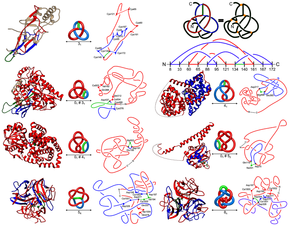
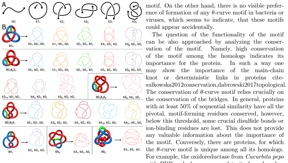
A θ-curve is an embedding of the letter θ in three dimensions: two trivalent vertices joined by three arcs. The load-bearing machinery is a pipeline that converts a protein into a spatial graph on Cα atoms (with edges for the backbone, disulfide bridges, ion-mediated contacts, and a chain-closure point at infinity), enumerates all cycles, and searches for an external arc connecting two residues of a cycle, which turns that cycle into a θ-curve. Each candidate is relaxed, simplified, and classified by three independent invariants: the Yamada polynomial of the spatial graph, the boundary-link invariant of θ-curves, and the constituent knots of the three arc pairs; the paper then assigns each motif a triple of knotoids, called its knotoid content, to distinguish different spatial realizations of the same topology.
What would settle it
Re-examine the human adiponectin structure (code 4dou) at higher resolution or in solution: if the Val-Ca contact and the cluster of four nearby ions prove to be crystallization or automated-assignment artifacts, then $\theta_{54}$ and $\theta_{8n}$ leave the census, and the number of observed θ-curve topologies falls from seven to five. The complementary check is to mutate the residues that coordinate the ions and observe whether the θ-curves disappear in a functional assay.
Extended reading notes
Core claim
On the paper's own terms, the central discovery is that the spatial graph formed by a protein main chain plus its disulfide bridges and ion-mediated contacts does contain non-trivial θ-curves, and that all known examples fall into seven topological classes: the prime $\theta_{31}$ and $\theta_{41}$, the composite $\theta_{01}\#3_1$, $\theta_{01}\#4_1$, $\theta_{01}\#5_2$, and two ion-mediated curves in human adiponectin, $\theta_{54}$ and an unnamed 8-crossing curve called $\theta_{8n}$. Most of these motifs sit in proteins whose backbone is unknotted, and several of them also appear as main-chain knots in other proteins. The authors classify each curve's spatial realization by its constituent knotoids, and they show that the function and organism of origin correlate with the motif type: roughly 70% of the host proteins are enzymes, and animal proteins show a preference for one handedness of the trefoil-based curves. They also report that formation of the knotted loop in the purely covalent example (horseshoe-crab coagulogen) is not the rate-limiting step of folding, unlike main-chain knots.
Load-bearing premise
The count of seven θ-curve topologies assumes that the metal ions used as junction points in some structures (calcium, magnesium, sodium) really are held there by the protein and are not crystallization or software artifacts; the paper itself flags one such contact, Val-Ca in 4dou, as highly dubious.
Editorial extensions
If this is right
- The census gives the first concrete answer to whether non-trivial θ-curves are possible in proteins: they are rare but real, appearing in 52 of more than 128,000 known chains.
- If the enzyme enrichment is not a detection artifact, θ-curves may rigidify part of the chain and help build active sites, a role already proposed for main-chain knots.
- For the covalent θ-curve in horseshoe-crab coagulogen, folding is not slowed by the knotted loop: the loop forms late (at native-contact fraction around 0.8) and is not tied to the main free-energy barrier.
- Bridge-removal simulations show the disulfide bridges that stabilize the protein are those that hold chain segments together; the knotted loop itself is not the stabilizing feature.
- Adding plausible new disulfide bridges to all known structures produces no new deterministic θ-curve topologies, but would create new probabilistic ones in some knotted proteins, such as $\theta_{66}$ or $\theta_{65}$ from the $6_1$-knotted hydrolase 3bjx.
Reading between the lines
- A direct extension of the census would be to redo the scan as more structures are deposited; if the ion-mediated θ-curves in 4dou disappear under better ion assignment, the topology count drops from seven to five, while a growing list would show the motifs are more common than this first pass suggests.
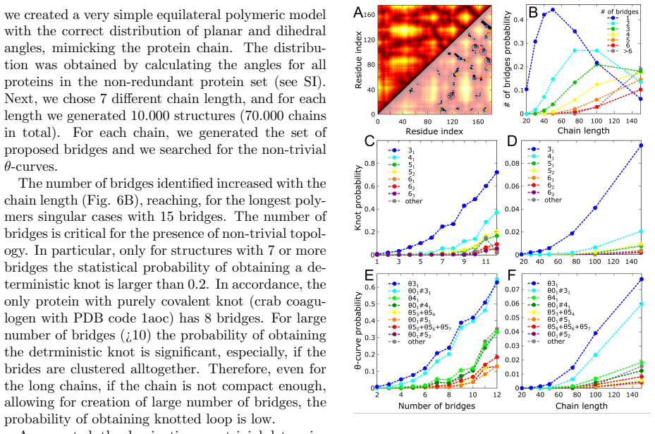
- The statistical model implies that engineered proteins with many clustered disulfide bonds (more than about ten) should be able to realize deterministic θ-curves that have never been observed; this is a concrete design target for de novo protein synthesis.
- Because the same θ-curve topology can carry different knotoid triples, knotoid content could be used as a finer evolutionary fingerprint, letting researchers ask whether functional convergence acts on the spatial realization rather than on the bare topology.
- The authors' closing suggestion to classify the full protein graph (all bridges at once) would generalize this work from θ-curves to arbitrary spatial graph motifs, and the Yamada polynomial used here is already the natural invariant for such a project.
Signed reviews
Editorial analysis
A structured set of objections, weighed in public.
Referee Report
Summary. The paper reports a systematic search for non-trivial θ-curve motifs in protein structures, treating the protein as a spatial graph with backbone edges, covalent (disulfide) bridges, ion-mediated interactions, and chain-closure edges. Applying three topological invariants (Yamada polynomial, Kauffman boundary links, constituent knots) implemented in the Topoly package to the PDB, the authors identify 52 non-redundant one-chain proteins and 4 multichain cases, which they group into 7 topological classes (9 including chirality). They further analyze the relation of these motifs to protein function and organism of origin, perform coarse-grained folding simulations of the covalent θ-curve protein coagulogen, quantify bridge-induced unfolding barriers, and use a polymer model to estimate which θ-curve topologies could plausibly form in designed or mutated proteins.
Significance. If the classification is robust, this is the first large-scale identification of non-trivial θ-curves in proteins and would substantially extend the known topological repertoire beyond main-chain knots, slipknots, and deterministic links. The paper's methodological strengths are its use of three independent topological invariants, its full-PDB scan, and its explicit local caveat about the dubious 4dou ion-mediated cases. The main limitation is that several of the reported classes rest on ion-mediated edges whose biological validity is not independently established; because the authors themselves discount the two classes unique to 4dou, the headline count of seven topologies is not yet on solid ground.
major comments (2)
- [Results, 'Types of θ-curves in proteins'; Table 1] The reported count of seven topologies includes θ54 and θ8n, each of which is supported solely by chain 4douA, in a structure whose ion-mediated interactions the authors themselves describe as 'highly dubious' and 'possibly assigned automatically'. Because these two classes are also the basis for the claim that a protein θ-curve can contain a constituent knot with unknotting number 2, the central classification is not robust to ion-assignment artifacts. The manuscript should either validate the 4dou ion coordination sites (e.g., via occupancy, B-factors, coordination geometry, or a stability calculation) or exclude these two classes and revise the counts and the unknotting-number claim accordingly.
- [Results, 'Types of θ-curves in proteins'; Table 2] The classes θ41 and θ01#52, which are not restricted to 4dou, nevertheless depend on a single ion-mediated edge (Mg498 in 3ulkA/4wkkA and Na331 in 3ihrA) in addition to chain closure. The paper provides no evidence that these ion-mediated edges are stable, biologically meaningful interactions rather than crystallization or automated-assignment artifacts. Since several of the seven classes share this dependency, the paper should report how the classification changes under stricter ion-bond criteria and, if possible, provide occupancy/B-factor or simulation-based support for the retained ion-mediated edges.
minor comments (6)
- [Throughout] There are multiple typos and grammatical slips, including 'travesting' for 'traversing', 'detrministic' for 'deterministic', 'priciple' for 'principle', 'bacterie' for 'bacteria', and 'reacher' for 'richer'; these should be corrected in a revision.
- [Results, 'Function, origin and conservation of the θ-curve motif'] The phrase 'cite-sulkowska2012conservation,dabrowski2017topological' is an unresolved LaTeX citation; the intended references should be inserted.
- [References] The entries for the Topoly package and the knotoid classification [30] and [36] contain unresolved '??' placeholders for journal and page information.
- [Table 2 caption] The caption does not define the symbols '...' and '↔', nor the meaning of 'Cls'; these are explained only in the main text and should be restated in the caption for readability.
- [Results, 'The algorithm'] The sentence 'we removed all structures with artificially long bonds or improbable gap filling' would benefit from concrete thresholds or a reference to the SI, since this filtering step affects the final counts.
- [Discussion] The statement that 'no new deterministic θ-curves may be found in proteins by introducing a bridge' is presented as a general result, but it depends on the contact-cutoff and representative-selection procedure described only in the SI; a brief restatement of the cutoff and a caveat about its dependence would help the reader assess that negative result.
Circularity Check
No circularity: θ-curve classification uses external invariants and the catalogue simulation is independent of the observed counts.
full rationale
The paper's central claim—that 52 non-redundant chains contain nontrivial θ-curves in seven topologies—is a data-classification result, not a derived prediction that reduces to its inputs. The classification pipeline starts from a spatial graph of Cα atoms and bridges, enumerates cycles and arcs, and then assigns topology by three external invariants (Yamada polynomial [31], Kauffman boundary link [32], and constituent knots) implemented in Topoly [30]; the invariants are cited to external mathematical sources, and the constituent-knot calculation is explicitly used only as validation because it cannot distinguish even θ31 from θ52. No parameter is fitted to the observed θ-curve counts and then repackaged as a prediction. The polymer-model catalogue in the last section uses a Ramachandran-like angle distribution measured independently from the PDB to generate random equilateral chains and then searches for θ-curves; the resulting θ31/θ01#31 dominance is an output of the simulation, not an input. The only flagged weakness—4dou's ion-mediated θ54 and θ8n being possibly automatic-assignment artifacts—is an explicit data-quality caveat, not a circularity: the authors do not use those cases to define the classification. Self-citations to Topoly and Knoto-ID are software/tool citations; the load-bearing mathematical content (Yamada invariant, knotoid theory, Moriuchi enumeration) is external. There is no equation or construction in which an input is identical to the claimed output.
Assumptions & free parameters
free parameters (4)
- Number of chain-closure trials =
100
- Contact cutoff for proposed bridges =
not stated in main text
- Backbone angle distribution for polymer model =
empirical distribution from non-redundant PDB set
- Cys-Cys interaction strength in folding simulations =
not specified numerically
assumptions (5)
- domain assumption Cα traces with straight-interval gap filling preserve the topology of the actual protein chain.
- domain assumption The dominant topology over 100 random chain closures represents the intrinsic topology of an open protein chain.
- domain assumption Ion-mediated interactions are stable, biologically meaningful edges in the protein graph.
- standard math The Yamada polynomial and Kauffman boundary link invariants distinguish the relevant θ-curve topologies.
- domain assumption The simplified equilateral polymer model with measured protein-like angle distributions is representative of possible protein conformations.
Cite this review
Pith. "Pith review of $\theta$-curves in proteins." pith.science (2026). https://pith.science/paper/X3I3H4YA
@misc{pith2026190805919,
  author       = {Pith},
  title        = {Pith review of: $\theta$-curves in proteins},
  year         = {2026},
  howpublished = {\url{https://pith.science/paper/X3I3H4YA}},
  note         = {Machine review of arXiv:1908.05919}
}
abstract
Apart from the knots formed by the main-chain, the proteins can form numerous topological structures, when included the covalent and ion-mediated interactions. In this work, we define the protein non-trivial $\theta$-curves and identify 7 different topologies in all structures known up to date. We study the correlation of the motif with the function and organism of origin, and pointing the similarity with main-chain knots, we show that some motifs may indeed be functional. We also analyze the folding and bridge-induced stability of an exemplary protein with $\theta$-curve motif and provide a catalogue of possible $\theta$-curves in proteins.
Figures


Figures from the paper (3 more)

Reference graph
Works this paper leans on
-
[1]
Mansfield, M. L. Are there knots in proteins? Nature Structural and Molecular Biology 1, 213 (1994)
work page 1994
-
[2]
Taylor, W. R. A deeply knotted protein struc- ture and how it might fold. Nature 406, 916 (2000)
work page 2000
-
[3]
Nureki, O. et al. An enzyme with a deep tre- foil knot for the active-site architecture. Acta Crystallographica Section D: Biological Crystal- lography 58, 1129–1137 (2002)
work page 2002
-
[4]
Nureki, O. et al. Deep knot structure for con- struction of active site and cofactor binding site of trna modification enzyme. Structure 12, 593– 602 (2004)
work page 2004
-
[5]
B¨ olinger, D.et al. A stevedore’s protein knot. PLoS computational biology 6, e1000731 (2010)
work page 2010
-
[6]
Virnau, P., Mirny, L. A. & Kardar, M. Intricate knots in proteins: Function and evolution. PLoS computational biology 2, e122 (2006)
work page 2006
-
[7]
Dabrowski-Tumanski, P. & Sulkowska, J. I. To tie or not to tie? that is the question. Polymers 9, 454 (2017)
work page 2017
-
[8]
King, N. P., Yeates, E. O. & Yeates, T. O. Iden- tification of rare slipknots in proteins and their implications for stability and folding. Journal of molecular biology 373, 153–166 (2007)
work page 2007
Show all 60 references
-
[9]
I., Rawdon, E
Su lkowska, J. I., Rawdon, E. J., Millett, K. C., Onuchic, J. N. & Stasiak, A. Conservation of complex knotting and slipknotting patterns in proteins. Proceedings of the National Academy of Sciences 109, E1715–E1723 (2012)
2012
-
[10]
& Sulkowska, J
Dabrowski-Tumanski, P., Niemyska, W., Pasznik, P. & Sulkowska, J. I. Lassoprot: server to analyze biopolymers with lassos. Nucleic acids research 44, W383–W389 (2016)
2016
-
[11]
& Orlandini, E
Caraglio, M., Micheletti, C. & Orlandini, E. Physical links: Defining and detecting inter- chain entanglement. Scientific Reports 7, 1156 (2017)
2017
-
[12]
Wikoff, W. R. et al. Topologically linked protein rings in the bacteriophage hk97 capsid. Science 289, 2129–2133 (2000)
2000
-
[13]
Niemyska, W. et al. Complex lasso: new en- tangled motifs in proteins. Scientific reports 6, 36895 (2016)
2016
-
[14]
Haglund, E. et al. Pierced lasso bundles are a new class of knot-like motifs. PLoS computa- tional biology 10, e1003613 (2014). 11
2014
-
[15]
J., Daly, N
Craik, D. J., Daly, N. L., Bond, T. & Waine, C. Plant cyclotides: A unique family of cyclic and knotted proteins that defines the cyclic cystine knot structural motif1. Journal of molecular bi- ology 294, 1327–1336 (1999)
1999
-
[16]
J., Daly, N
Craik, D. J., Daly, N. L. & Waine, C. The cystine knot motif in toxins and implications for drug design. Toxicon 39, 43–60 (2001)
2001
-
[17]
R., Cascio, D., Whitelegge, J., Perry, L
Boutz, D. R., Cascio, D., Whitelegge, J., Perry, L. J. & Yeates, T. O. Discovery of a thermophilic protein complex stabilized by topologically in- terlinked chains. Journal of molecular biology 368, 1332–1344 (2007)
2007
-
[18]
& Sulkowska, J
Dabrowski-Tumanski, P. & Sulkowska, J. I. Topological knots and links in proteins. Pro- ceedings of the National Academy of Sciences 201615862 (2017)
2017
-
[19]
& Mislow, K
Liang, C. & Mislow, K. Topological features of protein structures: knots and links. Journal of the American Chemical Society 117, 4201–4213 (1995)
1995
-
[20]
& Mislow, K
Liang, C. & Mislow, K. Knots in proteins. Journal of the American Chemical Society 116, 11189–11190 (1994)
1994
-
[21]
Christian, T. et al. Methyl transfer by substrate signaling from a knotted protein fold. Nature structural & molecular biology 23, 941 (2016)
2016
-
[22]
& Sulkowska, J
Dabrowski-Tumanski, P., Stasiak, A. & Sulkowska, J. I. In search of functional ad- vantages of knots in proteins. PloS one 11, e0165986 (2016)
2016
-
[23]
San Mart´ ın,´A. et al. Knots can impair protein degradation by atp-dependent proteases. Pro- ceedings of the National Academy of Sciences 114, 9864–9869 (2017)
2017
-
[24]
I., Su lkowski, P., Szymczak, P
Su lkowska, J. I., Su lkowski, P., Szymczak, P. & Cieplak, M. Stabilizing effect of knots on pro- teins. Proceedings of the National Academy of Sciences 105, 19714–19719 (2008)
2008
-
[25]
& Cieplak, M
Zhao, Y., Chwastyk, M. & Cieplak, M. Struc- tural entanglements in protein complexes. The Journal of Chemical Physics 146, 225102 (2017)
2017
-
[26]
Haglund, E., Pilko, A., Wollman, R., Jennings, P. A. & Onuchic, J. N. Pierced lasso topology controls function in leptin. The Journal of Phys- ical Chemistry B 121, 706–718 (2017)
2017
-
[27]
Haglund, E. et al. Uncovering the molecu- lar mechanisms behind disease-associated leptin variants. Journal of Biological Chemistry 293, 12919–12933 (2018)
2018
-
[28]
& Cieplak, M
Zhao, Y. & Cieplak, M. Stability of structurally entangled protein dimers. Proteins: Structure, Function, and Bioinformatics (2018)
2018
-
[29]
An enumeration of theta-curves with up to seven crossings
Moriuchi, H. An enumeration of theta-curves with up to seven crossings. Journal of Knot The- ory and Its Ramifications 18, 167–197 (2009)
2009
-
[30]
Dabrowski-Tumanski, P. et al. Topoly - a python package to analyze the topology of polymers. Bioinformatics ??, ?? (2019)
2019
-
[31]
An invariant of spatial graphs
Yamada, S. An invariant of spatial graphs. Jour- nal of Graph Theory 13, 537–551 (1989)
1989
-
[32]
& Zhao, P
Kauffman, L., Simon, J., Wolcott, K. & Zhao, P. Invariants of theta-curves and other graphs in 3-space. Topology and its Applications 49, 193–216 (1993)
1993
-
[33]
Turaev, V. et al. Knotoids. Osaka Journal of Mathematics 49, 195–223 (2012)
2012
-
[34]
& Stasiak, A
Goundaroulis, D., Dorier, J., Benedetti, F. & Stasiak, A. Studies of global and local entan- glements of individual protein chains using the concept of knotoids. Scientific reports 7, 6309 (2017)
2017
-
[35]
Goundaroulis, D. et al. Topological models for open-knotted protein chains using the concepts of knotoids and bonded knotoids. Polymers 9, 444 (2017). 12
2017
-
[36]
& Stasiak, A
Goundaroulis, D., Dorier, J. & Stasiak, A. Clas- sification of planar knotoids ??, ?? (2019)
2019
-
[37]
I., Su lkowski, P
Su lkowska, J. I., Su lkowski, P. & Onuchic, J. Dodging the crisis of folding proteins with knots. Proceedings of the National Academy of Sciences pnas–0811147106 (2009)
2009
-
[38]
& Faccioli, P
a Beccara, S., ˇSkrbi´ c, T., Covino, R., Micheletti, C. & Faccioli, P. Folding pathways of a knot- ted protein with a realistic atomistic force field. PLoS computational biology 9, e1003002 (2013)
2013
-
[39]
& Takada, S
Li, W., Wang, W. & Takada, S. Energy land- scape views for interplays among folding, bind- ing, and allostery of calmodulin domains. Pro- ceedings of the National Academy of Sciences 111, 10550–10555 (2014)
2014
-
[40]
& Cieplak, M
Chwastyk, M. & Cieplak, M. Cotranslational folding of deeply knotted proteins. Journal of Physics: Condensed Matter 27, 354105 (2015)
2015
-
[41]
& Sulkowska, J
Dabrowski-Tumanski, P., Piejko, M., Niewiecz- erzal, S., Stasiak, A. & Sulkowska, J. I. Pro- tein knotting by active threading of nascent polypeptide chain exiting from the ribosome exit channel. The Journal of Physical Chemistry B (2018)
2018
-
[42]
Lim, N. C. & Jackson, S. E. Mechanistic insights into the folding of knotted proteins in vitro and in vivo. Journal of molecular biology 427, 248– 258 (2015)
2015
-
[43]
Bergner, A. et al. Crystal structure of a coagu- logen, the clotting protein from horseshoe crab: a structural homologue of nerve growth factor. The EMBO journal 15, 6789–6797 (1996)
1996
-
[44]
& Micheletti, C
Polles, G., Marenduzzo, D., Orlandini, E. & Micheletti, C. Self-assembling knots of con- trolled topology by designing the geometry of patchy templates. Nature communications 6, 6423 (2015)
2015
-
[45]
& Micheletti, C
Marenda, M., Orlandini, E. & Micheletti, C. Discovering privileged topologies of molecular knots with self-assembling models. Nature com- munications 9, 3051 (2018)
2018
-
[46]
& Heller, G
Flapan, E. & Heller, G. Topological complexity in protein structures. Molecular Based Mathe- matical Biology 3 (2015)
2015
-
[47]
Crippen, G. M. Topology of globular pro- teins. Journal of theoretical biology 45, 327–338 (1974)
1974
-
[48]
Klapper, M. H. & Klapper, I. Z. The knotting- problem in proteins. loop penetration. Biochim- ica et Biophysica Acta (BBA)-Protein Structure 626, 97–105 (1980)
1980
-
[49]
Benham, C. J. & Saleet Jafri, M. Disulfide bond- ing patterns and protein topologies. Protein Sci- ence 2, 41–54 (1993)
1993
-
[50]
Weber, C., Carlen, M., Dietler, G., Rawdon, E. J. & Stasiak, A. Sedimentation of macro- scopic rigid knots and its relation to gel elec- trophoretic mobility of dna knots. Scientific re- ports 3, 1091 (2013)
2013
-
[51]
Vologodskii, A. V. et al. Sedimentation and electrophoretic migration of dna knots and cate- nanes1 (1998)
1998
-
[52]
& Uehara, E
Deguchi, T. & Uehara, E. Statistical and dy- namical properties of topological polymers with graphs and ring polymers with knots. Polymers 9, 252 (2017)
2017
-
[53]
& Sulkowska, J
Zhao, Y., Dabrowski-Tumanski, P., Niewiecz- erzal, S. & Sulkowska, J. I. The exclusive ef- fects of chaperonin on the behavior of proteins with 52 knot. PLoS computational biology 14, e1005970 (2018)
2018
-
[54]
A., Rey, A
Soler, M. A., Rey, A. & Fa´ ısca, P. F. Steric confinement and enhanced local flexibility as- sist knotting in simple models of protein fold- ing. Physical Chemistry Chemical Physics 18, 26391–26403 (2016)
2016
-
[55]
& Micheletti, C
DAdamo, G., Orlandini, E. & Micheletti, C. Linking of ring polymers in slit-like confinement. Macromolecules 50, 1713–1718 (2017). 13
2017
-
[56]
Freyd, P. et al. A new polynomial invariant of knots and links. Bulletin of the American Math- ematical Society 12, 239–246 (1985)
1985
-
[57]
Przytycki, J. H. & Traczyk, P. Invariants of links of conway type. arXiv preprint arXiv:1610.06679 (2016)
2016 arXiv
-
[58]
& Millett, K
Ewing, B. & Millett, K. C. A load balanced algorithm for the calculation of the polynomial knot and link invariants. In The mathematical heritage of CF Gauss, 225–266 (World Scientific, 1991)
1991
-
[59]
& Stasiak, A
Dorier, J., Goundaroulis, D., Benedetti, F. & Stasiak, A. Knoto-id: a tool to study the entan- glement of open protein chains using the concept of knotoids. Bioinformatics 1, 3 (2018)
2018
-
[60]
Pettersen, E. F. et al. Ucsf chimeraa visualiza- tion system for exploratory research and anal- ysis. Journal of computational chemistry 25, 1605–1612 (2004). 14
2004
Reviewed August 14, 2026 · model on record in the stance chip above.
Discussion (0). Continue with ORCID to comment.