REVIEW 3 major objections 8 minor 64 references
Immobility of isolated swarmer cells due to local liquid depletion
T0 review · 3 major / 8 minor · reviewed 2026-08-12 · deepseek-v4-flash
Pith's one-line read Isolated swarmer cells become immobile when the liquid film around them is locally depleted, and in those stalls their flagella are fully spread open.
desk verdict Solid observations, overstated title: the stall–open-flagella correlation is real, but the data do not show that liquid depletion causes immobility. read the letter →
The pith
A machine-rendered reading of the paper's core claim, the machinery that carries it, and where it could break.
The reading
What carries the argument
The load-bearing measurement is transmitted-light color-DIC microscopy with an added Rochon prism, in which the hue of each pixel is taken to be a monotonic function of the local slope of the specimen's upper surface, independent of material; a flat water layer therefore makes a covered cell nearly invisible, while a cell protruding from a shallow or absent film displays dry hue values. The other central object is the flagellar bundle, classified into three states—closed (bundle at one pole, 'run'), partially open ('tumble'), and open or spread-out ('stall')—whose lifetimes and transition matrix are measured by live fluorescence staining. Together these tools connect flagellar state, cell speed, and local liquid coverage.
What would settle it
A direct test would image the same cells with DIC and with a fluorescent dye dissolved in the swarm liquid, so that dye fluorescence measures actual water thickness without relying on slope; if cells that DIC classifies as liquid-depleted still fluoresce brightly around them, the central mechanism fails.
Extended reading notes
Core claim
The paper's central claim is that temporary immobility of isolated Bacillus subtilis swarmer cells is caused by local depletion of the thin liquid film covering the agar, rather than by fixed surface traps or simply by the absence of neighbors. In transmitted-light color-DIC microscopy, moving cells and bare agar share the same hue, while stationary cells show colors corresponding to exposed surface slopes; when a moving cell stops, the colors around it shift to the dry values, and when a stationary cell starts moving they shift back. Flagella in stalled cells are completely unbundled and spread out, while moving cells carry bundled flagella, and the transitions among run, tumble, and stall states follow a memoryless Markov chain. Liquid can flow around a stalled cell without moving it, as shown by beads dragged past stationary cells, indicating that the missing local film rather than the absence of ambient flow is the immediate obstacle. The paper explicitly notes that correlation alone cannot determine whether liquid depletion causes flagellar opening or the reverse.
Load-bearing premise
The whole argument rests on the Appendix 1 claim that color-DIC hue is a monotonic function only of the local slope of the upper surface, independent of material, and that a flat water layer makes cells invisible; if focus, cell height, or refractive-index differences alter the hue, then the liquid-depletion readout is not established.
Editorial extensions
If this is right
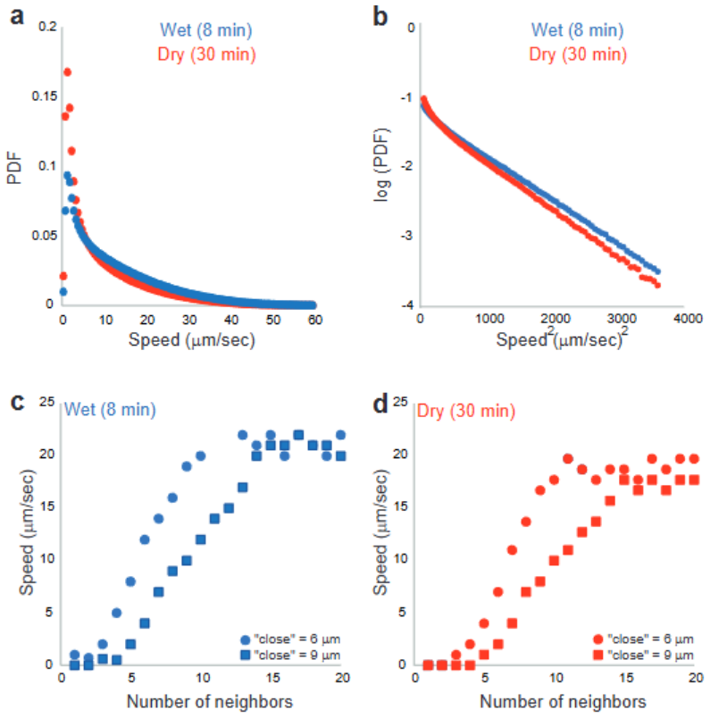
- On drier plates the fraction of temporarily stationary cells roughly doubles, from about 6% to 13% of bin-occupation time, while cells that do move fast enough keep nearly the same speed distribution; dryness acts mainly by stranding cells, not by slowing all motion.
- A cell with fully open flagella is almost never observed moving, and cells with closed or partially open flagella move with different speed statistics, so flagellar state and motility are tightly coupled.
- Liquid can flow around a stalled cell, as beads are dragged, yet the cell does not move; lack of a local film, not absence of ambient flow, is the immediate cause of immobility.
- The transitions among run, tumble, and stall match a memoryless Markov chain with rates roughly 4, 4.76, and 1.05 per second, so single-cell flagellar-state switching can be represented by constant rates in future models.
- Dry models of self-propelled rods that neglect liquid and hydrodynamic effects cannot reproduce the observed speed-density relation and the arrest of isolated cells.
Reading between the lines
- If the open-flagella stall is an active mechanism for extracting liquid from the substrate, then preventing flagellar unbundling should lengthen stalls or impair rehydration; this is a testable extension the paper does not perform.
- The neighbor-dependent speed data suggest a feedback loop: moving cells help maintain the local film, and cells in clusters keep one another wet, which could explain why speed increases with neighbor number; measuring film height around isolated versus clustered cells would test this.
- A minimal quantitative model coupling a thin-film equation for the liquid layer to a three-state Markov chain for flagellar state would use the paper's measured transition rates and predict cluster-edge speeds and the dry-plate stall fraction.
Editorial analysis
A structured set of objections, weighed in public.
Referee Report
Summary. This experimental paper addresses why isolated swarmer cells of Bacillus subtilis become temporarily immobile while cells in clusters move. Using DIC microscopy, the authors infer local liquid depletion around stationary cells; using fluorescent flagella staining, they find that stationary cells have completely un-bundled (open) flagella, while moving cells have bundled or partially open flagella. They also show that drying the agar increases the fraction of stationary cells, that bin-occupancy statistics rule out fixed surface traps, that average speed increases with the number of neighbors, and that flagellar-state transitions are statistically consistent with a continuous-time Markov chain. The paper concludes that immobility is related to local liquid depletion and that dry active-matter models are insufficient for swarming bacteria.
Significance. If the causal interpretation were supported, this would be a valuable contribution: it would link single-cell immobility to local hydration, flagellar arrangement, and collective motion, with implications for active-matter modeling of bacterial swarms. The paper contains several careful analyses: the bin-occupancy statistics convincingly show that the surface is not trapping cells; the Markov-chain analysis is a rigorous descriptive treatment of flagellar-state dynamics; and the MgO-bead experiments provide a clever control showing that liquid flow alone does not move cells. The main weakness is that the title and abstract claim a causal direction that the authors' own Discussion disavows, and the DIC-to-liquid calibration is asserted rather than quantitatively established.
major comments (3)
- [Title, Abstract, Fig. 5c-g, Discussion (last paragraph)] The central claim 'due to local liquid depletion' is not supported by the temporal ordering presented in the authors' own data. In Fig. 5c-g, the cell stops (c-d) before the DIC hue changes that indicate liquid loss (e-g), and Fig. 5h-l shows rewetting before acceleration. The Discussion explicitly states that 'our results cannot conclude on what is the cause and what is the effect.' Thus the title and abstract overstate the causal direction. Please either provide time-resolved evidence that liquid depletion precedes immobilization (e.g., faster DIC acquisition or controlled rewetting experiments) or reformulate the paper's claim as 'associated with' or 'correlated with' local liquid depletion. This is load-bearing because the title, abstract, and final conclusions all assert a causal link.
- [Appendix 1, Fig. 4d, Fig. 5] The inference that stationary cells reside in liquid-depleted regions rests entirely on the assertion in Appendix 1 that DIC hue is a monotonic function of only the local upper-surface slope, independent of material. No quantitative calibration relating hue to liquid film thickness is provided; the silica-bead experiment shows that a flat water layer makes a bead invisible, but it does not establish that the hue changes around cells are due specifically to changes in the liquid layer thickness rather than to focus drift, cell-surface topography, or refractive-index variations. Please provide a calibration (e.g., hue vs. known liquid wedge thickness) and appropriate controls, since the paper's central mechanism depends on this mapping.
- [Fig. 3b and Results (speed distributions)] The classification of cells as 'unaffected' by drying based on a speed threshold of 7 µm/s is applied to the response variable itself, so the conclusion that these cells have similar speed distributions in wet and dry cases is partly built into the analysis. A threshold-free comparison (e.g., a two-population fit of the full speed distribution, or a quantile regression) would more fairly support the claim that drying affects only the stationary and slow-moving population.
minor comments (8)
- [Introduction, second paragraph] The phrase 'isolated swarming cells do not move move' contains a duplicated word; please remove the extra 'move'.
- [Appendix 3] The term 'malowess' appears to be a typo; it should likely be 'lowess' or 'LOESS' (locally weighted scatterplot smoothing).
- [References] Reference 58 is incomplete: 'Purcell.Pdf' is not a proper citation and should be replaced with the full bibliographic details.
- [Appendix 1] The phrase 'adjutant points' is likely a typo for 'adjacent points' in the discussion of the lateral resolution of DIC.
- [Fig. 6d] The caption states 'Error bar equals 10 µm,' which is unclear; please specify what quantity the error bar represents. The inset referenced in the text should also be described in the caption.
- [Abstract and Results (flagellar states)] The terminology for the 'open' flagellar state is inconsistent: the abstract says 'completely spread-out,' the Results say 'completely unbundled,' and the Discussion says 'widely open.' Please unify the terminology.
- [Fig. 6h-j and Markov-chain analysis] The transition rates out of the closed, partial, and open states are quoted as 4, 4.76, and 1.05 s^-1, but the relationship between these rates and the average waiting times (0.25, 0.21, 0.95 s) should be made explicit, and confidence intervals for the rates should be reported.
- [Data availability] The statement 'All data will be available upon request' is weaker than current journal standards; please consider depositing the raw data and analysis code in a public repository.
Circularity Check
No significant circularity: the immobility–liquid-depletion association rests on an independent DIC calibration and external comparisons (Turner et al.), and the paper explicitly disavows causal ordering.
full rationale
The paper makes no prediction from a fitted parameter: the exponential waiting-time fits and the Markov-chain transition matrix are descriptive statistics of measured flagellar-state durations, and the two-step transition matrix check is a validation of Markovianity, not a model output forced to reproduce immobility. The central claim that immobile cells sit in liquid-depleted regions is inferred from a DIC hue-to-slope mapping that is argued and tested in Appendix 1 (including a silica-bead rewetting control), not imported from a self-citation; the citation to Be'er & Lereah (2002) for DIC slope sensitivity is background and the appendix supplies independent physical justification. The flagellar 'run/tumble/stall' vocabulary and the open-flagella-stall association are explicitly compared with Turner et al. (2010), an external dataset, so this is not a renamed known result or a self-citation chain. The manuscript's own Discussion states 'our results cannot conclude on what is the cause and what is the effect,' and Fig. 5c–g shows liquid loss after stopping; these are important limitations on the causal claim in the title, but they are evidentiary/correctness concerns, not circular reductions. No step in the paper reduces to its own input by construction, and no load-bearing premise is justified solely by self-citation. Score 1 reflects the non-load-bearing self-citations in the reference list rather than any circularity.
Assumptions & free parameters
free parameters (3)
- DIC hue classification thresholds =
hue <= 20 black; 21-24 blue; 25-27 green
- Speed threshold for 'unaffected' cells =
7 um/s
- Neighbor counting cutoff distance =
not specified (called 'max distance')
assumptions (3)
- domain assumption Color-DIC hue is a monotonic function of only the local slopes in the upper surface and not of the material through which the light is transmitted.
- domain assumption The flagellar staining procedure (cysteine mutation and Alexa 546 dye) does not alter swarming behavior or flagellar state statistics.
- domain assumption Cells at the colony edge at surface fraction rho=0.3 represent the low-density regime of interest and are in the swarming phenotype.
Cite this review
Pith. "Pith review of Immobility of isolated swarmer cells due to local liquid depletion." pith.science (2026). https://pith.science/paper/X5MKL7MM
@misc{pith2026241117842,
  author       = {Pith},
  title        = {Pith review of: Immobility of isolated swarmer cells due to local liquid depletion},
  year         = {2026},
  howpublished = {\url{https://pith.science/paper/X5MKL7MM}},
  note         = {Machine review of arXiv:2411.17842}
}
read the original abstract
Bacterial swarming is a complex phenomenon in which thousands of self-propelled rod-shaped cells move coherently on surfaces, providing an excellent example of active matter. However, bacterial swarming is different from most studied examples of active systems because single isolated cells do not move, while clusters do. The biophysical aspects underlying this behavior are unclear. In this work we explore the case of low local cell densities, where single cells become temporarily immobile. We show that immobility is related to local depletion of liquid. In addition, it is also associated with the state of the flagella. Specifically, the flagellar bundles at (temporarily) liquid depleted regions are completely spread-out. Our results suggest that dry models of self-propelled agents, which only consider steric alignments and neglect hydrodynamic effects, are oversimplified and are not sufficient to describe swarming bacteria.
Figures


Figures from the paper (3 more)


Reference graph
Works this paper leans on
-
[1]
Harshey, R. M. Bees aren’t the only ones: swarming in Gram‐negative bacteria. Mol. Microbiol. 13, 389–394 (1994)
work page 1994
-
[2]
Kearns, D. B. A field guide to bacterial swarming motility. Nat. Rev. Microbiol. 8, 634–644 (2010)
work page 2010
-
[3]
Harshey, R. M. Bacterial Motility on a Surface: Many Ways to a Common Goal. Annu. Rev. Microbiol. 57, 249–273 (2003)
work page 2003
-
[4]
Damton, N. C., Turner, L., Rojevsky, S. & Berg, H. C. Dynamics of bacterial swarming. Biophys. J. 98, 2082–2090 (2010)
work page 2010
-
[5]
Jeckel, H. et al. Learning the space-time phase diagram of bacterial swarm expansion. Proc. Natl. Acad. Sci. U. S. A. 116, 1489–1494 (2019)
work page 2019
-
[6]
Copeland, M. F. & Weibel, D. B. Bacterial swarming: A model system for studying dynamic self-assembly. Soft Matter 5, 1174–1187 (2009)
work page 2009
-
[7]
Kearns, D. B. & Losick, R. Swarming motility in undomesticated Bacillus subtilis. Mol. Microbiol. 49, 581–590 (2003)
work page 2003
-
[8]
Partridge, J. D. Surveying a Swarm: Experimental Techniques To Establish and Examine Bacterial Collective Motion. Appl. Environ. Microbiol. 88, (2022)
work page 2022
Show all 64 references
-
[9]
& Ariel, G
Be’er, A. & Ariel, G. A statistical physics view of swarming bacteria. Mov. Ecol. 7, 1– 17 (2019)
2019
-
[10]
Anyan, M. E. et al. Type IV pili interactions promote intercellular association and moderate swarming of Pseudomonas aeruginosa. Proc. Natl. Acad. Sci. U. S. A. 111, 18013–18018 (2014)
2014
-
[11]
Hefetz, I. et al. A reversible mutation in a genomic hotspot saves bacterial swarms from extinction. iScience 26, 106043 (2023)
2023
-
[12]
Li, H., Chaté, H., Sano, M., Shi, X. Q. & Zhang, H. P. Robust Edge Flows in Swarming Bacterial Colonies. Phys. Rev. X 14, 1–15 (2024)
2024
-
[13]
& Zafeiris, A
Vicsek, T. & Zafeiris, A. Collective motion. Phys. Rep. 517, 71–140 (2012)
2012
-
[14]
Coupling cell movement to multicellular development in myxobacteria
Kaiser, D. Coupling cell movement to multicellular development in myxobacteria. Nat. 18 Rev. Microbiol. 1, 45–54 (2003)
2003
-
[15]
Bacterial Swarming: A Re-examination of Cell-Movement Patterns
Kaiser, D. Bacterial Swarming: A Re-examination of Cell-Movement Patterns. Curr. Biol. 17, 561–570 (2007)
2007
-
[16]
& Mahadevan, L
Srinivasan, S., Nadir Kaplan, C. & Mahadevan, L. A multiphase theory for spreading microbial swarms and films. Elife 8, 1–28 (2019)
2019
-
[17]
& Be’er, A
Jose, A., Ariel, G. & Be’er, A. Physical characteristics of mixed-species swarming colonies. 064404, 1–8 (2022)
2022
-
[18]
M., Ariel, G
Natan, G., Worlitzer, V. M., Ariel, G. & Be’er, A. Mixed-species bacterial swarms show an interplay of mixing and segregation across scales. Sci. Rep. 12, 1–12 (2022)
2022
-
[19]
D., Ariel, G., Schvartz, O., Harshey, R
Partridge, J. D., Ariel, G., Schvartz, O., Harshey, R. M. & Be’er, A. The 3D architecture of a bacterial swarm has implications for antibiotic tolerance. Sci. Rep. 8, 1–11 (2018)
2018
-
[20]
T., Wang, Q
Butler, M. T., Wang, Q. & Harshey, R. M. Cell density and mobility protect swarming bacteria against antibiotics. Proc. Natl. Acad. Sci. U. S. A. 107, 3776–3781 (2010)
2010
-
[21]
Walker, D. M. Dead cells release a ‘necrosignal’ that activates antibiotic survival pathways in bacterial swarms. Nat. Commun. 1–12 doi:10.1038/s41467-020-17709-0
-
[22]
& Be’er, A
Benisty, S., Ben-Jacob, E., Ariel, G. & Be’er, A. Antibiotic-induced anomalous statistics of collective bacterial swarming. Phys. Rev. Lett. 114, 1–5 (2015)
2015
-
[23]
Be’er, A. et al. Paenibacillus dendritiformis bacterial colony growth depends on surfactant but not on bacterial motion. J. Bacteriol. 191, 5758–5764 (2009)
2009
-
[24]
& Asally, M
Grobas, I., Polin, M. & Asally, M. Swarming bacteria undergo localized dynamic phase transition to form stress-induced biofilms. Elife 10, 1–22 (2021)
2021
-
[25]
& Reynolds, A
Ariel, G., Be’er, A. & Reynolds, A. Chaotic Model for Lévy Walks in Swarming Bacteria. Phys. Rev. Lett. 118, 1–6 (2017)
2017
-
[26]
Ariel, G. et al. Swarming bacteria migrate by Lévy Walk. Nat. Commun. 6, (2015)
2015
-
[27]
Aranson, I. S. Bacterial active matter. Reports Prog. Phys. 85, (2022)
2022
-
[28]
Purcell, E. M. Purcell.Pdf. American Journal of Physics vol. 45 3–11 (1977)
1977
-
[29]
Elgeti, J., Winkler, R. G. & Gompper, G. Physics of microswimmers - Single particle 19 motion and collective behavior: A review. Reports Prog. Phys. 78, (2015)
2015
-
[30]
Wensink, H. H. et al. Meso-scale turbulence in living fluids. Proc. Natl. Acad. Sci. U. S. A. 109, 14308–14313 (2012)
2012
-
[31]
Be’er, A. et al. A phase diagram for bacterial swarming. Commun. Phys. 3, 1–8 (2020)
2020
-
[32]
P., Be’er, A., Florin, E.-L
Zhang, H. P., Be’er, A., Florin, E.-L. & Swinney, H. L. Collective motion and density fluctuations in bacterial colonies. Proc. Natl. Acad. Sci. 107, 13626–13630 (2010)
2010
-
[33]
& Aranson, I
Sokolov, A. & Aranson, I. S. Reduction of viscosity in suspension of swimming bacteria. Phys. Rev. Lett. 103, 2–5 (2009)
2009
-
[34]
& Aranson, I
Sokolov, A. & Aranson, I. S. Physical properties of collective motion in suspensions of bacteria. Phys. Rev. Lett. 109, 1–5 (2012)
2012
-
[35]
M., Gachelin, J., Douarche, C., Auradou, H
López, H. M., Gachelin, J., Douarche, C., Auradou, H. & Clément, E. Turning Bacteria Suspensions into Superfluids. Phys. Rev. Lett. 115, 1–5 (2015)
2015
-
[36]
G., Turner, L
Chen, B. G., Turner, L. & Berg, H. C. The wetting agent required for swarming in Salmonella enterica serovar typhimurium is not a surfactant. J. Bacteriol. 189, 8750– 8753 (2007)
2007
-
[37]
G., Tang, J
Ping, L., Wu, Y., Hosu, B. G., Tang, J. X. & Berg, H. C. Osmotic pressure in a bacterial swarm. Biophys. J. 107, 871–878 (2014)
2014
-
[38]
& Harshey, R
Be’er, A. & Harshey, R. M. Collective motion of surfactant-producing bacteria imparts superdiffusivity to their upper surface. Biophys. J. 101, 1017–1024 (2011)
2011
-
[39]
J., Hsueh, Y
Ke, W. J., Hsueh, Y. H., Cheng, Y. C., Wu, C. C. & Liu, S. T. Water surface tension modulates the swarming mechanics of Bacillus subtilis. Front. Microbiol. 6, 1–12 (2015)
2015
-
[40]
B., Ariel, G
Ilkanaiv, B., Kearns, D. B., Ariel, G. & Beer, A. Effect of Cell Aspect Ratio on Swarming Bacteria. Phys. Rev. Lett. 118, 1–5 (2017)
2017
-
[41]
Peled, S. et al. Heterogeneous bacterial swarms with mixed lengths. Phys. Rev. E 103, 1–7 (2021)
2021
-
[42]
& Be’er, A
Sidortsov, M., Morgenstern, Y. & Be’er, A. Role of tumbling in bacterial swarming. Phys. Rev. E 96, 1–7 (2017). 20
2017
-
[43]
& Harshey, R
Mariconda, S., Wang, Q. & Harshey, R. M. A mechanical role for the chemotaxis system in swarming motility. Mol. Microbiol. 60, 1590–1602 (2006)
2006
-
[44]
Yashunsky, V., Pearce, D. J. G., Ariel, G. & Be’er, A. Topological defects in multi- layered swarming bacteria. Soft Matter 20, 4237–4245 (2024)
2024
-
[45]
Hallatschek, O. et al. Proliferating active matter. doi:10.1038/s42254-023-00593-0
-
[46]
L., Sepúlveda, N
Pérez-Estay, B., Cordero, M. L., Sepúlveda, N. & Soto, R. Accumulation and depletion of E. coli in surfaces mediated by curvature. Phys. Rev. E 109, 1–9 (2024)
2024
-
[47]
& Bär, M
Peruani, F., Deutsch, A. & Bär, M. Nonequilibrium clustering of self-propelled rods. Phys. Rev. E - Stat. Nonlinear, Soft Matter Phys. 74, (2006)
2006
-
[48]
J., Schofield, J., Gaspard, P
Huang, M. J., Schofield, J., Gaspard, P. & Kapral, R. From single particle motion to collective dynamics in Janus motor systems. J. Chem. Phys. 150, (2019)
2019
-
[49]
R., Kyriakopoulos, N., Cenev, Z
Garza, R. R., Kyriakopoulos, N., Cenev, Z. M., Rigoni, C. & Timonen, J. V. I. Magnetic Quincke rollers with tunable single-particle dynamics and collective states. Sci. Adv. 9, 2–9 (2023)
2023
-
[50]
Zheng, X. et al. Non-Gaussian statistics for the motion of self-propelled Janus particles: Experiment versus theory. Phys. Rev. E - Stat. Nonlinear, Soft Matter Phys. 88, 1–11 (2013)
2013
-
[51]
Zheng, E. et al. Self-Oscillation and Synchronization Transitions in Elastoactive Structures. Phys. Rev. Lett. 130, 178202 (2023)
2023
-
[52]
& Peruani, F
Bär, M., Großmann, R., Heidenreich, S. & Peruani, F. Self-Propelled Rods: Insights and Perspectives for Active Matter. Annu. Rev. Condens. Matter Phys. 11, 441–466 (2020)
2020
-
[53]
The mechanics and statistics of active matter
Ramaswamy, S. The mechanics and statistics of active matter. Annu. Rev. Condens. Matter Phys. 1, 323–345 (2010)
2010
-
[54]
Cates, M. E. & Tailleur, J. Motility-induced phase separation. Annu. Rev. Condens. Matter Phys. 6, 219–244 (2015)
2015
-
[55]
Worlitzer, V. M. et al. Biophysics underlying the swarm to biofilm transition. 8152, 1– 9 (2022)
2022
-
[56]
& Ariel, G
Sorkin, B., Be’er, A., Diamant, H. & Ariel, G. Detecting and characterizing phase 21 transitions in active matter using entropy. Soft Matter 19, 5118–5126 (2023)
2023
-
[57]
Mezanges, X. et al. Modeling the role of water in Bacillus subtilis colonies. Phys. Rev. E - Stat. Nonlinear, Soft Matter Phys. 85, 1–9 (2012)
2012
-
[58]
& Lereah, Y
Be’er, A. & Lereah, Y. Time-resolved, three-dimensional quantitative microscopy of a droplet spreading on solid substrates. J. Microsc. 208, 148–152 (2002)
2002
-
[59]
& Berg, H
Zhang, R., Turner, L. & Berg, H. C. The upper surface of an Escherichia coli swarm is stationary. Proc. Natl. Acad. Sci. U. S. A. 107, 288–290 (2010)
2010
-
[60]
& Harshey, R
Wang, Q., Suzuki, A., Mariconda, S., Porwollik, S. & Harshey, R. M. Sensing wetness: A new role for the bacterial flagellum. EMBO J. 24, 2034–2042 (2005)
2005
-
[61]
F., Flickinger, S
Copeland, M. F., Flickinger, S. T., Tuson, H. H. & Weibel, D. B. Studying the dynamics of flagella in multicellular communities of Escherichia coli by using biarsenical dyes. Appl. Environ. Microbiol. 76, 1241–1250 (2010)
2010
-
[62]
D., Nhu, N
Partridge, J. D., Nhu, N. T. Q., Dufour, Y. S. & Harshey, R. M. Tumble suppression is a conserved feature of swarming motility. MBio 11, 1–5 (2020)
2020
-
[63]
Turner, L., Zhang, R., Darnton, N. C. & Berg, H. C. Visualization of flagella during bacterial swarming. J. Bacteriol. 192, 3259–3267 (2010)
2010
-
[64]
enhancement
Li, Y., Zhai, H., Sanchez, S., Kearns, D. B. & Wu, Y. Noncontact Cohesive Swimming of Bacteria in Two-Dimensional Liquid Films. Phys. Rev. Lett. 119, 1–6 (2017). 22 Supplementary Information Movie S1. A real-time, phase-contrast movie of swarming cells near the colony edge, wi...
2017
Reviewed August 12, 2026 · model on record in the stance chip above.
Discussion (0). Continue with ORCID to comment.