REVIEW 2 major objections 5 minor 30 references
High contrast dual-mode optical and 13C magnetic resonance imaging in diamond particles
T0 review · 2 major / 5 minor · reviewed 2026-08-14 · deepseek-v4-flash
Pith's one-line read Diamond particles with NV centers can be imaged by light and by MRI at the same time, from the same green beam.
desk verdict Solid dual-mode imaging proof of concept with a clean background-suppression demo; the Regime III acceleration claims rest on an equal-per-sample-cost assumption that does not survive realistic cost ratios. read the letter →
The pith
A machine-rendered reading of the paper's core claim, the machinery that carries it, and where it could break.
The reading
What carries the argument
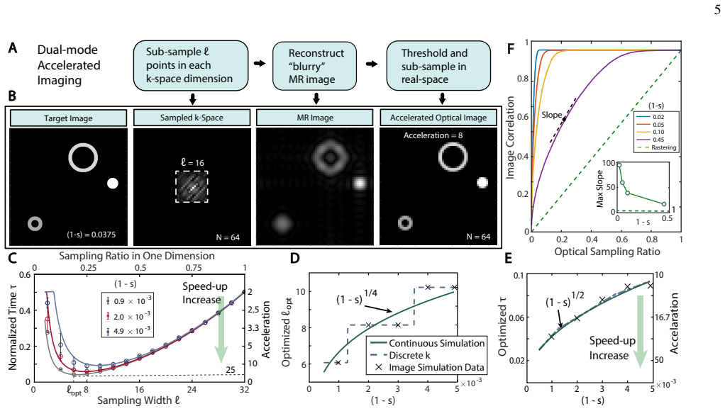
The load-bearing element is NV-mediated optical hyperpolarization of lattice $^{13}$C nuclei: sub-bandgap green light polarizes NV$^-$ electron spins, and microwave sweeps across the NV ESR spectrum drive Landau-Zener transitions that transfer polarization orientation-independently to the $^{13}$C bath, producing ~0.3% $^{13}$C polarization in 40 s at ~38 mT. A second mechanism carries the background-suppression claim: the NV fluorescence depends on the angle between the NV axis and an applied field (simulated by a seven-level kinetic model), so a pulsed field modulates the optical signal; and reversing the microwave sweep reverses the $^{13}$C hyperpolarization sign, providing full-contrast modulation in MRI. The acceleration argument rests on Fourier reciprocity: optics samples real space while MRI samples k-space, so a truncated k-space window gives a sinc-blurred real-space image that, after thresholding, confines the optical raster to a sparse subset of pixels.
What would settle it
Time the actual protocol on the demonstrated hardware: record total wall-clock acquisition for a sparse phantom with one MRI k-space line per ~40 s hyperpolarization cycle plus optical rastering, and compare with pure optical rastering at the same target resolution. If the measured speedup is far below $(1-s)^{-1/2}$, the equal-cost premise is the reason.
Extended reading notes
Core claim
The central claim is that a single material platform - diamond particles hosting NV centers - can be imaged simultaneously in the optical and MR domains, with each mode improving the other. Optically, the particles fluoresce brightly under 520 nm light; the same light polarizes the NV electron spins, and chirped microwave sweeps transfer that polarization to $^{13}$C nuclei, giving a hyperpolarized $^{13}$C MRI signal reported as over three orders of magnitude brighter than conventional MRI at low field (enhancement ~280 over thermal at 7 T, ~206 over 9.4 T). Since the NV fluorescence depends on applied magnetic field and the hyperpolarization sign depends on microwave sweep direction, both image modes can be modulated on demand, enabling lock-in background suppression in optics and difference imaging in MRI. The paper also proposes a third regime: a hybrid acquisition protocol that samples a few low-order k-space points by MRI, thresholds the resulting blurry image, and feeds that information forward to restrict real-space optical scanning to promising pixels; at high sparsity $s$ this yields acceleration scaling as $(1-s)^{-1/2}$, with an optimal k-space sample count scaling as $(1-s)^{1/4}$.
Load-bearing premise
The acceleration calculation assumes every extra sample costs the same time in optics and in MRI, even though one MRI k-space sample can require a fresh hyperpolarization cycle while an optical pixel is read in milliseconds; if that ratio is large, the headline speedup shrinks.
Editorial extensions
If this is right
- Diamond particles can be tracked by optical microscopy and by MRI from the same illumination source, so scattering environments that blind optics can be cross-checked with MRI and vice versa.
- On-demand modulation of both modes enables background-free imaging: lock-in suppression recovers diamond signals under a dye that is twice as bright, and difference imaging cancels a $^{13}$C-methanol background five times stronger.
- In sparse-imaging settings, the hybrid k-space/real-space protocol promises more than an order-of-magnitude acquisition speedup with a matching reduction in delivered optical power.
- Because polarization is replenished continuously at low field and is detection-field agnostic, low-field MRI becomes practical: the agent's brightness does not depend on the detection magnet.
- MRI resolution can be pushed toward optical resolution by rastering a focused beam to hyperpolarize one pixel at a time, trading power density for pixel size.
Reading between the lines
- If the equal per-sample cost assumption is replaced by a realistic ratio where one MRI k-space line requires a fresh ~40 s hyperpolarization cycle, the optimal k-space sample count and the $(1-s)^{-1/2}$ acceleration both shift; the practical speedup will depend on that cost ratio and may be much smaller than plotted.
- The same Fourier-reciprocity trick could be combined with compressed sensing: a random k-space subsample plus convex reconstruction could produce the feed-forward mask, potentially improving the blurry-image quality and pushing the protocol to lower sparsity.
- The 0.3% polarization number was obtained in 200 micrometer particles; the paper itself notes roughly 10^-2 lower hyperpolarizability for <100 nm particles, so a key extension is to verify whether material improvements (annealing, $^{13}$C enrichment) close that gap before clinical-scale agents are practical.
- The background-suppression scheme is generic: any agent whose fluorescence or hyperpolarization can be switched on demand could use the same lock-in/difference logic, so the method may transfer to other optically polarizable spin labels.
Editorial analysis
A structured set of objections, weighed in public.
Referee Report
Summary. The paper reports dual-mode imaging of diamond microparticles using NV-center fluorescence and optically pumped 13C hyperpolarization. The authors demonstrate about 0.3% 13C polarization in 40 s under 1 W green illumination, corresponding to roughly 280-fold enhancement over thermal 13C at 7 T, and use it for FLASH MRI of a ring phantom. They also demonstrate optical and MR background suppression by field modulation and MW-sweep reversal, and they propose a 'Regime III' hybrid protocol that samples low-order k-space with MRI and feeds the thresholded result forward to guide real-space optical rastering. Simulations of this protocol are used to claim acquisition acceleration and optical power reduction scaling as (1-s)^-1/2 at high sparsity s, with 'more than an order of magnitude' and 'as much as two orders of magnitude' speedups in sparse-imaging scenarios.
Significance. The experimental part is a useful proof of concept: the same green illumination both fluoresces and hyperpolarizes, and the demonstrated lock-in and sweep-reversal cancellation give concrete, quantified background suppression. The use of measured rate constants and experimental parameters (e.g., Ref. [20] kinetics, T1 data) in the supporting analysis is a strength. However, the paper's headline quantitative claims for Regime III are not yet supported: the acceleration and power-reduction scalings assume equal per-sample time costs for optics and MRI and omit the fixed 40 s hyperpolarization overhead. Until those claims are re-derived or substantially qualified, the paper's significance is prospective rather than established.
major comments (2)
- [Main text, 'Accelerated conjugate-space imaging'; SI §III.A, Eq. (8)] The central Regime III claim of order-of-magnitude time savings and up to two orders of magnitude acceleration (Fig. 4E, Abstract) rests on the assumption that 'the time cost to be accrued per sample (pixel) is identical for both optical and MR imaging dimensions.' In the demonstrated setup this is not the case: each MR acquisition requires a 40 s hyperpolarization step (main text, Results; Fig. 1B) followed by a FLASH train with TR = 6 ms, while an optical pixel can be read in milliseconds. The MR term in Eq. (8) is l^2/N^2 with no cost ratio; scaling it by eta gives l_opt proportional to eta^{-1/4}(1-s)^{1/4} and tau_opt proportional to eta^{1/2}(1-s)^{1/2}. For eta values of order 10^3-10^4, representative of the 40 s hyperpolarization overhead, the advertised savings vanish and the protocol can be slower than full rastering. The paper notes that results can be scaled by eta but never computes the regime in which the headline numbers survive; this is load-bearing for the abstract and Table I.
- [Table I, Regime III row; main text, 'Finally, we comment that imaging acceleration results in a lower total optical…] The claim of power reduction by the same factor as the acceleration omits the optical power spent on hyperpolarization. The experimental hyperpolarization consumes 1 W for 40 s (Fig. 1B), a fixed energy cost per MR acquisition. At high sparsity, where the raster energy is reduced by the acceleration factor, this fixed cost can dominate; the power-reduction factors of 14-25 quoted in the text and Table I are therefore not the total optical power budget. A full energy accounting including the hyperpolarization laser, or a clear statement that the quoted reduction applies only to the raster component, is needed.
minor comments (5)
- [Abstract and main text (Fig. 1C)] The abstract states the particles are 'over three-orders of magnitude brighter than in conventional MRI,' but the measured enhancement is 280-fold over thermal 13C at 7 T; specify the reference field or revise the wording.
- [Fig. 4B and surrounding text] The text quotes '16-fold' and '~14 times' acceleration, while the figure panel says 'Acceleration = 8'; clarify what is being counted (reduction in real-space samples vs total acquisition time).
- [SI §III.A and Fig. S2] The analytic model uses r0 = 0.75, selected by thresholding the sinc shoulder at 0.3; a sensitivity analysis or a principled selection rule for r0 would strengthen the scaling results.
- [Fig. 2D and definition of Delta] For full sign reversal the denominator I+I in the modulation contrast is zero; state how the 194% contrast is computed, e.g., using absolute values or a noise floor.
- [References] Several references are duplicated or repeated (e.g., Refs. [8] and [64], [13] and [67]); consolidate to avoid confusion.
Circularity Check
No circular reduction found: experimental dual-mode results are measured against external benchmarks, and the Regime III acceleration analysis is an explicitly conditional analytic model rather than a fitted prediction.
full rationale
The paper's load-bearing experimental assertions are direct measurements: approximately 0.3% 13C polarization under 1 W illumination in 40 s (Fig. 1B-C), 10% optical and 194% MR modulation contrast (Fig. 2), and background suppression by factors of 2 and 5 (Fig. 3). These do not reduce to definitions or to fitted parameters. The NV fluorescence simulation uses the externally measured 7-level rate constants of Ref. [20], and the hyperpolarization mechanism is supported by the independently published, experimentally grounded work in Refs. [8,11]; self-citation here is enabling background, not an unverified uniqueness claim. The Regime III acceleration scalings (l_opt proportional to (1-s)^(1/4), tau proportional to (1-s)^(1/2)) are derived in SI Eq. (8) from a sinc-blur model, with r0 = 0.75 set by the stated 0.3 threshold rather than fitted to simulation outputs; the agreement with the paper's own simulations is a consistency check, not a fit renamed as prediction. The paper explicitly flags the equal per-sample time-cost assumption and says results can be scaled by an appropriate cost ratio eta. That caveat is a modeling limitation affecting quantitative applicability of the acceleration numbers, but it is not a circular reduction of the conclusion to its inputs. No self-definitional, fitted-input, or imported-uniqueness step was found.
Assumptions & free parameters
free parameters (3)
- r0 (sinc-shoulder threshold radius) =
0.75
- optical sampling threshold =
0.1 times mean pixel value
- gamma_0 (optical/MR SNR ratio at depth 0) =
15
assumptions (6)
- standard math k-space truncation to an l x l window produces real-space convolution with a sinc kernel (Fourier convolution theorem)
- domain assumption A seven-level NV rate model with transition rates from Ref [20] describes powder-averaged fluorescence response to magnetic field
- domain assumption Randomly oriented particle ensemble samples all NV axis angles uniformly
- domain assumption Scattering media are homogeneous with Gaussian angular spreading, so a Wigner-function slice model gives resolution that degrades linearly with depth
- domain assumption Per-pixel imaging time cost is identical for optical and MR sampling
- domain assumption 13C hyperpolarization is detection-field agnostic and T1 is long enough for a roughly one-second shuttle to 9.4T
Cite this review
Pith. "Pith review of High contrast dual-mode optical and 13C magnetic resonance imaging in diamond particles." pith.science (2026). https://pith.science/paper/YHE6VCYX
@misc{pith2026190908064,
  author       = {Pith},
  title        = {Pith review of: High contrast dual-mode optical and 13C magnetic resonance imaging in diamond particles},
  year         = {2026},
  howpublished = {\url{https://pith.science/paper/YHE6VCYX}},
  note         = {Machine review of arXiv:1909.08064}
}
read the original abstract
Multichannel imaging -- the ability to acquire images of an object through more than one imaging mode simultaneously -- has opened interesting new perspectives in areas ranging from astronomy to medicine. Visible optics and magnetic resonance imaging (MRI) offer complementary advantages of resolution, speed and depth of penetration, and as such would be attractive in combination. In this paper, we take first steps towards marrying together optical and MR imaging in a class of biocompatible particulate materials constructed out of diamond. The particles are endowed with a high density of quantum defects (Nitrogen Vacancy centers) that under optical excitation fluoresce brightly in the visible, but also concurrently electron spin polarize. This allows the hyperpolarization of lattice 13C nuclei to make the particles over three-orders of magnitude brighter than in conventional MRI. Dual-mode optical and MR imaging permits immediate access to improvements in resolution and signal-to-noise especially in scattering environments. We highlight additional benefits in background-free imaging, demonstrating lock-in suppression by factors of 2 and 5 in optical and MR domains respectively. Ultimate limits could approach as much as two orders of magnitude in each domain. Finally, leveraging the ability of optical and MR imaging to simultaneously probe Fourier-reciprocal domains (real and k-space), we elucidate the ability to employ hybrid sub-sampling in both conjugate spaces to vastly accelerate dual-image acquisition, by as much as two orders of magnitude in practically relevant sparse-imaging scenarios. This is accompanied by a reduction in optical power by the same factor. Our work suggests interesting possibilities for the simultaneous optical and low-field MR imaging of targeted diamond nanoparticles.
Figures


Figures from the paper (1 more)
Reference graph
Works this paper leans on
-
[20]
Similarities Apart from the differences described above, CS and dual- mode accelerated protocol share some similarities in the follow- ing ways. Subsampling – Both methods exploit subsampling to avoid scanning across the entire imaging space and therefore gain time saving. CS MRI subsamples k-space while our protocol subsam- ples both Fourier conjugate sp...
-
[1]
Compressed sensing 14
-
[2]
Accelerated dual-mode protocol 14
-
[3]
Similarities 15 IV . Imaging efficiency of two modalities 15 A. Imaging efficiency of optics 15 B. Imaging efficiency of MRI 16 C. Estimation of imaging SNR in scattering media 16 D. Estimation of imaging resolution in scattering media 16 V . Materials 17 A. Diamond particles 17 B. T1 relaxation in diamond 17 C. Hyperpolarization in 13C enriched diamond 18 D...
-
[4]
Red fluorescence loss. Optical imaging incurs “round-trip” losses due to the need to also collect NV fluorescence. Similar to Step 1 above, one can estimate these attenuation and scattering losses. We take the attenuation coefficient at 650nm as∼1 cm−1, and the scattering coefficient as ∼ 12.1cm−1 (calculated based on fatty tissue data in [33]). For diamonds ...
-
[5]
Geometric effects in light collection. The finite numerical aperture (NA) of the detection optics restricts the total number of photons that can be collected. Only fluorescence light within a solid angle Ω can be captured by the objective lens. We have: Ω = 2π(1− cos(ϑmax)), (13) whereϑmax = arcsin(NA/nd). Assuming that the fluorescence emission is initially...
-
[6]
Signal amplitude reduction by lock-in detection. Finally, we consider the amplitude loss caused by lock-in detection in practi- cal scenarios. Lock-in techniques are normally used when optical background presents, and the signal of interest can be discerned by the method. However, such benefit of background suppression 16 comes at the cost of reduced overa...
-
[18]
CS is the origin of a major evolution in signal processing [36, 37]
Compressed sensing Let us first briefly review basics of CS, in order to better com- pare it with our protocol. CS is the origin of a major evolution in signal processing [36, 37]. It is widely applied in photography, medical imaging, as well as astronomy, because of its ability to recover the signal from very few linear measurements. Operating regime – CS,...
Show all 30 references
-
[19]
generated
Accelerated dual-mode protocol The accelerated dual-mode protocol proposed in this work takes sub-sampled k-space data by MR as a reference to guide real-space measurement by optics. This protocol warrants acqui- sition time saving concurrently with optical power reduction. Op...
-
[21]
Scattering and attenuation lead to loss of photon flux
Green photon scattering and loss at media. Scattering and attenuation lead to loss of photon flux. The attenuation loss can be evaluated by Beer’s law:φp φm = exp{ ∫d0 0 −[α(d) +µ(d)]· dd}, whereφp is the photon flux of the incident pump beam, andφm is the part that reaches diam...
-
[22]
In fact, during optical il- lumination, only a small portion of the incident green photons finally convert to red photons by NV centers
Green-to-red photon conversion. In fact, during optical il- lumination, only a small portion of the incident green photons finally convert to red photons by NV centers. We now estimate this conversion rate ηo,2 based on Ref [38] . A waveguide is employed on a 0.1 ppm NV diamond...
-
[23]
NV centers emit in red, and a portion of the emission light can be restricted within the diamond because of total internal reflection (TIR)
Total internal reflection loss. NV centers emit in red, and a portion of the emission light can be restricted within the diamond because of total internal reflection (TIR). The critical angle of TIR for a diamond-media interface is ϑc = arcsin (nm/nd) = 24.6◦. We calculate ηo,3 ...
-
[27]
Green laser loss at media. Similar to the optical imaging case, since the hyperpolarization is optically induced, we con- tinue to incur one-way optical scattering and attenuation losses through the imaging media, corresponding to a factor ηm,1 = ηo,1 = 9.6× 10⁻³ as above
-
[28]
Let us now estimate efficiency of the 13C hyperpolarization process
Quantum efficiency of 13C hyperpolarization. Let us now estimate efficiency of the 13C hyperpolarization process. In Fig- ure 1C, the hyperpolarization signal is obtained by 1W laser irra- diation with a duration of 40s; as a result, the 10mg natural abun- dance diamond sample i...
-
[29]
The sample filling factor in MR imaging plays a similar role as the geometric fac- tors related to finite numerical aperture in optical imaging
Geometric factors related to detection. The sample filling factor in MR imaging plays a similar role as the geometric fac- tors related to finite numerical aperture in optical imaging. In our experiments, the volume of the coil is approximately 2400 mm3, and the typical volume o...
-
[30]
object region
Detection frequency. We note that the hyperpolarization process delivers 13C polarization that is agnostic to detection field. However, the choice of detection field does play a role in determining the final obtained SNR, scaling as ∝ (Qω0)1/2 [27], whereQ is the quality factor o...
-
[58]
Zhang, R
X.-Q. Zhang, R. Lam, X. Xu, E. K. Chow, H.-J. Kim, and D. Ho, Advanced materials 23, 4770 (2011)
2011
-
[59]
G. Xi, E. Robinson, B. Mania-Farnell, E. F. Vanin, K.-W. Shim, T. Takao, E. V . Allender, C. S. Mayanil, M. B. Soares, D. Ho,et al., Nanomedicine: Nanotechnology, Biology and Medicine 10, 381 (2014)
2014
-
[60]
E. K. Chow, X.-Q. Zhang, M. Chen, R. Lam, E. Robinson, H. Huang, D. Schaffer, E. Osawa, A. Goga, and D. Ho, Science translational medicine 3, 73ra21 (2011)
2011
-
[61]
Ho, Clinical Trials (2020)
D. Ho, Clinical Trials (2020)
2020
-
[62]
P. W. Goodwill and S. M. Conolly, IEEE transactions on medical imaging 29, 1851 (2010)
2010
-
[63]
Zurbuchen, F
U. Zurbuchen, F. Poch, O. Gemeinhardt, M. E. Kreis, S. M. Niehues, J. L. Vahldieck, and K. S. Lehmann, Acta Radiologica 58, 164 (2017)
2017
-
[64]
A. Ajoy, K. Liu, R. Nazaryan, X. Lv, P. R. Zangara, B. Safvati, G. Wang, D. Arnold, G. Li, A. Lin, et al., Science Advances 4, eaar5492 (2018)
2018
-
[65]
M. L. Denton, M. S. Foltz, L. E. Estlack, D. J. Stolarski, G. D. Noojin, R. J. Thomas, D. Eikum, and B. A. Rockwell, Investigative ophthalmology & visual science 47, 3065 (2006)
2006
-
[66]
Takegoshi and C
K. Takegoshi and C. McDowell, Chemical physics letters 116, 100 (1985)
1985
-
[67]
M. A. Frey, M. Michaud, J. N. VanHouten, K. L. Insogna, J. A. Madri, and S. E. Barrett, Proceedings of the National Academy of Sciences 109, 5190 (2012)
2012
-
[68]
E. Y . Chang, J. Du, and C. B. Chung, Journal of magnetic reso- nance imaging 41, 870 (2015)
2015
-
[69]
Thorlabs, Apd410x operation manual, Tech. Rep. (2018)
2018
-
[70]
Lightning-link rapid alexa fluor 647 conjugate data sheet , Tech. Rep. (2018)
2018
-
[71]
Dual mode
A. S. Merbach, L. Helm, and E. Toth, The chemistry of con- trast agents in medical magnetic resonance imaging (John Wiley & Sons, 2013). 9 Supplementary Information Background-free dual-mode optical and 13C magnetic resonance imaging in diamond particles X. Lv,1 J. H. Walton,2...
2013
Reviewed August 14, 2026 · model on record in the stance chip above.
Discussion (0). Continue with ORCID to comment.