REVIEW 3 major objections 6 minor 58 references
Chemotaxis-Driven Instabilities Govern Size, Shape and Migration Efficiency of Multicellular Clusters
T0 review · 3 major / 6 minor · reviewed 2026-08-07 · deepseek-v4-flash
Pith's one-line read Chemotaxing cell clusters above a threshold gradient elongate perpendicular to it and fragment; the breakup caps cluster size and is where forward migration is fastest.
desk verdict A plausible new instability mechanism for chemotactic cluster breakup, but the quantitative match to experiments rests on an uncalibrated gradient mapping that a referee should pin down. read the letter →
The pith
A machine-rendered reading of the paper's core claim, the machinery that carries it, and where it could break.
The reading
What carries the argument
The load-bearing object is the open-arc local gradient vector $\vec{g}_i$ (Eq. 5): each cell senses the chemoattractant only through the stretches of its boundary not covered by neighbors, so a flank cell that “peeks” around the cell ahead of it suddenly gains a strong directional signal. That local gradient enters the propulsion direction (Eq. 3) together with persistence and neighbor alignment, while contact inhibition of locomotion (Eq. 2) makes rim cells ($p_{\text{rim}}=8$) far more forceful than core cells ($p_{\text{core}}=1.5$). Comparing the magnitudes of the alignment term $\alpha\hat{V}$ and the gradient term $\beta\vec{g}_i\chi(y_i)$ yields two analytical thresholds, $g_{\text{fb}}\approx0.002$ (flocking to breaking) and $g_{\text{bs}}\approx0.013$ (breaking to solid), and the shape–velocity feedback loop between these thresholds is what produces perpendicular elongation and fragmentation. Cluster geometry is tracked through the gyration-tensor aspect ratio $\sqrt{\lambda_1/\lambda_2}$ and orientation angle $\theta$, and internal fluidity through the weighted mixing entropy $G$.
What would settle it
Measure breakup probability, aspect ratio, and orientation angle for JVM3 lymphocyte clusters over a finely stepped series of CCL19 gradients (roughly 10–200 ng/ml/mm) and record the results by cluster size. The paper predicts that breakup probability and perpendicular elongation rise with both gradient and cluster size and that forward speed peaks in the breaking regime; observing breakup that is independent of cluster size, or elongation aligned with the gradient, would falsify the mechanism. In a cell type lacking chemorepulsion, a rigid rotating solid state should appear at very high gradients; if no gradient produces it, the predicted $g_{\text{bs}}$ transition is wrong.
Extended reading notes
Core claim
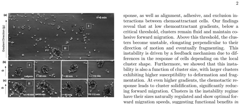
The central claim is that a migrating chemotactic cluster is intrinsically prone to a shape-driven instability: at low gradients the cluster stays fluid and moves cohesively, but above a threshold the response to the gradient generates velocity differentials that depend on local cluster shape, driving elongation perpendicular to the gradient and eventual fragmentation. The mechanism, as the paper states it, is that rim cells propel faster than core cells (contact inhibition of locomotion); front cells persist up the gradient, flank cells slip forward into the open space beside the leading edge and flatten it, and the high-curvature side cells then experience stronger local gradient exposure and pull outward, stretching the cluster into wing-like shapes until cohesion fails. Using the weighted mixing entropy the authors sort the dynamics into a fluid regime (low gradient, fully mixed, cohesive), a breaking regime (intermediate gradient, partial mixing, fragmentation), and a solid regime (high gradient, rigid and rotating, translation suppressed); they note the solid phase was not observed experimentally and may be masked by chemorepulsion at high concentrations. They further claim that breakup probability rises with gradient strength and cluster size, that elongated clusters align nearly perpendicular to the direction of motion, and that velocity in the gradient direction is highest in the breaking regime—read as a natural size limit and an efficiency benefit of the instability.
Load-bearing premise
The quantitative agreement with experiment rests on assuming that the simulation's dimensionless gradient parameter $g_r$ maps monotonically onto the experimental CCL19 gradient in ng/ml/mm, with $g_r=0.006$ standing for the 0–100 ng/ml/mm condition; the paper does not calibrate this mapping independently, so if the mapping is wrong the reported match for breakup probability, aspect ratio, and orientation is not established.
Editorial extensions
If this is right
- Above the threshold $g_{\text{fb}}$, clusters elongate perpendicular to the gradient and fragment, so chemotaxis does not guarantee cohesion: steep gradients actively break clusters apart.
- Breakup probability rises with both gradient strength and cluster size, so the instability sets an upper bound on the size of a cluster that can migrate cohesively.
- Clusters in the breaking regime show the highest velocity along the gradient direction, so fragmentation coincides with—and may enable—optimal forward migration and dispersal.
- For gradients above $g_{\text{bs}}$ the model predicts a solid, rotating, non-mixing cluster with suppressed translation; the paper flags that this phase was not seen experimentally and may be hidden by chemorepulsion at high CCL19 concentrations.
- Because the model is built from generic ingredients (active propulsion, contact inhibition of locomotion, adhesion, alignment, gradient sensing), the same instability is expected in other living or synthetic active clusters, not just lymphocytes.
Reading between the lines
- The paper's own mechanism implies a tuning relation it does not test: raising cohesion (for example, the spring constant $k_s$ or the Lennard-Jones depth $\epsilon$) should raise the critical breakup gradient, so measuring that shift would let experimenters control the instability directly.
- The breakup can be read as the chemotactic counterpart of motility-induced phase separation run in reverse: the gradient feedback arrests coarsening and should set a steady cluster-size distribution, and measuring how that distribution's width varies with gradient in vitro would test this reading.
- The optimal-speed claim suggests cells could tune receptor density or chemotactic sensitivity (the parameter $\beta$) to sit at the edge of the breaking regime; comparing CCR7 expression across the cell population with breakup frequency would connect the mechanism to receptor-level biology.
- The predicted solid regime should be visible in cells that do not show high-concentration chemorepulsion, which the paper names as the likely reason the phase was missed; a systematic gradient scan in such a system is the direct way to confirm or refute it.
Editorial analysis
A structured set of objections, weighed in public.
Referee Report
Summary. The paper develops a two-dimensional agent-based model of chemotactic cell clusters, extending an earlier model (Copenhagen et al., 2018) with contact-inhibition-of-locomotion propulsion, velocity alignment, adhesion, and a Langmuir-type chemotactic response. It identifies three regimes in gradient strength—fluid, breaking, and solid—using a mixing-entropy measure (Fig. 3), and describes a feedback instability in the intermediate regime that elongates clusters perpendicular to the gradient and causes fragmentation (Figs. 5-7). The paper claims quantitative agreement with in vitro CCL19 cluster data from a previous publication [20] for breakup probability versus size, aspect-ratio/orientation behavior, and the size dependence of the forward migration index, and argues that the breaking regime confers optimal migration efficiency and size control.
Significance. If the central claims hold, the paper contributes a generic instability mechanism for chemotactic clusters: shape-dependent velocity differentials produce lateral elongation, breakup, and a size-selection effect, with potential functional benefits in a defined gradient range. The model is not fitted to the new experimental data; its parameters are inherited from an earlier validated model [19], which reduces overfitting risk. The qualitative prediction that elongated clusters orient orthogonal to the gradient is striking and testable, and the qualitative trend that larger clusters break more readily matches experiment. The comparison with the prior experimental videos [20] is a useful re-analysis. However, the quantitative validation is not yet established because the simulation gradient is not calibrated to experimental chemokine units, and the regime boundaries depend on an unexamined 10% entropy-threshold choice. The solid phase is admittedly not observed experimentally, so only the fluid/breaking portion of the phase diagram is empirically supported.
major comments (3)
- [§III.E, §III.F, Eq. (7), Appendix A] The central quantitative comparisons in Fig. 5(e) and Fig. 7(h) identify the simulation gradient g_r=0.006 with the experimental 0–100 ng/ml/mm CCL19 condition, but this mapping is never derived or calibrated. In Eq. (7), g_r is a dimensionless slope whose scale is set by y0=250 and c0=40, while Appendix A converts simulation time to minutes using cell speed and size; neither provides a relation between g_r and ng/ml/mm. If g_r=0.006 does not correspond to the stated experimental gradient, the claimed agreement for breakup probability, aspect-ratio/orientation, and the FMI-size trend is unsupported. Please provide an independent calibration of g_r against chemokine concentration or a sensitivity analysis over plausible g_r values showing that the conclusions are robust.
- [§III.A, Fig. 3(c,d)] The boundaries between the fluid, breaking, and solid regimes are set by defining critical transitions as 10% of the maximum saturated mixing entropy and 10% of the minimum mixing entropy, with an ad hoc baseline δ. These entropy-derived boundaries are then used throughout (Figs. 3(d,i,j), 5(d), 7(f)) and also motivate the choice of g_r=0.006 as the representative breaking-regime point. Because the paper's phase-transition claim and the selection of the comparison gradient depend on this 10% threshold, the robustness of the regimes to the threshold fraction (e.g., 5%, 20%) and to the δ baseline should be tested, and the relationship between these entropy contours and the analytic thresholds of Appendix D (g_fb≈0.002, g_bs≈0.013) should be reconciled.
- [§III.C, Fig. SI2] The claim that the FMI–size trend in the breaking regime at g_r=0.006 aligns closely with the experimentally observed size dependence is supported only by a citation to Supplementary Figure SI2(Dii) of reference [20]; the experimental curve is not reproduced and no quantitative measure of agreement (slope, correlation, residuals) is provided. Since this agreement is used to support the functional-benefit and optimal-migration interpretation, the experimental FMI–size data should be shown and the comparison quantified.
minor comments (6)
- [Abstract and §III.E] The abstract states that above a threshold gradient clusters display an instability and break apart, but the model predicts solidification at high gradients, not breakup; the statement should be qualified to the intermediate gradient range.
- [Appendix D] The numerical evaluation of g_bs is inconsistent with the stated α=6: the formula 40α/(20000−250α) gives 240/18500≈0.0130, not 280/18250. The rounded conclusion is unaffected, but the arithmetic should be corrected.
- [Eqs. (2) and (11)] The symbol p_i denotes both the propulsion speed in Eq. (2) and the bin probability p_i(j) in Eq. (11); please rename one to avoid ambiguity.
- [Fig. 5(e)] The simulation quantity called 'average breakup probability' is not defined; please specify how clusters are tracked, how a breakup event is identified, and how the small/medium/large size bins are chosen.
- [Materials and Methods] The experimental section reports the original protocol of reference [20] but does not state how many clusters were analyzed in the present comparison or provide error bars; please report these details.
- [Appendix D] The analytic threshold derivation assumes |g_i|≈1 and neglects the LJ and spring forces; since g_i is a model output that depends on local geometry, this assumption should be justified or its sensitivity checked.
Circularity Check
No significant circularity: model and parameters are inherited from prior published work, and the quantitative comparisons are tests rather than reductions, though the g_r-to-experimental-gradient mapping is an uncalibrated assumption.
full rationale
The derivation chain is not circular. The agent-based model (Eqs. 1-10) and all force parameters (alpha=6, epsilon=18, p_core=1.5, p_rim=8, beta=80, c0=40) are taken from the authors' prior published model [19], which was validated against independent running/rotating phase observations; this is inherited external support, not a premise defined by the present target results. The experimental data analyzed are from [20], a different prior study, and no experimental data from [20] are used to fit the model parameters in this paper. The comparisons in Figs. 5(e) and 7(h) use a chosen dimensionless gradient g_r=0.006 to represent the 0-100 ng/ml/mm CCL19 condition; this mapping is assumed rather than derived, which weakens the quantitative validation, but it is a calibration/correctness limitation, not a reduction of the prediction to the input: the model could have failed the aspect-ratio, orientation, and breakup-size trends even after choosing g_r=0.006. The analytic thresholds in Appendix D are consistency estimates computed from the same model parameters, not independent first-principles constraints, so they add no circularity. The solid-phase prediction is explicitly not observed and is presented as a theoretical possibility, so no result is made true by definition. No self-citation chain forces the central claim.
Assumptions & free parameters
free parameters (9)
- Propulsion speed differential (p_rim, p_core) =
p_rim=8, p_core=1.5
- Chemotactic response strength beta =
80
- Alignment strength alpha =
6
- Langmuir half-saturation c0 =
40
- Background concentration offset y0 =
250
- Simulation-to-experiment gradient mapping =
g_r=0.006 chosen to represent 0-100 ng/ml/mm; no scaling formula given
- Regime threshold fraction =
10% of max/min mixing entropy
- Mixing entropy baseline delta =
0.5
- Time conversion factor =
1 simulation step = 30 s
assumptions (7)
- domain assumption Cells are represented as overdamped self-propelled Brownian particles with instantaneous force balance (Eq. 1).
- domain assumption Contact inhibition of locomotion makes propulsion depend linearly on neighbor count, with rim cells faster than core cells (Eq. 2).
- domain assumption Each cell senses the chemical gradient through the open arcs between neighboring cells (Eq. 5, Fig. 2c).
- domain assumption Chemokine receptor occupancy follows single-site Langmuir adsorption (Eq. 6).
- domain assumption Lennard-Jones pair forces plus second-neighbor springs maintain cluster cohesion (Eqs. 8-9).
- domain assumption Experimental CCL19 gradients are linear and stable over the assay.
- domain assumption Clusters are defined as groups containing more than three particles (Section III E).
Cite this review
Pith. "Pith review of Chemotaxis-Driven Instabilities Govern Size, Shape and Migration Efficiency of Multicellular Clusters." pith.science (2026). https://pith.science/paper/ZLEUXUNZ
@misc{pith2026250600310,
  author       = {Pith},
  title        = {Pith review of: Chemotaxis-Driven Instabilities Govern Size, Shape and Migration Efficiency of Multicellular Clusters},
  year         = {2026},
  howpublished = {\url{https://pith.science/paper/ZLEUXUNZ}},
  note         = {Machine review of arXiv:2506.00310}
}
read the original abstract
The collective chemotaxis of multicellular clusters is an important phenomenon in various physiological contexts, ranging from embryonic development to cancer metastasis. Such clusters often display interesting shape dynamics and instabilities, but their physical origin, functional benefits, and role in overall chemotactic migration remain unclear. Here, we combine computational modeling and experimental observations of malignant lymphocyte cluster migration in vitro to understand how these dynamics arise from an interplay of chemotactic response and inter-cellular interactions. Our cell-based computational model incorporates active propulsion of cells, contact inhibition of locomotion, chemoattractant response, as well as alignment, adhesive, and exclusion interactions between cells. We find that clusters remain fluid and maintain cohesive forward migration in low chemoattractant gradients. However, above a threshold gradient, clusters display an instability driven by local cluster-shape dependent velocity differentials that causes them to elongate perpendicular to the gradient and eventually break apart. Comparison with our in vitro data shows the predicted transition to the cluster instability regime with increased gradient, as well as quantitative agreement with key features such as cluster aspect ratio, orientation, and breaking frequency. This instability naturally limits the size of multicellular aggregates, and, in addition, clusters in the instability regime display optimal forward migration speeds, suggesting functional implications in vivo. Our work provides valuable insights into generic instabilities of chemotactic clusters, elucidates physical factors that could contribute to metastatic spreading, and can be extended to other living or synthetic systems of active clusters.
Figures


Figures from the paper (4 more)


Reference graph
Works this paper leans on
-
[20]
A. Be’er and G. Ariel, A statistical physics view of swarming bacteria, Movement Ecology7, 9 (2019)
work page 2019
-
[19]
C. K. Hemelrijk and H. Hildenbrandt, Schools of fish and flocks of birds: Their shape and internal structure by self- organization, Interface Focus3, 20120067 (2012)
work page 2012
-
[1]
Entropy of the Completely Unmixed Case When the three color particle layers are completely unmixed, the particles remain confined to their specific layers. In this scenario, the probabilityp i for each parti- cle to be in its designated layer is 1, and the probability of being in any other layer is 0. Using the entropy formula: S=−k B X i pi ln(pi) (C1) S...
-
[2]
Entropy of the Completely Mixed Case When the three color particle layers arecompletely mixed, the particles are spread out evenly across all three layers. In this scenario, the probabilityp i for any particle to be in a particular layer is equal for all layers. Since there are three layers, each particle has ap i = 1 3 chance of being in any given layer....
-
[3]
High Gradient Regime(g r > gbs) As we lower the value ofg r from very high values, a first critical point occurs when the alignment termα ˆV and the gradient term balanceβ⃗ giχ(yi), i.e., when: α ˆV≈β⃗ giχ(yi). Using the values quoted above and solving for the crit- ical gradientg bs, we obtain: 80 250·g bs 250·g bs + 40 =α, gbs = 40(α) 20000−250(α) = 280...
-
[4]
Again, solving for the critical gradientg f b, we find: gf b= 40 19750 ≈0.002
Low Gradient, Fluid and Breaking Regime (gr < gbs) As we reduce the gradient below this breaking-solid threshold,g r < gbs, the next critical point occurs when the self-persistence term and the gradient term balance: ˆvi(t−∆t)≈β⃗ giχ(yi). Again, solving for the critical gradientg f b, we find: gf b= 40 19750 ≈0.002. Thus, we identify three regions based o...
-
[5]
Vicsek and A
T. Vicsek and A. Zafeiris, Collective motion, Physics re- ports517, 71 (2012)
2012
-
[6]
A. Shellard and R. Mayor, Rules of collective migra- tion: from the wildebeest to the neural crest, Philosoph- ical Transactions of the Royal Society B375, 20190387 (2020)
work page 2020
Show all 58 references
-
[7]
McMillen and M
P. McMillen and M. Levin, Collective intelligence: A uni- fying concept for integrating biology across scales and substrates, Communications Biology7, 378 (2024)
2024
-
[8]
Gompper, H
G. Gompper, H. A. Stone, C. Kurzthaler, D. Saintillan, F. Peruani, D. A. Fedosov, T. Auth, C. Cottin-Bizonne, C. Ybert, E. Cl´ ement, T. Darnige, A. Lindner, R. E. Goldstein, B. Liebchen, J. Binysh, A. Souslov, L. Isa, R. di Leonardo, G. Frangipane, H. Gu, B. J. Nelson, F. Bra...
2025
-
[9]
Dreyer, A
T. Dreyer, A. Haluts, A. Korman, N. Gov, E. Fonio, and O. Feinerman, Comparing cooperative geometric puzzle solving in ants versus humans, Proceedings of the Na- tional Academy of Sciences122, e2414274121 (2025)
2025
-
[10]
Feinerman, I
O. Feinerman, I. Pinkoviezky, A. Gelblum, E. Fonio, and N. S. Gov, The physics of cooperative transport in groups of ants, Nature Physics14, 683 (2018)
2018
-
[11]
Elgeti, R
J. Elgeti, R. G. Winkler, and G. Gompper, Physics of mi- croswimmers—single particle motion and collective be- havior: a review, Reports on Progress in Physics78, 056601 (2015)
2015
-
[12]
Tunstrøm, Y
K. Tunstrøm, Y. Katz, C. C. Ioannou, C. Huepe, M. J. Lutz, and I. D. Couzin, Collective states, multistability and transitional behavior in schooling fish, PLoS compu- tational biology9, e1002915 (2013)
2013
-
[13]
Gopinathan and N
A. Gopinathan and N. S. Gov, Cell cluster migration: Connecting experiments with physical models, Seminars in Cell and Developmental Biology93, 77 (2019)
2019
-
[14]
W. Shi, S. Gupta, C. Copos, and A. Mogilner, Collective mechanics of small migrating cell groups, Seminars in Cell & Developmental Biology166, 1 (2025). 18
2025
-
[15]
Bhattacharjee, D
T. Bhattacharjee, D. B. Amchin, R. Alert, J. A. Ott, and S. S. Datta, Chemotactic smoothing of collective migra- tion, eLife11, e71226 (2022)
2022
-
[16]
Hakim and P
V. Hakim and P. Silberzan, Collective cell migration: a physics perspective, Reports on Progress in Physics80, 076601 (2017)
2017
-
[17]
Lopez, J
U. Lopez, J. Gautrais, I. D. Couzin, and G. Theraulaz, From behavioural analyses to models of collective motion in fish schools, Interface focus2, 693 (2012)
2012
-
[18]
Toner, Y
J. Toner, Y. Tu, and S. Ramaswamy, Hydrodynamics and phases of flocks, Annals of Physics318, 170 (2005)
2005
-
[21]
Friedl and D
P. Friedl and D. Gilmour, Collective cell migration in morphogenesis, regeneration and cancer, Nature reviews Molecular cell biology10, 445 (2009)
2009
-
[22]
B. A. Camley and W.-J. Rappel, Physical models of collective cell motility: From cell to tissue, Journal of Physics D: Applied Physics50, 113002 (2017)
2017
-
[23]
Copenhagen, G
K. Copenhagen, G. Malet-Engra, W. Yu, G. Scita, N. Gov, and A. Gopinathan, Frustration-induced phases in migrating cell clusters, Science advances4, eaar8483 (2018)
2018
-
[24]
Malet-Engra, W
G. Malet-Engra, W. Yu, A. Oldani, J. Rey-Barroso, N. S. Gov, G. Scita, and L. Dupr´ e, Collective Cell Motility Pro- motes Chemotactic Prowess and Resistance to Chemore- pulsion, Current Biology25, 242 (2015)
2015
-
[25]
Rørth, Collective cell migration, Annual review of cell and developmental25, 407 (2009)
P. Rørth, Collective cell migration, Annual review of cell and developmental25, 407 (2009)
2009
-
[26]
Spatarelu, H
C.-P. Spatarelu, H. Zhang, D. T. Nguyen, X. Han, R. Liu, Q. Guo, J. Notbohm, J. Fan, L. Liu, and Z. Chen, Biome- chanics of collective cell migration in cancer progression: experimental and computational methods, ACS bioma- terials science & engineering5, 3766 (2019)
2019
-
[27]
E. S. Colizzi, R. M. Vroomans, and R. M. Merks, Evolu- tion of multicellularity by collective integration of spatial information, eLife9, e56349 (2020), publisher: eLife Sci- ences Publications, Ltd
2020
-
[28]
Amintas, A
S. Amintas, A. Bedel, F. Moreau-Gaudry, J. Boutin, L. Buscail, J.-P. Merlio, V. Vendrely, S. Dabernat, and E. Buscail, Circulating tumor cell clusters: united we stand divided we fall, International journal of molecular sciences21, 2653 (2020)
2020
-
[29]
Varennes, B
J. Varennes, B. Han, and A. Mugler, Collective chemo- taxis through noisy multicellular gradient sensing, Bio- physical journal111, 640 (2016)
2016
-
[30]
Serra, S
M. Serra, S. Streichan, M. Chuai, C. J. Weijer, and L. Mahadevan, Dynamic morphoskeletons in develop- ment, Proceedings of the National Academy of Sciences 117, 11444 (2020), publisher: Proceedings of the Na- tional Academy of Sciences
2020
-
[31]
T. H. Tan, A. Mietke, J. Li, Y. Chen, H. Higinbotham, P. J. Foster, S. Gokhale, J. Dunkel, and N. Fakhri, Odd dynamics of living chiral crystals, Nature607, 287 (2022)
2022
-
[32]
S.-Z. Lin, D. Bi, B. Li, and X.-Q. Feng, Dynamic in- stability and migration modes of collective cells in chan- nels, Journal of The Royal Society Interface16, 20190258 (2019)
2019
-
[33]
Dowdell, P
A. Dowdell, P. I. Paschke, P. A. Thomason, L. Tweedy, and R. H. Insall, Competition between chemoattractants causes unexpected complexity and can explain negative chemotaxis, Current Biology33, 1704 (2023)
2023
-
[34]
Th¨ uroff, A
F. Th¨ uroff, A. Goychuk, M. Reiter, and E. Frey, Bridging the gap between single-cell migration and collective dy- namics, eLife8, e46842 (2019), publisher: eLife Sciences Publications, Ltd
2019
-
[35]
Rappel and L
W.-J. Rappel and L. Edelstein-Keshet, Mechanisms of cell polarization, Current Opinion in Systems Biology3, 43 (2017)
2017
-
[36]
Cristini, H
V. Cristini, H. B. Frieboes, R. Gatenby, S. Caserta, M. Ferrari, and J. Sinek, Morphologic instability and can- cer invasion, Clinical Cancer Research11, 6772 (2005)
2005
-
[37]
Friedl, J
P. Friedl, J. Locker, E. Sahai, and J. E. Segall, Classifying collective cancer cell invasion, Nature cell biology14, 777 (2012)
2012
-
[38]
Poujade, E
M. Poujade, E. Grasland-Mongrain, A. Hertzog, J. Jouanneau, P. Chavrier, B. Ladoux, A. Buguin, and P. Silberzan, Collective migration of an epithelial mono- layer in response to a model wound, Proceedings of the National Academy of Sciences104, 15988 (2007)
2007
-
[39]
B. A. Camley, Collective gradient sensing and chemo- taxis: Modeling and recent developments, Journal of Physics: Condensed Matter30, 223001 (2018)
2018
-
[40]
Alert and X
R. Alert and X. Trepat, Physical models of collective cell migration, Annual Review of Condensed Matter Physics 11, 77 (2020)
2020
-
[41]
Sunyer, V
R. Sunyer, V. Conte, J. Escribano, A. Elosegui-Artola, A. Labernadie, L. Valon, D. Navajas, J. M. Garc ´ ıa- Aznar, J. J. Mu˜ noz, P. Roca-Cusachs,et al., Collective cell durotaxis emerges from long-range intercellular force transmission, Science353, 1157 (2016)
2016
-
[42]
Ilina, P
O. Ilina, P. G. Gritsenko, S. Syga, J. Lippoldt, C. A. La Porta, O. Chepizhko, S. Grosser, M. Vullings, G.-J. Bakker, J. Starruß,et al., Cell–cell adhesion and 3d ma- trix confinement determine jamming transitions in breast cancer invasion, Nature cell biology22, 1103 (2020)
2020
-
[43]
W. Wang, R. A. Law, E. Perez Ipi˜ na, K. Konstantopou- los, and B. A. Camley, Confinement, Jamming, and Ad- hesion in Cancer Cells Dissociating from a Collectively Invading Strand, PRX Life3, 013012 (2025), publisher: American Physical Society
2025
-
[44]
Haeger, K
A. Haeger, K. Wolf, M. M. Zegers, and P. Friedl, Collec- tive cell migration: guidance principles and hierarchies, Trends in cell biology25, 556 (2015)
2015
-
[45]
Copos, Y.-H
C. Copos, Y.-H. Sun, K. Zhu, Y. Zhang, B. Reid, B. Draper, F. Lin, H. Yue, Y. Bernadskaya, M. Zhao, et al., Galvanotactic directionality of cell groups depends on group size, Proceedings of the National Academy of Sciences122, e2416440122 (2025)
2025
-
[46]
Pi-Jauma, R
I. Pi-Jauma, R. Alert, and J. Casademunt, Collective durotaxis of cohesive cell clusters on a stiffness gradient, The European Physical Journal E45, 1 (2022)
2022
-
[47]
Camley, J
B. Camley, J. Zimmermann, H. Levine, and W.-J. Rap- pel, Emergent collective chemotaxis without single-cell gradient sensing, Biophysical Journal110, 306a (2016)
2016
-
[48]
M. C. Poznansky, I. T. Olszak, R. Foxall, R. H. Evans, A. D. Luster, and D. T. Scadden, Active movement of t cells away from a chemokine, Nature medicine6, 543 (2000)
2000
-
[49]
J.-A. Park, J. H. Kim, D. Bi, J. A. Mitchel, N. T. Qazvini, K. Tantisira, C. Y. Park, M. McGill, S.-H. Kim, B. Gweon,et al., Unjamming and cell shape in the asthmatic airway epithelium, Nature materials14, 1040 (2015). 19
2015
-
[50]
Garcia, E
S. Garcia, E. Hannezo, J. Elgeti, J.-F. Joanny, P. Sil- berzan, and N. S. Gov, Physics of active jamming during collective cellular motion in a monolayer, Proceedings of the National Academy of Sciences112, 15314 (2015)
2015
-
[51]
G. S. Redner, A. Baskaran, and M. F. Hagan, Reentrant phase behavior in active colloids with attraction, Phys- ical Review E—Statistical, Nonlinear, and Soft Matter Physics88, 012305 (2013)
2013
-
[52]
A. M. Tayar, F. Caballero, T. Anderberg, O. A. Saleh, M. Cristina Marchetti, and Z. Dogic, Controlling liquid– liquid phase behaviour with an active fluid, Nature Ma- terials22, 1401 (2023)
2023
-
[53]
M. E. Cates and J. Tailleur, Motility-induced phase sepa- ration, Annu. Rev. Condens. Matter Phys.6, 219 (2015)
2015
-
[54]
Sanoria, R
M. Sanoria, R. Chelakkot, and A. Nandi, Influence of in- teraction softness on phase separation of active particles, Physical Review E103, 052605 (2021)
2021
-
[55]
Sanoria, R
M. Sanoria, R. Chelakkot, and A. Nandi, Percolation transition in phase-separating active fluid, Physical Re- view E106, 034605 (2022)
2022
-
[56]
M. N. Van Der Linden, L. C. Alexander, D. G. Aarts, and O. Dauchot, Interrupted motility induced phase sep- aration in aligning active colloids, Physical review letters 123, 098001 (2019)
2019
-
[57]
Alert, C
R. Alert, C. Blanch-Mercader, and J. Casademunt, Ac- tive fingering instability in tissue spreading, Physical re- view letters122, 088104 (2019)
2019
-
[58]
K. J. Cheung and A. J. Ewald, A collective route to metastasis: Seeding by tumor cell clusters, Science352, 167 (2016)
2016
Reviewed August 7, 2026 · model on record in the stance chip above.
Discussion (0). Sign in to comment.