RAM-W600: A Multi-Task Wrist Dataset and Benchmark for Rheumatoid Arthritis
Pith reviewed 2026-05-19 05:54 UTC · model grok-4.3
pith:SQ6ACUT5 Add to your LaTeX paper
What is a Pith Number?\usepackage{pith}
\pithnumber{SQ6ACUT5}
Prints a linked pith:SQ6ACUT5 badge after your title and writes the identifier into PDF metadata. Compiles on arXiv with no extra files. Learn more
The pith
A new public dataset supplies pixel-level annotations for wrist bone segmentation and Sharp/van der Heijde bone erosion scores in rheumatoid arthritis radiographs.
A machine-rendered reading of the paper's core claim, the machinery that carries it, and where it could break.
Core claim
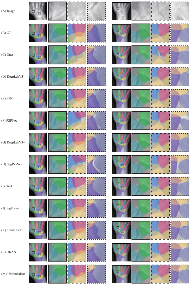
The authors establish that a multi-center collection of 1048 wrist radiographs, annotated at the pixel level for bone instances on 618 cases and scored for bone erosion on 800 cases, constitutes the first openly available resource for wrist bone instance segmentation and simultaneously supports Sharp/van der Heijde bone erosion scoring in rheumatoid arthritis.
What carries the argument
The RAM-W600 dataset, which pairs conventional wrist radiographs with pixel-level instance segmentation masks and SvdH bone erosion scores.
If this is right
- The annotations enable training of models for automated joint space narrowing measurement across multiple centers.
- The scores support development of algorithms that detect and quantify bone erosion progression.
- The same images can be used to evaluate bone deformity and osteophyte formation.
- The resource extends to non-RA wrist tasks such as locating carpal bone fractures.
Where Pith is reading between the lines
- Models trained on this data could be tested for cross-center robustness using the multi-site collection.
- Automated scoring pipelines built from these labels might reduce the time experts spend on routine RA monitoring.
- Future releases could add follow-up images to support longitudinal studies of disease progression.
Load-bearing premise
The provided pixel-level segmentation masks and SvdH scores are sufficiently consistent and accurate to function as reliable ground truth even though small bones, narrow spaces, overlaps, and disease changes make expert annotation difficult.
What would settle it
Re-annotation of a random subset of the images by independent rheumatology experts that produces substantially different bone boundaries or erosion scores from the released labels would undermine the dataset's value as ground truth.
Figures


read the original abstract
Rheumatoid arthritis (RA) is a common autoimmune disease that has been the focus of research in computer-aided diagnosis (CAD) and disease monitoring. In clinical settings, conventional radiography (CR) is widely used for the screening and evaluation of RA due to its low cost and accessibility. The wrist is a critical region for the diagnosis of RA. However, CAD research in this area remains limited, primarily due to the challenges in acquiring high-quality instance-level annotations. (i) The wrist comprises numerous small bones with narrow joint spaces, complex structures, and frequent overlaps, requiring detailed anatomical knowledge for accurate annotation. (ii) Disease progression in RA often leads to osteophyte, bone erosion (BE), and even bony ankylosis, which alter bone morphology and increase annotation difficulty, necessitating expertise in rheumatology. This work presents a multi-task dataset for wrist bone in CR, including two tasks: (i) wrist bone instance segmentation and (ii) Sharp/van der Heijde (SvdH) BE scoring, which is the first public resource for wrist bone instance segmentation. This dataset comprises 1048 wrist conventional radiographs of 388 patients from six medical centers, with pixel-level instance segmentation annotations for 618 images and SvdH BE scores for 800 images. This dataset can potentially support a wide range of research tasks related to RA, including joint space narrowing (JSN) progression quantification, BE detection, bone deformity evaluation, and osteophyte detection. It may also be applied to other wrist-related tasks, such as carpal bone fracture localization. We hope this dataset will significantly lower the barrier to research on wrist RA and accelerate progress in CAD research within the RA-related domain.
Editorial analysis
A structured set of objections, weighed in public.
Referee Report
Summary. The paper introduces the RAM-W600 dataset, a multi-task collection of 1048 wrist conventional radiographs from 388 patients across six medical centers. It provides pixel-level instance segmentation annotations for 618 images and Sharp/van der Heijde (SvdH) bone erosion (BE) scores for 800 images. The central claim is that this is the first public resource for wrist bone instance segmentation in conventional radiography, intended to support CAD research on rheumatoid arthritis tasks including BE detection, joint space narrowing quantification, and related applications.
Significance. If the ground-truth annotations prove reliable, the dataset would represent a meaningful contribution by filling a gap in public resources for wrist-specific RA imaging analysis. The multi-center design and dual-task structure (segmentation plus scoring) could facilitate development of generalizable models and support downstream tasks such as osteophyte detection or carpal fracture localization.
major comments (1)
- [Annotation Protocol / Methods] Annotation Protocol / Methods section: The manuscript acknowledges that accurate pixel-level annotation of wrist bones requires detailed anatomical and rheumatology expertise due to small bones, narrow joint spaces, overlaps, and RA-induced morphological changes, yet provides no quantitative inter-annotator agreement statistics (e.g., mean Dice or IoU across multiple experts) for the 618 segmentation masks. Without such metrics or a description of the adjudication process, the reliability of the instance-segmentation ground truth remains unsubstantiated and directly undermines the dataset's utility as a benchmark.
minor comments (2)
- [Abstract] The abstract and introduction would benefit from explicit clarification on the overlap between the 618 segmentation-annotated images and the 800 scored images, including whether any images carry both labels.
- [Dataset Statistics] Figure captions and dataset statistics tables should include the exact number of images per center and per task to allow readers to assess potential center-specific biases.
Simulated Author's Rebuttal
We thank the referee for their thoughtful review and for identifying a key area where the manuscript can be strengthened. We address the major comment below and outline the revisions we will make.
read point-by-point responses
-
Referee: [Annotation Protocol / Methods] Annotation Protocol / Methods section: The manuscript acknowledges that accurate pixel-level annotation of wrist bones requires detailed anatomical and rheumatology expertise due to small bones, narrow joint spaces, overlaps, and RA-induced morphological changes, yet provides no quantitative inter-annotator agreement statistics (e.g., mean Dice or IoU across multiple experts) for the 618 segmentation masks. Without such metrics or a description of the adjudication process, the reliability of the instance-segmentation ground truth remains unsubstantiated and directly undermines the dataset's utility as a benchmark.
Authors: We agree that quantitative measures of annotation reliability are essential for establishing the dataset as a robust benchmark. The current manuscript describes the general annotation challenges and the involvement of experts with anatomical and rheumatology knowledge but does not report inter-annotator agreement or detail the adjudication steps. In the revised manuscript we will expand the Methods section to include: (i) the number of annotators and their specific qualifications, (ii) a description of the multi-stage annotation and adjudication workflow, and (iii) quantitative agreement statistics (mean Dice and IoU) computed on a representative subset of images that were annotated independently by multiple experts. These additions will directly address the concern and strengthen the evidence for ground-truth quality. revision: yes
Circularity Check
Dataset release paper with no derivations or predictions exhibits no circularity
full rationale
This is a dataset introduction paper whose central claims concern the composition, scale, and public availability of a new multi-task wrist radiograph collection (1048 images, 618 with instance segmentation, 800 with SvdH scores). No mathematical derivations, equations, fitted parameters, or predictive models are present. The claims rest directly on the described data acquisition and annotation process rather than any self-referential reduction, self-citation chain, or renaming of prior results. The paper is therefore self-contained against external benchmarks of dataset utility, warranting a score of 0 with no circular steps.
Axiom & Free-Parameter Ledger
axioms (1)
- domain assumption Accurate wrist bone instance segmentation in conventional radiographs requires detailed anatomical knowledge and rheumatology expertise because of small bones, narrow joint spaces, complex structures, overlaps, and disease-induced changes such as osteophytes and bone erosion.
Lean theorems connected to this paper
-
IndisputableMonolith/Foundation/RealityFromDistinction.leanreality_from_one_distinction unclear?
unclearRelation between the paper passage and the cited Recognition theorem.
This work presents a multi-task dataset for wrist bone in CR, including two tasks: (i) wrist bone instance segmentation and (ii) Sharp/van der Heijde (SvdH) BE scoring... 618 images... 800 images.
-
IndisputableMonolith/Cost/FunctionalEquation.leanwashburn_uniqueness_aczel unclear?
unclearRelation between the paper passage and the cited Recognition theorem.
We provide high-quality pixel-level annotations... benchmark for wrist bone instance segmentation and SvdH BE scoring.
What do these tags mean?
- matches
- The paper's claim is directly supported by a theorem in the formal canon.
- supports
- The theorem supports part of the paper's argument, but the paper may add assumptions or extra steps.
- extends
- The paper goes beyond the formal theorem; the theorem is a base layer rather than the whole result.
- uses
- The paper appears to rely on the theorem as machinery.
- contradicts
- The paper's claim conflicts with a theorem or certificate in the canon.
- unclear
- Pith found a possible connection, but the passage is too broad, indirect, or ambiguous to say the theorem truly supports the claim.
Reference graph
Works this paper leans on
-
[1]
Iftekharul Abedeen, Md Ashiqur Rahman, Fatema Zohra Prottyasha, Tasnim Ahmed, Tareque Mohmud Chowdhury, and Swakkhar Shatabda. Fracatlas: A dataset for fracture classification, localization and segmentation of musculoskeletal radiographs.Scientific data, 10(1):521, 2023
work page 2023
-
[2]
Diagnosis and management of rheumatoid arthritis: a review.Jama, 320(13):1360–1372, 2018
Daniel Aletaha and Josef S Smolen. Diagnosis and management of rheumatoid arthritis: a review.Jama, 320(13):1360–1372, 2018
work page 2018
-
[3]
Douglas G Altman and J Martin Bland. Diagnostic tests. 1: Sensitivity and specificity.BMJ: British Medical Journal, 308(6943):1552, 1994
work page 1994
-
[4]
Emran Mohammad Abu Anas, Abtin Rasoulian, Alexander Seitel, Kathryn Darras, David Wil- son, Paul St John, David Pichora, Parvin Mousavi, Robert Rohling, and Purang Abolmaesumi. Automatic segmentation of wrist bones in ct using a statistical wrist shape + pose model.IEEE transactions on medical imaging, 35(8):1789–1801, 2016
work page 2016
-
[5]
Radiographic imaging of the wrist
Anil K Bhat, Bhaskaranand Kumar, and Ashwath Acharya. Radiographic imaging of the wrist. Indian journal of plastic surgery: official publication of the Association of Plastic Surgeons of India, 44(2):186, 2011. 10
work page 2011
-
[6]
Deep learning models to automate the scoring of hand radiographs for rheumatoid arthritis
Zhiyan Bo, Laura C Coates, and Bartłomiej W Papie˙z. Deep learning models to automate the scoring of hand radiographs for rheumatoid arthritis. InAnnual Conference on Medical Image Understanding and Analysis, pages 398–413. Springer, 2024
work page 2024
-
[7]
Convolutional kolmogorov-arnold networks.arXiv preprint arXiv:2406.13155, 2024
Alexander Dylan Bodner, Antonio Santiago Tepsich, Jack Natan Spolski, and Santiago Pourteau. Convolutional kolmogorov-arnold networks.arXiv preprint arXiv:2406.13155, 2024
-
[8]
The balanced accuracy and its posterior distribution
Kay Henning Brodersen, Cheng Soon Ong, Klaas Enno Stephan, and Joachim M Buhmann. The balanced accuracy and its posterior distribution. In2010 20th international conference on pattern recognition, pages 3121–3124. IEEE, 2010
work page 2010
-
[9]
Karin Bruynesteyn, Désirée van der Heijde, Maarten Boers, Ariane Saudan, Paul Peloso, Harold Paulus, Harry Houben, Bridget Griffiths, John Edmonds, Barry Bresnihan, et al. Determination of the minimal clinically important difference in rheumatoid arthritis joint damage of the sharp/van der heijde and larsen/scott scoring methods by clinical experts and co...
work page 2002
-
[10]
Fei Cao, HK Huang, Ewa Pietka, and Vicente Gilsanz. Digital hand atlas and web-based bone age assessment: system design and implementation.Computerized medical imaging and graphics, 24(5):297–307, 2000
work page 2000
-
[11]
TransUNet: Transformers Make Strong Encoders for Medical Image Segmentation
Jieneng Chen, Yongyi Lu, Qihang Yu, Xiangde Luo, Ehsan Adeli, Yan Wang, Le Lu, Alan L Yuille, and Yuyin Zhou. Transunet: Transformers make strong encoders for medical image segmentation.arXiv preprint arXiv:2102.04306, 2021
work page internal anchor Pith review Pith/arXiv arXiv 2021
-
[12]
Rethinking Atrous Convolution for Semantic Image Segmentation
Liang-Chieh Chen, George Papandreou, Florian Schroff, and Hartwig Adam. Rethinking atrous convolution for semantic image segmentation.arXiv preprint arXiv:1706.05587, 2017
work page internal anchor Pith review Pith/arXiv arXiv 2017
-
[13]
Encoder-decoder with atrous separable convolution for semantic image segmentation
Liang-Chieh Chen, Yukun Zhu, George Papandreou, Florian Schroff, and Hartwig Adam. Encoder-decoder with atrous separable convolution for semantic image segmentation. In Proceedings of the European conference on computer vision (ECCV), pages 801–818, 2018
work page 2018
-
[14]
Nancy Chinchor and Beth M Sundheim. Muc-5 evaluation metrics. InFifth Message Under- standing Conference (MUC-5): Proceedings of a Conference Held in Baltimore, Maryland, August 25-27, 1993, 1993
work page 1993
-
[15]
Measures of the amount of ecologic association between species.Ecology, 26(3):297–302, 1945
Lee R Dice. Measures of the amount of ecologic association between species.Ecology, 26(3):297–302, 1945
work page 1945
-
[16]
Hongbo Du, Hai Wang, Chunlai Yang, Luyando Kabalata, Henian Li, and Changfu Qiang. Hand bone extraction and segmentation based on a convolutional neural network.Biomedical Signal Processing and Control, 89:105788, 2024
work page 2024
-
[17]
Anatomy, biomechanics, and loads of the wrist joint.Life, 12(2):188, 2022
Jörg Eschweiler, Jianzhang Li, Valentin Quack, Björn Rath, Alice Baroncini, Frank Hildebrand, and Filippo Migliorini. Anatomy, biomechanics, and loads of the wrist joint.Life, 12(2):188, 2022
work page 2022
-
[18]
Fatemeh Ezzati and Parham Pezeshk. Radiographic findings of inflammatory arthritis and mimics in the hands.Diagnostics, 12(9):2134, 2022
work page 2022
-
[19]
Lukas Folle, David Simon, Koray Tascilar, Gerhard Krönke, Anna-Maria Liphardt, Andreas Maier, Georg Schett, and Arnd Kleyer. Deep learning-based classification of inflammatory arthritis by identification of joint shape patterns—how neural networks can tell us where to “deep dive” clinically.Frontiers in Medicine, 9:850552, 2022
work page 2022
-
[20]
Brent Foster, Anand A Joshi, Marissa Borgese, Yasser Abdelhafez, Robert D Boutin, and Abhijit J Chaudhari. Wrist: A wrist image segmentation toolkit for carpal bone delineation from mri.Computerized Medical Imaging and Graphics, 63:31–40, 2018
work page 2018
-
[21]
The elements of statistical learning: Data mining, inference, and prediction
Jerome Friedman. The elements of statistical learning: Data mining, inference, and prediction. (No Title), 2009. 11
work page 2009
-
[22]
Afina S Glas, Jeroen G Lijmer, Martin H Prins, Gouke J Bonsel, and Patrick MM Bossuyt. The diagnostic odds ratio: a single indicator of test performance.Journal of clinical epidemiology, 56(11):1129–1135, 2003
work page 2003
-
[23]
Levit: a vision transformer in convnet’s clothing for faster inference
Benjamin Graham, Alaaeldin El-Nouby, Hugo Touvron, Pierre Stock, Armand Joulin, Hervé Jégou, and Matthijs Douze. Levit: a vision transformer in convnet’s clothing for faster inference. InProceedings of the IEEE/CVF international conference on computer vision, pages 12259– 12269, 2021
work page 2021
-
[24]
The rsna pediatric bone age machine learning challenge.Radiology, 290(2):498– 503, 2019
Safwan S Halabi, Luciano M Prevedello, Jayashree Kalpathy-Cramer, Artem B Mamonov, Alexander Bilbily, Mark Cicero, Ian Pan, Lucas Araújo Pereira, Rafael Teixeira Sousa, Nitamar Abdala, et al. The rsna pediatric bone age machine learning challenge.Radiology, 290(2):498– 503, 2019
work page 2019
-
[25]
Deep residual learning for image recognition
Kaiming He, Xiangyu Zhang, Shaoqing Ren, and Jian Sun. Deep residual learning for image recognition. InProceedings of the IEEE conference on computer vision and pattern recognition, pages 770–778, 2016
work page 2016
-
[26]
Evaluation method of rheumatoid arthritis by the x-ray photograph using deep learning
Yuri Hioki, Koji Makino, Kensuke Koyama, Hirotaka Haro, and Hidetsugu Terada. Evaluation method of rheumatoid arthritis by the x-ray photograph using deep learning. In2021 IEEE 3rd Global Conference on Life Sciences and Technologies (LifeTech), pages 444–447. IEEE, 2021
work page 2021
-
[27]
T Hirano, M Nishide, N Nonaka, J Seita, K Ebina, K Sakurada, et al. Development and validation of a deep-learning model for scoring of radiographic finger joint destruction in rheumatoid arthritis. rheumatol adv pract. 2019. available from:; 3
work page 2019
-
[28]
Toru Hirano, Masayuki Nishide, Naoki Nonaka, Jun Seita, Kosuke Ebina, Kazuhiro Sakurada, and Atsushi Kumanogoh. Development and validation of a deep-learning model for scoring of radiographic finger joint destruction in rheumatoid arthritis.Rheumatology advances in practice, 3(2):rkz047, 2019
work page 2019
-
[29]
Stefanie Hirsiger, Andreas Schweizer, Junichi Miyake, Ladislav Nagy, and Philipp Fürnstahl. Corrective osteotomies of phalangeal and metacarpal malunions using patient-specific guides: Ct-based evaluation of the reduction accuracy.Hand, 13(6):627–636, 2018
work page 2018
-
[30]
Jan Lucas Hoving, Rachelle Buchbinder, Stephen Hall, Gary Lawler, Peter Coombs, Stephen McNealy, Paul Bird, and David Connell. A comparison of magnetic resonance imaging, sonography, and radiography of the hand in patients with early rheumatoid arthritis.The Journal of rheumatology, 31(4):663–675, 2004
work page 2004
-
[31]
MobileNets: Efficient Convolutional Neural Networks for Mobile Vision Applications
Andrew G Howard. Mobilenets: Efficient convolutional neural networks for mobile vision applications.arXiv preprint arXiv:1704.04861, 2017
work page internal anchor Pith review Pith/arXiv arXiv 2017
-
[32]
Yinghe Huo, Koen L Vincken, Desiree Van Der Heijde, Maria JH De Hair, Floris P Lafeber, and Max A Viergever. Automatic quantification of radiographic wrist joint space width of patients with rheumatoid arthritis.IEEE Transactions on Biomedical Engineering, 64(11):2695–2703, 2017
work page 2017
-
[33]
Predictors of radiographic joint damage in patients with early rheumatoid arthritis
LMA Jansen, IE Van der Horst-Bruinsma, D Van Schaardenburg, PD Bezemer, and BAC Dijkmans. Predictors of radiographic joint damage in patients with early rheumatoid arthritis. Annals of the rheumatic diseases, 60(10):924–927, 2001
work page 2001
-
[34]
Bo-kyeong Kang, Yelin Han, Jaehoon Oh, Jongwoo Lim, Jongbin Ryu, Myeong Seong Yoon, Juncheol Lee, and Soorack Ryu. Automatic segmentation for favourable delineation of ten wrist bones on wrist radiographs using convolutional neural network.Journal of Personalized Medicine, 12(5):776, 2022
work page 2022
-
[35]
Kathryn M Kingsmore, Christopher E Puglisi, Amrie C Grammer, and Peter E Lipsky. An introduction to machine learning and analysis of its use in rheumatic diseases.Nature Reviews Rheumatology, 17(12):710–730, 2021. 12
work page 2021
-
[36]
Alexander Kirillov, Eric Mintun, Nikhila Ravi, Hanzi Mao, Chloe Rolland, Laura Gustafson, Tete Xiao, Spencer Whitehead, Alexander C Berg, Wan-Yen Lo, et al. Segment anything. In Proceedings of the IEEE/CVF international conference on computer vision, pages 4015–4026, 2023
work page 2023
-
[37]
Hyungeun Lee, Ung Hwang, Seungwon Yu, Chang-Hun Lee, and Kijung Yoon. Osteoporosis prediction from hand and wrist x-rays using image segmentation and self-supervised learning. arXiv preprint arXiv:2311.06834, 2023
-
[38]
U-kan makes strong backbone for medical image segmentation and generation
Chenxin Li, Xinyu Liu, Wuyang Li, Cheng Wang, Hengyu Liu, Yifan Liu, Zhen Chen, and Yixuan Yuan. U-kan makes strong backbone for medical image segmentation and generation. InProceedings of the AAAI Conference on Artificial Intelligence, volume 39, pages 4652–4660, 2025
work page 2025
-
[39]
Yanyu Li, Geng Yuan, Yang Wen, Ju Hu, Georgios Evangelidis, Sergey Tulyakov, Yanzhi Wang, and Jian Ren. Efficientformer: Vision transformers at mobilenet speed.Advances in Neural Information Processing Systems, 35:12934–12949, 2022
work page 2022
-
[40]
Chung-Yueh Lien, Hao-Jan Wang, Cheng-Kai Lu, Tzu-Hsuan Hsu, Woei-Chyn Chu, and Chien- Chih Lai. Deep learning with an attention mechanism for enhancing automated modified total sharp/van der heijde scoring of hand x-ray images in rheumatoid arthritis.Journal of Medical and Biological Engineering, pages 1–9, 2025
work page 2025
-
[41]
Feature pyramid networks for object detection
Tsung-Yi Lin, Piotr Dollár, Ross Girshick, Kaiming He, Bharath Hariharan, and Serge Belongie. Feature pyramid networks for object detection. InProceedings of the IEEE conference on computer vision and pattern recognition, pages 2117–2125, 2017
work page 2017
-
[42]
Swin-umamba: Mamba-based unet with imagenet-based pretraining
Jiarun Liu, Hao Yang, Hong-Yu Zhou, Yan Xi, Lequan Yu, Cheng Li, Yong Liang, Guangming Shi, Yizhou Yu, Shaoting Zhang, et al. Swin-umamba: Mamba-based unet with imagenet-based pretraining. InInternational Conference on Medical Image Computing and Computer-Assisted Intervention, pages 615–625. Springer, 2024
work page 2024
-
[43]
Segment anything in medical images.Nature Communications, 15:654, 2024
Jun Ma, Yuting He, Feifei Li, Lin Han, Chenyu You, and Bo Wang. Segment anything in medical images.Nature Communications, 15:654, 2024
work page 2024
-
[44]
U-Mamba: Enhancing Long-range Dependency for Biomedical Image Segmentation
Jun Ma, Feifei Li, and Bo Wang. U-mamba: Enhancing long-range dependency for biomedical image segmentation.arXiv preprint arXiv:2401.04722, 2024
work page internal anchor Pith review Pith/arXiv arXiv 2024
-
[45]
Krzysztof Maziarz, Anna Krason, and Zbigniew Wojna. Deep learning for rheumatoid arthritis: Joint detection and damage scoring in x-rays.arXiv preprint arXiv:2104.13915, 2021
-
[46]
Sachin Mehta and Mohammad Rastegari. Mobilevit: light-weight, general-purpose, and mobile- friendly vision transformer.arXiv preprint arXiv:2110.02178, 2021
-
[47]
Kazuki Miyama, Ryoma Bise, Satoshi Ikemura, Kazuhiro Kai, Masaya Kanahori, Shinkichi Arisumi, Taisuke Uchida, Yasuharu Nakashima, and Seiichi Uchida. Deep learning-based automatic-bone-destruction-evaluation system using contextual information from other joints. Arthritis Research & Therapy, 24(1):227, 2022
work page 2022
-
[48]
Douglas C Moore, Joseph J Crisco, Theodore G Trafton, and Evan L Leventhal. A digital database of wrist bone anatomy and carpal kinematics.Journal of biomechanics, 40(11):2537– 2542, 2007
work page 2007
-
[49]
3d mri brain tumor segmentation using autoencoder regularization
Andriy Myronenko. 3d mri brain tumor segmentation using autoencoder regularization. In International MICCAI brainlesion workshop, pages 311–320. Springer, 2018
work page 2018
-
[50]
Triquetrum fracture with pisiform dislocation.Orthopedic Reviews, 14(2):32339, 2022
Arjun Nanduri, Alison Kim, Carolyn Nolan, Jesse Dubey, and Andrew Barbera. Triquetrum fracture with pisiform dislocation.Orthopedic Reviews, 14(2):32339, 2022
work page 2022
-
[51]
Stanislav Nikolov, Sam Blackwell, Alexei Zverovitch, Ruheena Mendes, Michelle Livne, Jeffrey De Fauw, Yojan Patel, Clemens Meyer, Harry Askham, Bernadino Romera-Paredes, et al. Clinically applicable segmentation of head and neck anatomy for radiotherapy: deep learning algorithm development and validation study.Journal of medical Internet research, 23(7):e...
work page 2021
-
[52]
Yafei Ou, Prasoon Ambalathankandy, Ryunosuke Furuya, Seiya Kawada, Tianyu Zeng, Yujie An, Tamotsu Kamishima, Kenichi Tamura, and Masayuki Ikebe. A sub-pixel accurate quantifi- cation of joint space narrowing progression in rheumatoid arthritis.IEEE Journal of Biomedical and Health Informatics, 27(1):53–64, 2023
work page 2023
-
[53]
Benjamin Schultz Overgaard, Anders Bossel Holst Christensen, Lene Terslev, Thiusius Rajeeth Savarimuthu, and Søren Andreas Just. Artificial intelligence model for segmentation and severity scoring of osteophytes in hand osteoarthritis on ultrasound images.Frontiers in Medicine, 11:1297088, 2024
work page 2024
-
[54]
Raj Ponnusamy, Ming Zhang, Zhiheng Chang, Yue Wang, Carmine Guida, Samantha Kuang, Xinyue Sun, Jordan Blackadar, Jeffrey B Driban, Timothy McAlindon, et al. Automatic measuring of finger joint space width on hand radiograph using deep learning and conventional computer vision methods.Biomedical signal processing and control, 84:104713, 2023
work page 2023
-
[55]
Karl Ludger Radke, Lena Marie Wollschläger, Sven Nebelung, Daniel Benjamin Abrar, Christoph Schleich, Matthias Boschheidgen, Miriam Frenken, Justus Schock, Dirk Klee, Jens Frahm, et al. Deep learning-based post-processing of real-time mri to assess and quantify dynamic wrist movement in health and disease.Diagnostics, 11(6):1077, 2021
work page 2021
-
[56]
Stefan Raith, Matthias Deitermann, Tobias Pankert, Jianzhang Li, Ali Modabber, Frank Hölzle, Frank Hildebrand, and Jörg Eschweiler. Multi-label segmentation of carpal bones in mri using expansion transfer learning.Physics in Medicine and Biology, 2025
work page 2025
-
[57]
Eric EJ Raven, Michel PJ van den Bekerom, Annechien Beumer, and C Niek van Dijk. Ra- diocarpal and midcarpal instability in rheumatoid patients: a systematic review.The Open Orthopaedics Journal, 9:246, 2015
work page 2015
-
[58]
U-net: Convolutional networks for biomedical image segmentation
Olaf Ronneberger, Philipp Fischer, and Thomas Brox. U-net: Convolutional networks for biomedical image segmentation. InMedical image computing and computer-assisted intervention–MICCAI 2015: 18th international conference, Munich, Germany, October 5-9, 2015, proceedings, part III 18, pages 234–241. Springer, 2015
work page 2015
-
[59]
Maja Schlereth, Melek Yalcin Mutlu, Jonas Utz, Sara Bayat, Tobias Heimann, Jingna Qiu, Chris Ehring, Chang Liu, Michael Uder, Arnd Kleyer, et al. Deep learning-based classification of erosion, synovitis and osteitis in hand mri of patients with inflammatory arthritis.RMD open, 10(2):e004273, 2024
work page 2024
-
[60]
Ronnie Sebro and Cynthia De la Garza-Ramos. Machine learning for opportunistic screening for osteoporosis from ct scans of the wrist and forearm.Diagnostics, 12(3):691, 2022
work page 2022
-
[61]
Kassem Sharif, Alaa Sharif, Fareed Jumah, Rod Oskouian, and R Shane Tubbs. Rheumatoid arthritis in review: Clinical, anatomical, cellular and molecular points of view.Clinical Anatomy, 31(2):216–223, 2018
work page 2018
-
[62]
Marina Sokolova and Guy Lapalme. A systematic analysis of performance measures for classification tasks.Information processing & management, 45(4):427–437, 2009
work page 2009
-
[63]
Berend C Stoel, Marius Staring, Monique Reijnierse, and Annette HM van der Helm-van Mil. Deep learning in rheumatological image interpretation.Nature Reviews Rheumatology, 20(3):182–195, 2024
work page 2024
-
[64]
Dongmei Sun, Thanh M Nguyen, Robert J Allaway, Jelai Wang, Verena Chung, Thomas V Yu, Michael Mason, Isaac Dimitrovsky, Lars Ericson, Hongyang Li, et al. A crowdsourcing approach to develop machine learning models to quantify radiographic joint damage in rheumatoid arthritis.JAMA network open, 5(8):e2227423–e2227423, 2022
work page 2022
-
[65]
Abdel Aziz Taha and Allan Hanbury. Metrics for evaluating 3d medical image segmentation: analysis, selection, and tool.BMC medical imaging, 15:1–28, 2015
work page 2015
-
[66]
Bachir Taouli, Souhil Zaim, Charles G Peterfy, John A Lynch, Alexander Stork, Ali Guermazi, Bo Fan, Kenneth H Fye, and Harry K Genant. Rheumatoid arthritis of the hand and wrist: comparison of three imaging techniques.American Journal of Roentgenology, 182(4):937–943, 2004. 14
work page 2004
-
[67]
EHS Teule, N Lessmann, EPA van der Heijden, and S Hummelink. Automatic segmentation and labelling of wrist bones in four-dimensional computed tomography datasets via deep learning. Journal of Hand Surgery (European Volume), 49(4):507–509, 2024
work page 2024
-
[68]
Kemal Üreten, Hasan Erbay, and Hadi Hakan Mara¸ s. Detection of rheumatoid arthritis from hand radiographs using a convolutional neural network.Clinical rheumatology, 39:969–974, 2020
work page 2020
-
[69]
How to read radiographs according to the sharp/van der heijde method
DMFM Van der Heijde. How to read radiographs according to the sharp/van der heijde method. The Journal of rheumatology, 27(1):261–263, 2000
work page 2000
-
[70]
DMFM Van der Heijde, T Dankert, F Nieman, R Rau, and M Boers. Reliability and sensitivity to change of a simplification of the sharp/van der heijde radiological assessment in rheumatoid arthritis.Rheumatology, 38(10):941–947, 1999
work page 1999
-
[71]
Hao-Jan Wang, Chi-Ping Su, Chien-Chih Lai, Wun-Rong Chen, Chi Chen, Liang-Ying Ho, Woei-Chyn Chu, and Chung-Yueh Lien. Deep learning-based computer-aided diagnosis of rheumatoid arthritis with hand x-ray images conforming to modified total sharp/van der heijde score.Biomedicines, 10(6):1355, 2022
work page 2022
-
[72]
Bls-gan: A deep layer separation framework for eliminating bone overlap in conventional radiographs
Haolin Wang, Yafei Ou, Prasoon Ambalathankandy, Gen Ota, Pengyu Dai, Masayuki Ikebe, Kenji Suzuki, and Tamotsu Kamishima. Bls-gan: A deep layer separation framework for eliminating bone overlap in conventional radiographs. InProceedings of the AAAI Conference on Artificial Intelligence, volume 39, pages 7674–7681, 2025
work page 2025
-
[73]
Haolin Wang, Yafei Ou, Prasoon Ambalathankandy, Gen Ota, Pengyu Dai, Masayuki Ikebe, Kenji Suzuki, and Tamotsu Kamishima. Layer separation: Adjustable joint space width images synthesis in conventional radiography.arXiv preprint arXiv:2502.01972, 2025
-
[74]
Haolin Wang, Yafei Ou, Wanxuan Fang, Prasoon Ambalathankandy, Naoto Goto, Gen Ota, Taichi Okino, Jun Fukae, Kenneth Sutherland, Masayuki Ikebe, et al. A deep registration method for accurate quantification of joint space narrowing progression in rheumatoid arthritis. Computerized Medical Imaging and Graphics, 108:102273, 2023
work page 2023
-
[75]
Enze Xie, Wenhai Wang, Zhiding Yu, Anima Anandkumar, Jose M Alvarez, and Ping Luo. Segformer: Simple and efficient design for semantic segmentation with transformers.Advances in neural information processing systems, 34:12077–12090, 2021
work page 2021
-
[76]
Fan Yang, Xin Weng, Yuehong Miao, Yuhui Wu, Hong Xie, and Pinggui Lei. Deep learning approach for automatic segmentation of ulna and radius in dual-energy x-ray imaging.Insights into Imaging, 12:1–9, 2021
work page 2021
-
[77]
Shunhan Yao, Yuanxiang Huang, Xiaoyu Wang, Yiwen Zhang, Ian Costa Paixao, Zhikang Wang, Charla Lu Chai, Hongtao Wang, Dinggui Lu, Geoffrey I Webb, et al. A radiograph dataset for the classification, localization, and segmentation of primary bone tumors.Scientific Data, 12(1):88, 2025
work page 2025
-
[78]
Chungwun Yiu, James Francis Griffith, Fan Xiao, Lin Shi, Bingjing Zhou, Su Wu, and Lai-Shan Tam. Automated quantification of wrist bone marrow oedema, pre-and post-treatment, in early rheumatoid arthritis.Rheumatology Advances in Practice, 8(3):rkae073, 2024
work page 2024
-
[79]
Medmamba: Vision mamba for medical image classification
Yubiao Yue and Zhenzhang Li. Medmamba: Vision mamba for medical image classification. arXiv preprint arXiv:2403.03849, 2024
-
[80]
Hengshuang Zhao, Jianping Shi, Xiaojuan Qi, Xiaogang Wang, and Jiaya Jia. Pyramid scene parsing network. InProceedings of the IEEE conference on computer vision and pattern recognition, pages 2881–2890, 2017
work page 2017
discussion (0)
Sign in with ORCID, Apple, or X to comment. Anyone can read and Pith papers without signing in.