REVIEW 3 major objections 5 minor 16 references
A Digital Twin for Robotic Post Mortem Tissue Sampling using Virtual Reality
T0 review · 3 major / 5 minor · reviewed 2026-08-05 · deepseek-v4-flash
Pith's one-line read A virtual-reality digital twin lets pathologists plan and supervise robotic post mortem biopsies from a separate room; 132 insertions landed within 5.30 ± 3.25 mm off-axis.
desk verdict A solid integrated feasibility study: remote VR planning plus robotic post mortem biopsy works on cadavers, but the headline accuracy number is softer than it looks because excluded insertions and possible target displacement are both in play. read the letter →
The pith
A machine-rendered reading of the paper's core claim, the machinery that carries it, and where it could break.
The reading
What carries the argument
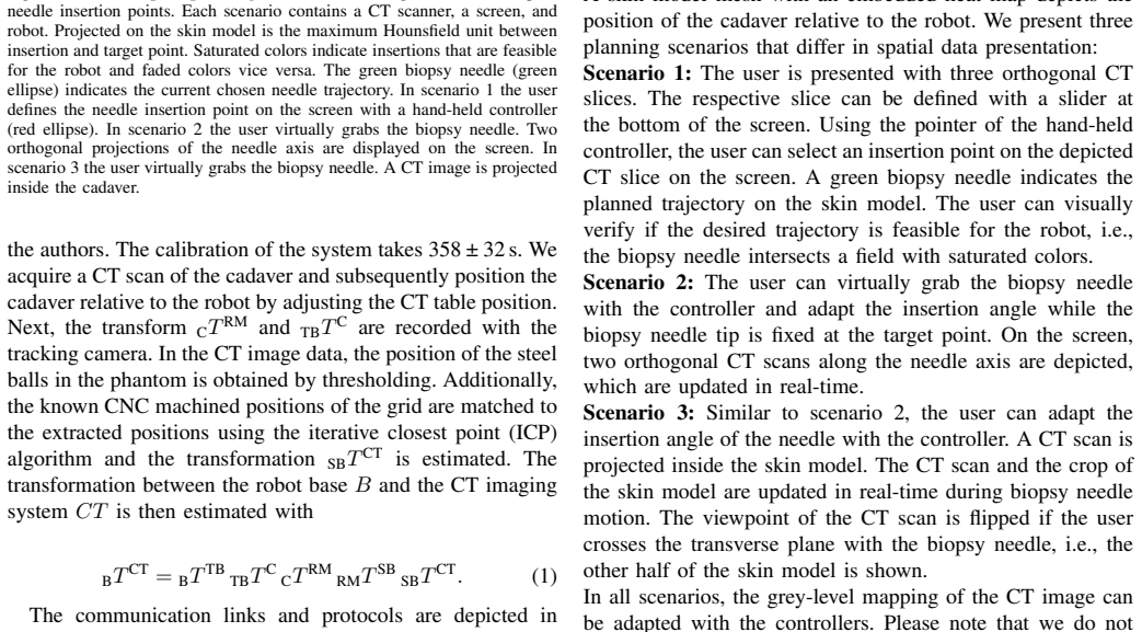
The load-bearing mechanism is the digital twin's single shared coordinate frame. A chain of rigid transformations—robot base, tracking camera, phantom on the cadaver's thorax, and CT volume—maps a trajectory drawn in VR into robot motion in the intervention room. In the VR scene, a CT-derived skin mesh is overlaid with a color map of the maximum Hounsfield unit along candidate needle paths, so the user immediately sees which insertions would hit bone and whether the robot's reach and joint limits make the trajectory feasible. The three planning scenarios differ only in how the needle path is specified; the underlying registration and feasibility checks are identical.
What would settle it
Compare the planning CT and a post-insertion CT using radio-opaque markers embedded in the phantom and at the target sites: if internal targets shift relative to the strapped phantom, the registered needle-tip error should decrease once that displacement is compensated. If it does not, the 5.30 mm off-axis error is genuine placement accuracy.
Extended reading notes
Core claim
The central claim is that a VR digital twin can support fully remote planning and execution of robotic post mortem biopsies with acceptable accuracy and usability. The paper demonstrates this by combining a CT-guided registration chain (robot base to tracking camera to a strapped phantom to the CT volume) with a VR application in which the user sees a life-size skin model, the robot, and CT slices in a shared coordinate frame. Users choose insertion points and needle angles in one of three interaction modes; the system immediately marks which trajectories are robot-feasible by overlaying a maximum-Hounsfield-unit color map on the skin. In the cadaver study, the mean off-axis error was 5.30 ±
Load-bearing premise
The accuracy numbers assume the cadaver's skin and internal organs stay fixed relative to the tracked phantom between the planning CT and each insertion; if the body shifts under the tension straps, the reported error could partly be target displacement rather than robot placement error.
Editorial extensions
If this is right
- Remote planning and execution: a physician can run the whole biopsy workflow from a control room, eliminating direct contact with the cadaver and reducing infection risk.
- No robotics expertise required: novice and expert users planned feasible trajectories after a five-minute introduction, and planning times dropped 33–53% across repeated insertions.
- Systematic sampling becomes practical: because the robot places each needle consistently, the workflow suits large multi-target collections such as national autopsy registries.
- Accuracy is comparable to prior robotic PM biopsy systems (target error 7.35 vs 7.19 mm), while surface puncture error is lower (2.62 mm).
- Scalability to infectious disease outbreaks: the system is mobile and can be deployed where autopsies are otherwise avoided.
Reading between the lines
- Because the reported error is computed against the planning CT, part of the 5.30 mm off-axis value may reflect target displacement (e.g., tissue shifting after earlier insertions) rather than pure robot placement error; a marker-based repeat-CT experiment could separate the two.
- The 65% histology confirmation rate suggests the limiting factor for small targets is not placement error alone but the biopsy needle and target size; a different needle design or intraprocedural confirmation could plausibly raise the hit rate.
- The same digital-twin interaction could be adapted to living patients if respiratory and cardiac motion are added to the registration and planning, but that would require motion compensation the current post mortem setting does not need.
- The strong preference for the 3D grab-and-orient modes over the conventional triplanar screen mode hints that pathologists may accept VR planning more readily if the interaction matches how they think about anatomy, not how legacy software displays slices.
Signed reviews
Editorial analysis
A structured set of objections, weighed in public.
Referee Report
Summary. The paper presents a mobile robotic post-mortem biopsy system in which a physician plans needle trajectories in a virtual-reality digital twin and the robot executes the insertions in a physically separate intervention room. The system is evaluated in a usability study with 21 participants across three VR planning scenarios and in a clinical-feasibility study on three cadavers. The authors report 132 needle insertions with a mean off-axis error of 5.30±3.25 mm, a mean target error of 7.35±4.10 mm, and histopathological confirmation of 65% of tissue samples. The central claims are that fully remote VR planning is feasible and usable and that the system achieves clinically acceptable placement accuracy.
Significance. If the accuracy figure is valid, the system is a meaningful step toward reducing infection risk during post-mortem tissue sampling and enabling remote systematic biopsy collection. The paper provides a concrete and largely transparent evaluation protocol: an explicit registration chain (Eq. 1), three separate error measures, a multi-user usability study with physicians, and histopathological follow-up. The disclosure of excluded needles and the acknowledgment that targets can be displaced by prior insertions are also to the authors' credit. However, the central accuracy claim is threatened by the conflation of needle placement error with target motion, and the abstract overstates the histopathological result. The paper's contribution is promising, but the accuracy evidence needs strengthening before the headline number can be accepted at face value.
major comments (3)
- [Section II.F / Section III.B, Eq. (1)] The headline off-axis error of 5.30±3.25 mm is computed for all 132 insertions, including the second and third insertion to the same target, by comparing the needle axis annotated in the post-insertion CT against the target position from the planning CT. The paper itself states in Section II.F that 'targets can be displaced due to previous needle insertions,' and indeed only tissue samples for the first insertion are extracted to avoid this bias. This creates a direct confound: for later insertions, the deviation between the annotated needle axis and the planned target may reflect target displacement rather than robot needle-placement error. Please report the error measures separately for first insertions only, or otherwise correct for target motion (e.g., by registering post-insertion CT to planning CT and updating target coordinates). Without this, the central accuracy claim is not sup
- [Abstract / Section III.B] The abstract states 'Tissue samples were successfully biopsied and histopathologically verified,' which implies that the extracted samples were all confirmed. The results in Section III.B report histopathological confirmation in only 65% of samples, with the majority of misses for small structures such as the coronary artery. This is a substantial understatement of a limitation and should be corrected. Please report the 65% confirmation rate, or an equivalent qualification, in the abstract so that the summary matches the evidence.
- [Section III.B (needle exclusion)] Five needles were removed from the study because they did not stay fixed after insertion, and one more was removed because it was touched by the robot EEF. Removing failure cases from the accuracy analysis can bias the reported error downward, especially because the five loose needles are attributed to the low-BMI cadaver (cadaver 2), making the exclusion potentially systematic rather than random. The disclosure is commendable, but the paper should quantify the sensitivity of the reported error to these exclusions, e.g., by reporting the error with the excluded needles included (with reasonable handling of their positions) or by reporting per-cadaver breakdowns that make the effect of the exclusion explicit.
minor comments (5)
- [Discussion, comparison to [9]] The comparison of the target point error with Neidhardt et al. [9] (7.19±4.22 mm vs. 7.35±4.10 mm) is presented without any statistical test or confidence interval. Please clarify whether this is an informal comparison, and avoid drawing conclusions about equivalence without a quantitative basis.
- [Fig. 6 caption] The caption appears truncated: 'In red the learning curve is indicated' and then the sentence about users being tasked is incomplete. Please rewrite for clarity.
- [Section II.F] The text reads 'Gauge 13 biopsy needle'; standard notation is '13 Gauge' or '13G'. Also, please specify the needle diameter and length for reproducibility.
- [Section II.B] The calibration time 358±32 s is reported, but it is not clear whether this includes the CT scan and table positioning or only the hand-eye calibration. Please clarify what the reported interval covers.
- [Section II.C] The term 'digital twin' is used to describe the VR representation, but no formal definition is given. A brief statement of what aspects of the physical system are mirrored (robot kinematics, CT geometry, skin model) and which are not (e.g., tissue deformation, needle-tissue interaction) would help set expectations.
Circularity Check
No significant circularity: the central claims are empirical measurements of system accuracy and usability, not derivations from fitted inputs or self-cited premises.
full rationale
The paper is an empirical systems evaluation. The headline accuracy figures (target error 7.35±4.10 mm, off-axis error 5.30±3.25 mm, surface point error 2.62±1.76 mm) are measured quantities obtained by annotating needle axes in post-insertion CT and comparing them to planned target positions from the planning CT. This is an external measurement, not a prediction derived from a fitted parameter. Eq. (1), B_T_CT = B_T_TB · T_B_T_C · C_T_RM · R_M_T_SB · S_B_T_CT, is a calibration chain composed of independently estimated rigid transformations (hand-eye calibration, tracked marker positions, phantom geometry, CT segmentation); it is not constructed to reproduce the target error or any other reported outcome. The usability study compares three VR interaction methods with user ratings and planning times; these are direct empirical observations. The only self-citation to prior work [9] is used to compare a similar target error (7.19±4.22 mm vs. 7.35±4.10 mm) and to justify the phantom design as 'similar to [9]'; this comparison is not an input to the reported error and does not function as a load-bearing derivation. The limitation that targets may be displaced by previous insertions, and the corresponding decision to extract tissue only for first insertions, is a validity concern about whether the measured off-axis error conflates target displacement with placement error. That is a correctness/interpretation issue, not circularity: the error is still measured, not defined into existence by the paper's assumptions. Similarly, the 65% histopathological confirmation rate is an empirical outcome explicitly discussed with plausible causes (needle type, small targets, autolysis). No fitted input is renamed as a prediction, no uniqueness theorem is imported from the authors' prior work, and no ansatz is smuggled in via citation. The paper's central claims are therefore self-contained empirical evaluations rather than circular derivations.
Assumptions & free parameters
free parameters (2)
- Needle insertion depth offset =
15 mm subtracted from insertion depth
- Feasibility sampling spacing =
10 mm
assumptions (3)
- domain assumption Cadavers are static during the procedure, with no respiratory or cardiac motion.
- domain assumption The rigid transforms of Eq. (1) remain valid between registration, planning, and insertion.
- domain assumption The annotated needle in the post-insertion CT represents the placed needle, and the planned target remains at its pre-insertion CT location.
Cite this review
Pith. "Pith review of A Digital Twin for Robotic Post Mortem Tissue Sampling using Virtual Reality." pith.science (2026). https://pith.science/paper/7TBP2QSS
@misc{pith2026250902760,
  author       = {Pith},
  title        = {Pith review of: A Digital Twin for Robotic Post Mortem Tissue Sampling using Virtual Reality},
  year         = {2026},
  howpublished = {\url{https://pith.science/paper/7TBP2QSS}},
  note         = {Machine review of arXiv:2509.02760}
}
read the original abstract
Studying tissue samples obtained during autopsies is the gold standard when diagnosing the cause of death and for understanding disease pathophysiology. Recently, the interest in post mortem minimally invasive biopsies has grown which is a less destructive approach in comparison to an open autopsy and reduces the risk of infection. While manual biopsies under ultrasound guidance are more widely performed, robotic post mortem biopsies have been recently proposed. This approach can further reduce the risk of infection for physicians. However, planning of the procedure and control of the robot need to be efficient and usable. We explore a virtual reality setup with a digital twin to realize fully remote planning and control of robotic post mortem biopsies. The setup is evaluated with forensic pathologists in a usability study for three interaction methods. Furthermore, we evaluate clinical feasibility and evaluate the system with three human cadavers. Overall, 132 needle insertions were performed with an off-axis needle placement error of 5.30+-3.25 mm. Tissue samples were successfully biopsied and histopathologically verified. Users reported a very intuitive needle placement approach, indicating that the system is a promising, precise, and low-risk alternative to conventional approaches.
Figures


Figures from the paper (5 more)



Reference graph
Works this paper leans on
-
[9]
Robotic tissue sampling for safe post-mortem biopsy in infectious corpses,
M. Neidhardt et al., “Robotic tissue sampling for safe post-mortem biopsy in infectious corpses,” IEEE T-MRB, vol. 4, pp. 94–105, 2022
work page 2022
-
[1]
Contamination of personal protective equipment during covid-19 autopsies,
J. M. Brandner et al., “Contamination of personal protective equipment during covid-19 autopsies,” Virchows Archiv, vol. 480, no. 3, pp. 519– 528, 2022
work page 2022
-
[2]
T. Lahmer et al., “Postmortem minimally invasive autopsy in critically ill covid-19 patients at the bedside: A proof-of-concept study at the icu,” Diagnostics, vol. 14, no. 3, p. 294, 2024
work page 2024
-
[3]
A. Terence Azeke et al., “Minimally invasive tissue sampling via post mortem ultrasound: A feasible tool (not only) in infectious diseases—a case report,” Diagnostics, vol. 13, no. 16, p. 2643, 2023
work page 2023
-
[4]
Clinical evaluation of a robotic system for precise ct- guided percutaneous procedures,
S. Levy et al., “Clinical evaluation of a robotic system for precise ct- guided percutaneous procedures,” Abdominal radiology, vol. 46, no. 10, pp. 5007–5016, 2021
work page 2021
-
[5]
S. Bodard et al., “Percutaneous liver interventions with robotic systems: a systematic review of available clinical solutions,” Brit. J. Radiol., vol. 96, no. 1152, p. 20230620, 2023
work page 2023
-
[6]
C. Moser et al., “A novel laser navigation system reduces radiation exposure and improves accuracy and workflow of ct-guided spinal interventions: a prospective, randomized, controlled, clinical trial in comparison to conventional freehand puncture,” European Journal of Radiology, vol. 82, no. 4, pp. 627–632, 2013
work page 2013
-
[7]
Collaborative robotic biopsy with trajectory guidance and needle tip force feedback,
R. Mieling et al., “Collaborative robotic biopsy with trajectory guidance and needle tip force feedback,” in 2023 IEEE International Conference on Robotics and Automation (ICRA). IEEE, 2023, pp. 6893–6900
work page 2023
Show all 16 references
-
[8]
Virtobot–a multi-functional robotic system for 3d surface scanning and automatic post mortem biopsy,
L. C. Ebert et al., “Virtobot–a multi-functional robotic system for 3d surface scanning and automatic post mortem biopsy,” Int J Med Robot Comput Assist Surg, vol. 6, no. 1, pp. 18–27, 2010
2010
-
[10]
Augmented reality for surgical navigation: A review of advanced needle guidance systems for percutaneous tumor ablation,
M. Evans et al., “Augmented reality for surgical navigation: A review of advanced needle guidance systems for percutaneous tumor ablation,” Radiology: Imaging Cancer, vol. 7, no. 1, p. e230154, 2025
2025
-
[11]
Diagnostic performance of a next-generation virtual/augmented reality headset: A pilot study of diverticulitis on ct,
P. M. Murphy et al., “Diagnostic performance of a next-generation virtual/augmented reality headset: A pilot study of diverticulitis on ct,” JIIM, pp. 1–8, 2024
2024
-
[12]
Slicervr for medical intervention training and planning in immersive virtual reality,
C. Pinter et al., “Slicervr for medical intervention training and planning in immersive virtual reality,” IEEE T-MRB, vol. 2, no. 2, pp. 108–117, 2020
2020
-
[13]
Non-orthogonal tool/flange and robot/world calibration,
F. Ernst et al., “Non-orthogonal tool/flange and robot/world calibration,” Int J Med Robot Comput Assist Surg, vol. 8, no. 4, pp. 407–420, 2012
2012
-
[14]
I. A. Sucan and S. Chitta, “Moveit,” 2012. [Online]. Available: moveit.ros.org
2012
-
[15]
Assessment of the dimensions of coronary arteries for the manifestation of coronary artery disease,
M. Muneeb et al., “Assessment of the dimensions of coronary arteries for the manifestation of coronary artery disease,” Cureus, vol. 15, 2023
2023
-
[16]
First report from the german covid-19 autopsy registry,
S. von Stillfried et al., “First report from the german covid-19 autopsy registry,” The Lancet Regional Health–Europe, vol. 15, 2022
2022
Reviewed August 5, 2026 · model on record in the stance chip above.
Discussion (0). Continue with ORCID to comment.