Deep learning-based color holographic microscopy
Pith reviewed 2026-05-24 21:01 UTC · model grok-4.3
The pith
A GAN reconstructs high-fidelity color images from one three-wavelength hologram.
A machine-rendered reading of the paper's core claim, the machinery that carries it, and where it could break.
Core claim
A generative adversarial network trained on hologram-color image pairs learns to remove missing-phase artifacts and to perform an accurate color transformation, enabling high-fidelity color reconstruction from a single hologram of a sample illuminated simultaneously at three wavelengths; the approach is shown on lung and prostate tissue sections carrying different histological stains.
What carries the argument
A generative adversarial network that maps a single three-wavelength hologram to a full-color image while correcting phase artifacts and color balance.
If this is right
- Only a single hologram exposure is required for accurate color imaging of stained tissue.
- Throughput of coherent microscopy systems increases because sequential wavelength captures are eliminated.
- The method applies to point-of-care histopathology on differently stained sections.
- Color transformation and artifact removal are learned jointly rather than handled by separate post-processing steps.
Where Pith is reading between the lines
- The same network architecture could be retrained on other coherent imaging modalities that combine multiple wavelengths in one shot.
- Hardware designs that illuminate with three wavelengths simultaneously become more attractive once reconstruction is reliable.
- Extension to dynamic or live specimens would require only that new training pairs be collected under the same illumination geometry.
Load-bearing premise
The trained network generalizes to new tissue samples and illumination conditions without adding artifacts or color errors.
What would settle it
Reconstruction of a fresh set of lung or prostate sections never seen during training that shows visible color shifts or phase-induced artifacts would falsify the claim.
Figures


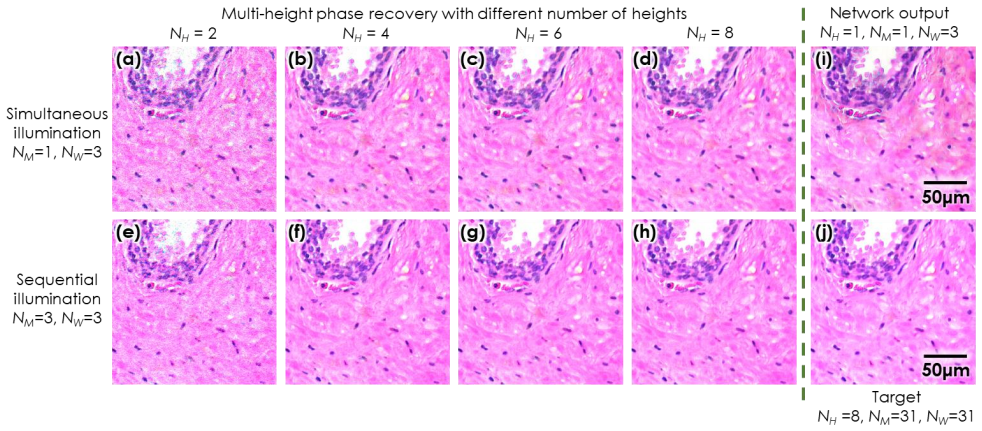
read the original abstract
We report a framework based on a generative adversarial network (GAN) that performs high-fidelity color image reconstruction using a single hologram of a sample that is illuminated simultaneously by light at three different wavelengths. The trained network learns to eliminate missing-phase-related artifacts, and generates an accurate color transformation for the reconstructed image. Our framework is experimentally demonstrated using lung and prostate tissue sections that are labeled with different histological stains. This framework is envisaged to be applicable to point-of-care histopathology, and presents a significant improvement in the throughput of coherent microscopy systems given that only a single hologram of the specimen is required for accurate color imaging.
Editorial analysis
A structured set of objections, weighed in public.
Referee Report
Summary. The paper claims a GAN-based framework for high-fidelity color holographic image reconstruction from a single multi-wavelength hologram (three simultaneous illuminations), where the network learns to remove missing-phase artifacts and apply accurate color transformation. The approach is experimentally demonstrated on lung and prostate tissue sections labeled with different histological stains, with the goal of enabling higher-throughput coherent microscopy for point-of-care histopathology.
Significance. If the generalization and artifact-free performance hold, the result would be significant for computational imaging by reducing the number of required holograms from multiple to one while producing color output, directly addressing throughput limitations in holographic microscopy. The experimental use of real stained histological samples provides a concrete demonstration of the method on relevant data.
major comments (2)
- [Abstract / experimental demonstration] Abstract and experimental section: the central claim of applicability to point-of-care histopathology for arbitrary samples rests on generalization of the GAN mapping, yet the reported experiments are restricted to lung and prostate sections with specific stains; no cross-validation, hold-out tissue types, or stain-variation tests are described to rule out memorization of training-distribution color statistics or hologram patterns.
- [Abstract] Abstract: the soundness of the high-fidelity claim cannot be assessed because no quantitative validation metrics (e.g., PSNR, SSIM, color error), error analysis, or training/validation split details are provided, leaving the weakest assumption (generalization without new artifacts on unseen samples) untested.
Simulated Author's Rebuttal
We thank the referee for the constructive comments and positive assessment of the work's significance. We provide point-by-point responses to the major comments below and have revised the manuscript to address concerns where feasible by adding clarifications and quantitative details.
read point-by-point responses
-
Referee: [Abstract / experimental demonstration] Abstract and experimental section: the central claim of applicability to point-of-care histopathology for arbitrary samples rests on generalization of the GAN mapping, yet the reported experiments are restricted to lung and prostate sections with specific stains; no cross-validation, hold-out tissue types, or stain-variation tests are described to rule out memorization of training-distribution color statistics or hologram patterns.
Authors: The experiments are restricted to lung and prostate sections with different histological stains, which provide variation in color mapping. We have revised the abstract and added a discussion section clarifying the scope of the demonstrations and explicitly noting that the applicability to arbitrary samples is envisaged rather than fully validated. The use of multiple stains helps mitigate concerns of pure memorization, but we acknowledge the absence of hold-out tissue-type testing. revision: partial
-
Referee: [Abstract] Abstract: the soundness of the high-fidelity claim cannot be assessed because no quantitative validation metrics (e.g., PSNR, SSIM, color error), error analysis, or training/validation split details are provided, leaving the weakest assumption (generalization without new artifacts on unseen samples) untested.
Authors: We agree that quantitative metrics strengthen the claims. The revised manuscript now includes PSNR, SSIM, and color error metrics, along with error analysis and explicit details on the training/validation splits (e.g., data partitioning from the acquired holograms). These were computed on the existing experimental data to support the high-fidelity assessment. revision: yes
- Additional cross-validation or tests on hold-out tissue types and further stain variations, as this would require new experimental acquisitions beyond the current dataset.
Circularity Check
No circularity: standard supervised GAN training against external ground truth
full rationale
The paper presents a GAN framework for reconstructing color images from single multi-wavelength holograms. The approach relies on supervised training where the network learns a mapping from hologram inputs to color outputs using paired experimental data from lung and prostate sections. No equations, parameters, or claims reduce the output to a fitted input by construction, nor do any load-bearing steps depend on self-citations or imported uniqueness theorems. The central result is an empirical demonstration of the trained model's performance on the reported samples, which is self-contained and falsifiable against held-out measurements.
Axiom & Free-Parameter Ledger
free parameters (1)
- GAN network weights and hyperparameters
Lean theorems connected to this paper
-
IndisputableMonolith/Cost/FunctionalEquation.leanwashburn_uniqueness_aczel unclear?
unclearRelation between the paper passage and the cited Recognition theorem.
We report a framework based on a generative adversarial network (GAN) that performs high-fidelity color image reconstruction using a single hologram of a sample that is illuminated simultaneously by light at three different wavelengths.
-
IndisputableMonolith/Foundation/ArithmeticFromLogic.leanLogicNat.induction unclear?
unclearRelation between the paper passage and the cited Recognition theorem.
The trained network learns to eliminate missing-phase-related artifacts, and generates an accurate color transformation for the reconstructed image.
What do these tags mean?
- matches
- The paper's claim is directly supported by a theorem in the formal canon.
- supports
- The theorem supports part of the paper's argument, but the paper may add assumptions or extra steps.
- extends
- The paper goes beyond the formal theorem; the theorem is a base layer rather than the whole result.
- uses
- The paper appears to rely on the theorem as machinery.
- contradicts
- The paper's claim conflicts with a theorem or certificate in the canon.
- unclear
- Pith found a possible connection, but the passage is too broad, indirect, or ambiguous to say the theorem truly supports the claim.
Reference graph
Works this paper leans on
-
[1]
Color management in digital pathology,
W. C. Revie, M. Shires, P. Ja ckson, D. Brettle, R. Cochrane, and D. Treanor, "Color management in digital pathology," Analytical Cellular Pathology 2014, (2014)
work page 2014
-
[2]
Sixty - five thousand shades of gra y: importance of color in surgical pathology diagnoses,
W. S. Campbell, G. A. Talmon, K. W. Foster, S. M. Lele, J. A. Kozel, and W. W. West, "Sixty - five thousand shades of gra y: importance of color in surgical pathology diagnoses," Human p a- thology 46, 1945–1950 (2015)
work page 1945
-
[3]
Color standardization in whole slide imaging using a color calibration slide,
P. A. Bautista, N. Hashimoto, and Y. Yagi, "Color standardization in whole slide imaging using a color calibration slide," Journal of pathology informatics 5, (2014)
work page 2014
-
[4]
Color accuracy and reproducibility in whole slide imaging scanners,
P. Shrestha and B. Hulsken, "Color accuracy and reproducibility in whole slide imaging scanners," Journal of Medical Imaging 1, 027501 (2014)
work page 2014
-
[5]
Wavelength selection for true -color holography,
M. S. Peercy and L. Hesselink, "Wavelength selection for true -color holography," Applied optics 33, 6811–6817 (1994)
work page 1994
-
[6]
A review of rgb color spaces... from xyy to r’g’b’,
D. Pascale, "A review of rgb color spaces... from xyy to r’g’b’," Babel Color 18, 136–152 (2003)
work page 2003
-
[7]
Y. Zhang, T. Liu, Y. Huang, D. Teng, Y. Bian, Y. Wu, Y. Rivenson, A. Feizi, and A. Ozcan, "A c- curate color imaging of pathology slides using holography and absorbance spectrum estimation of histochemical stains," Journal of Biophotonics e201800335 (2018)
work page 2018
-
[8]
Hyperspectral digital holography of microob- jects,
S. G. Kalenkov, G. S. Kalenkov, and A. E. Shtanko, "Hyperspectral digital holography of microob- jects," in Practical Holography XXIX: Materials and Applications (International Society for Optics and Photonics, 2015), Vol. 9386, p. 938604
work page 2015
-
[9]
Digital Holography Using Spe ctral Estimation Technique,
P. Xia, Y. Ito, Y. Shimozato, T. Tahara, T. Kakue, Y. Awatsuji, K. Nishio, S. Ura, T. Kubota, and O. Matoba, "Digital Holography Using Spe ctral Estimation Technique," J. Display Technol., JDT 10, 235–242 (2014)
work page 2014
-
[10]
Imaging without lenses: achievements and remaining challenges of wide -field on- chip microscopy,
A. Greenbaum, W. Luo, T.-W. Su, Z. Göröcs, L. Xue, S. O. Isikman, A. F. Coskun, O. Mudanyali, and A. Ozcan, "Imaging without lenses: achievements and remaining challenges of wide -field on- chip microscopy," Nat. Methods 9, 889–895 (2012)
work page 2012
-
[11]
Phase retrieval from series of images obtained by defocus variation,
L. J. Allen and M. P. Oxley, "Phase retrieval from series of images obtained by defocus variation," Optics Communications 199, 65–75 (2001)
work page 2001
-
[12]
Wide - field computational imaging of pathology slides using lens -free on -chip microscopy,
A. Greenbaum, Y. Zhang, A. Feizi, P. -L. Chung, W. Luo, S. R. Kandukuri, and A. Ozcan, "Wide - field computational imaging of pathology slides using lens -free on -chip microscopy," Science Translational Medicine 6, 267ra175-267ra175 (2014)
work page 2014
-
[13]
A. Greenbaum and A. Ozcan, "Maskless imaging of dense samples using pixel super -resolution based multi-height lensfree on-chip microscopy," Opt. Express, OE 20, 3129–3143 (2012)
work page 2012
-
[14]
A. Greenbaum, U. Sikora, and A. Ozcan, "Field -portable wide-field microscopy of dense samples using multi -height pixel super -resolution based lensfree imaging," Lab Chip 12, 1242 –1245 (2012)
work page 2012
-
[15]
Sparsity -based multi-height phase recovery in holographic microscopy,
Y. Rivenson, Y. Wu, H. Wang, Y. Zhang, A. Feizi, and A. Ozcan, "Sparsity -based multi-height phase recovery in holographic microscopy," Scientific Reports 6, 37862 (2016)
work page 2016
-
[16]
Synthetic aperture -based on-chip microscopy,
W. Luo, A. Greenbaum, Y. Zhang, and A. Ozcan, "Synthetic aperture -based on-chip microscopy," Light: Science & Applications 4, e261 (2015)
work page 2015
-
[17]
Pixel super -resolution using wavelength scanning,
W. Luo, Y. Zhang, A. Feizi, Z. Göröcs, and A. Ozcan, "Pixel super -resolution using wavelength scanning," Light Sci Appl. 5, e16060 (2016)
work page 2016
-
[18]
Lensfree on -chip microscopy over a wide field-of-view using pixel super-resolution,
W. Bishara, T. -W. Su, A. F. Coskun, and A. Ozcan, "Lensfree on -chip microscopy over a wide field-of-view using pixel super-resolution," Optics Express 18, 11181 (2010)
work page 2010
-
[19]
W. Bishara, U. Sikora, O. Mudanyali, T. -W. Su, O. Yaglidere, S. Luckhart, and A. Ozcan, "Hol o- graphic pixel super -resolution in portable lensless on -chip microscopy using a fiber -optic array," Lab on a Chip 11, 1276 (2011)
work page 2011
-
[20]
A. Greenbaum and A. Ozcan, "Maskl ess imaging of dense samples using pixel super -resolution based multi-height lensfree on-chip microscopy," Optics Express 20, 3129 (2012)
work page 2012
-
[21]
A. Greenbaum, U. Sikora, and A. Ozcan, "Field -portable wide-field microscopy of dense samples using multi-height pixel super-resolution based lensfree imaging," Lab on a Chip 12, 1242 (2012)
work page 2012
-
[22]
S. O. Isikman, W. Bishara, and A. Ozcan, "Lensfree On-chip Tomographic Microscopy Employing Multi-angle Illumination and Pixel Super-resolution," Journal of Visualized Experiments (2012)
work page 2012
-
[23]
Field -Portable Pixel Super - Resolution Colour Microscope,
A. Greenbaum, N. Akbari, A. Feizi, W. Luo, and A. Ozcan, "Field -Portable Pixel Super - Resolution Colour Microscope," PLoS ONE 8, e76475 (2013)
work page 2013
-
[24]
Wide -field computational color imaging using pixel super-resolved on-chip microscopy,
A. Greenbaum, A. Feizi, N. Akbari, and A. Ozcan, "Wide -field computational color imaging using pixel super-resolved on-chip microscopy," Optics Express 21, 12469 (2013)
work page 2013
-
[25]
Increased space-bandwidth product in pixel super -resolved lensfree on -chip microscopy ,
A. Greenbaum, W. Luo, B. Khademhosseinieh, T. -W. Su, A. F. Coskun, and A. Ozcan, "Increased space-bandwidth product in pixel super -resolved lensfree on -chip microscopy ," Sci. Rep. 3, (2013)
work page 2013
-
[26]
Demosaiced pixel super -resolution for multiplexed holographic color imaging,
Y. Wu, Y. Zhang, W. Luo, and A. Ozcan, "Demosaiced pixel super -resolution for multiplexed holographic color imaging," Sci Rep 6, (2016)
work page 2016
-
[27]
Image quality assessment: from error visibility to structural similarity,
Z. Wang, A. C. Bovik, H. R. Sheikh, and E. P. Simoncelli, "Image quality assessment: from error visibility to structural similarity," IEEE transactions on image processing 13, 600–612 (2004)
work page 2004
-
[28]
B. Hill, T. Roger, and F. W. Vorhagen, "Comparative analysis of the quantization of color spaces on the basis of the CIELAB color -difference formula," ACM Transactions on Graphics (TOG) 16, 109–154 (1997)
work page 1997
-
[29]
J. W. Goodman, Introduction to Fourier Optics (Roberts and Company Publishers, 2005)
work page 2005
-
[30]
Edge sparsity criterion for robust ho l- ographic autofocusing,
Y. Zhang, H. Wang, Y. Wu, M. Tamamitsu, and A. Ozcan, "Edge sparsity criterion for robust ho l- ographic autofocusing," Optics Letters 42, 3824 (2017)
work page 2017
-
[31]
M. Tamamitsu, Y. Zhang, H. Wang, Y. Wu, and A. Ozcan, "Comparison of Gini index and Tam u- ra coefficient for holographic autofocusing based on the edge sparsity of the complex optical wavefront," arXiv:1708.08055 [physics.optics] (2017)
work page internal anchor Pith review Pith/arXiv arXiv 2017
-
[32]
I. Goodfellow, J. Pouget -Abadie, M. Mirza, B. Xu, D. Warde -Farley, S. Ozair, A. Courville, and Y. Bengio, "Generative adversarial nets," in Advances in Neural Information Processing Systems (2014), pp. 2672–2680
work page 2014
-
[33]
Deep learning enables cross -modality super-resolution in fluorescence microscopy,
H. Wang, Y. Rivenson, Y. Jin, Z. Wei, R. Gao, H. Günaydın, L. A. Bentolila, C. Kural, and A. O z- can, "Deep learning enables cross -modality super-resolution in fluorescence microscopy," Nature Methods 16, 103–110 (2019)
work page 2019
-
[34]
Deep learning -based super-resolution in coherent imaging systems,
T. Liu, K. de Haan, Y. Rivenson, Z. Wei, X. Zeng, Y. Zhang, and A. Ozcan, "Deep learning -based super-resolution in coherent imaging systems," Scientific reports 9, 3926 (2019)
work page 2019
-
[35]
Resolution enhancement in sca n- ning electron microscopy using deep learning,
K. de Haan, Z. S. Ballard, Y. Rivenson, Y. Wu, and A. Ozcan, "Resolution enhancement in sca n- ning electron microscopy using deep learning," arXiv:1901.11094 [physics] (2019)
-
[36]
Y. Wu, Y. Luo, G. Chaudhari, Y. Rivenson, A. Calis, K. de Haan, and A. Ozcan, "Bright -field ho- lography: cross-modality deep learning enables snapshot 3D imaging with bright -field contrast us- ing a single hologram," Light: Science & Applications 8, 25 (2019)
work page 2019
-
[37]
Virtual histological staining of unlabelled tissue -autofluorescence images via deep learning,
Y. Rivenson, H. Wang, Z. Wei, K. de Haan, Y. Zhang, Y. Wu, H. Günaydın, J. E. Zuckerman, T. Chong, and A. E. Sisk, "Virtual histological staining of unlabelled tissue -autofluorescence images via deep learning," Nature Biomedical Engineering 1 (2019)
work page 2019
-
[38]
U-Net: Convolutional Networks for Biomedical Image Segmentation
O. Ronneberger, P. Fischer, and T. Brox, "U -Net: Convolutional Networks for Biomedical Image Segmentation," arXiv:1505.04597 [cs] (2015)
work page internal anchor Pith review Pith/arXiv arXiv 2015
-
[39]
Phase recovery and holographic image reconstruction using deep learning in neural networks,
Y. Rivenson, Y. Zhang, H. Güna ydın, D. Teng, and A. Ozcan, "Phase recovery and holographic image reconstruction using deep learning in neural networks," Light: Science & Applications 7, 17141 (2018)
work page 2018
-
[40]
Understanding the difficulty of training deep feedforwa rd neural ne t- works. 2010,
X. Glorot and Y. Bengio, "Understanding the difficulty of training deep feedforwa rd neural ne t- works. 2010," in Internaional Conference on Artificial Intelligence and Statistics (n.d.)
work page 2010
-
[41]
Adam: A Method for Stochastic Optimization
D. P. Kingma and J. Ba, "Adam: A method for stochastic optimization," arXiv preprint arXiv:1412.6980 (2014)
work page internal anchor Pith review Pith/arXiv arXiv 2014
-
[42]
Globally optimal stitching of tiled 3D microscopic image acquisitions,
S. Preibisch, S. Saalfeld, and P. Tomancak, "Globally optimal stitching of tiled 3D microscopic image acquisitions," Bioinformatics 25, 1463–1465 (2009). Figure 1. Comparison between the traditional hyperspectral imaging and the proposed neural network -based approaches for the recon- struction of accurate color images. NH is the number of sample-to-senso...
work page 2009
discussion (0)
Sign in with ORCID, Apple, or X to comment. Anyone can read and Pith papers without signing in.