REVIEW 4 major objections 6 minor 12 references
End-to-End Boundary Aware Networks for Medical Image Segmentation
T0 review · 4 major / 6 minor · reviewed 2026-08-14 · deepseek-v4-flash
Pith's one-line read A boundary-aware network with an edge-loss branch segments brain tumors more accurately than U-Net or V-Net, lifting Dice from 0.731/0.769 to 0.822.
desk verdict A plausible boundary-aware segmentation mechanism undermined by an internally inconsistent results table and an under-specified evaluation protocol. read the letter →
The pith
A machine-rendered reading of the paper's core claim, the machinery that carries it, and where it could break.
The reading
What carries the argument
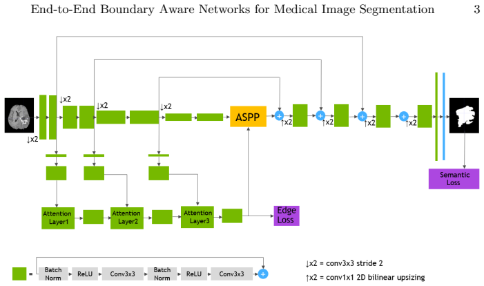
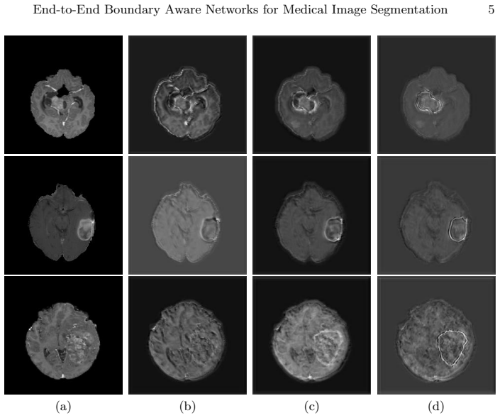
The mechanism is a two-stream encoder-decoder: a main residual-block segmentation stream plus a shape stream that receives encoder feature maps at each resolution, applies a $1\times 1$ convolution, and passes them through attention layers that compute $\alpha_l = \sigma(C_{1\times1}(s_l \| m_l))$ and gate the shape features by element-wise multiplication. The shape stream's boundary prediction is supervised by a generalized Dice loss plus a weighted binary cross-entropy edge loss, and its last attention output is concatenated with the encoder output and fed into dilated spatial pyramid pooling. The edge loss is the load-bearing component: with it, Dice reaches 0.822; without it, the boundary stream adds nothing and the model behaves like V-Net.
What would settle it
Retrain the full model and the no-edge-loss ablation on complete 3D BraTS volumes using the standard training/validation split and compare whole-tumor-core Dice; if the gap between the two models falls to near zero, the reported edge-loss benefit is an artifact of the 2D slice selection rather than a genuine segmentation improvement.
Extended reading notes
Core claim
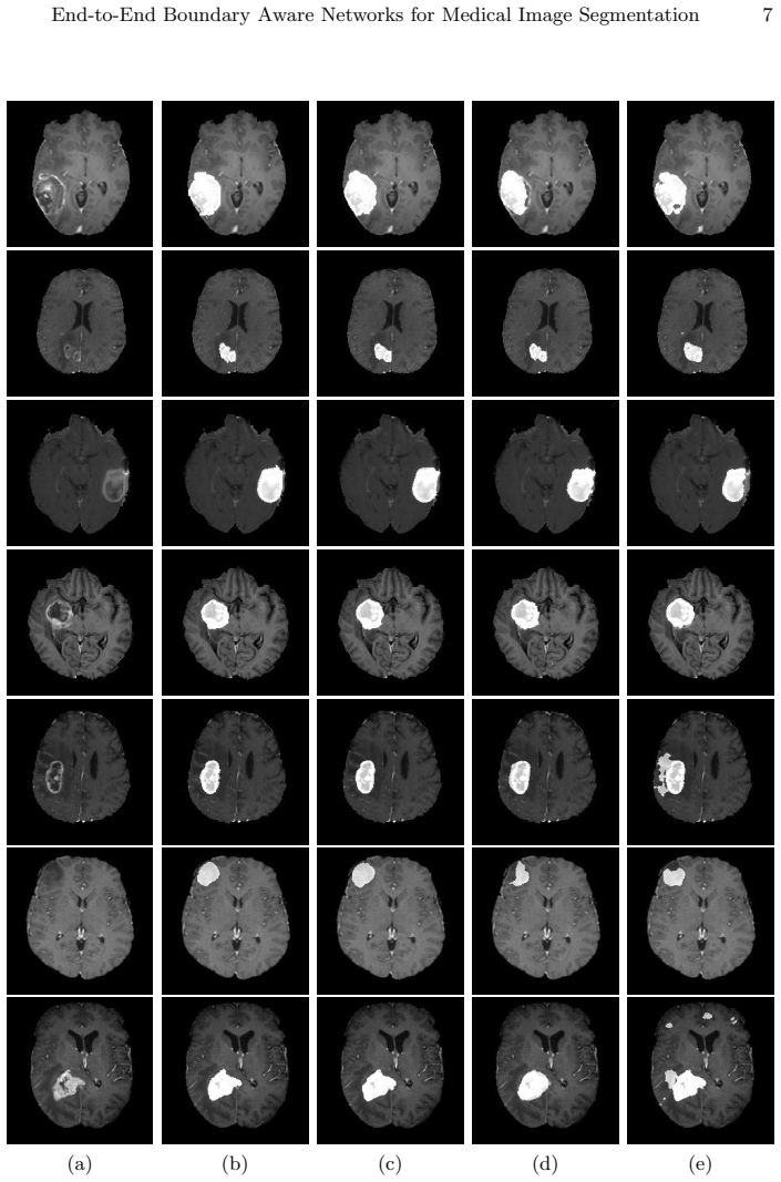
The central discovery is that supervising an auxiliary edge-stream branch with a weighted binary cross-entropy edge loss, combined with Dice losses on both the main segmentation output and the edge output, regularizes the shared encoder so that it produces finer boundaries and fewer false positives. The authors claim that their boundary-aware network outperforms U-Net and V-Net across Dice score, Jaccard index, and Hausdorff distance on the tumor-core segmentation task, and that the ablation without the edge loss performs nearly identically to V-Net (0.768 versus 0.769 Dice), pinning the improvement on the edge-aware supervision rather than on the extra parameters of the shape stream.
Load-bearing premise
The evaluation rests on 2D axial slices extracted from 3D BraTS volumes without a described train/test split or slice count, so the reported Dice gains assume those slices represent realistic full-volume tumor segmentation.
Editorial extensions
If this is right
- The edge-aware loss is the active ingredient: the same architecture without it gives a Dice of 0.768, essentially matching V-Net's 0.769.
- Boundary-aware training cuts false positives and sharpens edges, improving all three reported metrics over both baselines.
- The gain is available under a simplified setting (single T1c modality, single tumor core region, 2D slices), so it does not depend on using all four BraTS modalities.
- Since the edge stream is supervised mainly during training, adding it does not complicate inference compared with the baseline encoder-decoder.
Reading between the lines
- A natural extension the paper leaves untested is applying the same edge-supervised stream to full 3D volumes; because the paper deliberately reduces BraTS to 2D slices centered on the tumor, the reported gains might shrink or grow when evaluated on whole-volume segmentation.
- The texture-versus-shape motivation suggests the edge-aware loss might also improve robustness to image appearance shifts, a property that could be tested by evaluating on a different MRI protocol or on natural images.
- The absence of a described train/test split and slice count means the quantitative advantage should be re-measured on a standard 3D BraTS validation split before relying on the exact numbers; this is an editorial caution, not a claim in the paper.
- One could combine the edge-loss idea with other shape-bias mechanisms, such as explicit boundary alignment, to see whether the gains accumulate.
Signed reviews
Editorial analysis
A structured set of objections, weighed in public.
Referee Report
Summary. The paper proposes an end-to-end trainable boundary-aware convolutional network for medical image segmentation. The architecture augments an encoder-decoder segmentation stream with a shape stream that receives features from each encoder resolution, applies attention layers, and produces boundary predictions; the training loss combines Dice losses on the segmentation and boundary outputs with a weighted binary cross-entropy edge loss. The method is evaluated on a 2D axial-slice dataset built from BraTS 2018 volumes for tumor core segmentation and compared with U-Net and V-Net. The authors report Dice, Jaccard, and Hausdorff distances and conclude that the boundary-aware network outperforms the baselines, with an ablation showing that the edge loss is responsible for most of the gain.
Significance. The idea of explicitly supervising an edge stream alongside a segmentation stream is sensible and has been explored in related work; the paper's contribution is the specific architecture (attention layers, dilated spatial pyramid pooling, combined losses) and its application to BraTS. Strengths include an end-to-end trainable design and a direct ablation of the proposed edge loss, which is a legitimate comparison. However, the quantitative evidence is currently unreliable: the reported Dice/Jaccard pairs are mathematically inconsistent, the evaluation protocol lacks a described train/test split and patient-level separation, and no significance tests are reported. If corrected, the ablation could still support the mechanism, but the paper as written does not establish the claimed margin over U-Net and V-Net.
major comments (4)
- [Table 1, §3.2 Eq. (4)] The reported Dice and Jaccard values are mutually incompatible under the standard definitions used in the paper. For any two sets, Dice >= Jaccard because |A∪B| <= |A|+|B|; however, Table 1 reports Dice 0.731, 0.769, 0.768, 0.822 with Jaccard 0.805, 0.837, 0.832, 0.861, so every Jaccard value exceeds its corresponding Dice. Since this relation is impossible, at least one metric was computed with a nonstandard definition or the entries are misreported. The central comparison and the edge-loss ablation both depend on these numbers and must be corrected or recomputed.
- [§3.1, Table 1] The evaluation protocol is underspecified and likely biased. The authors state that "several axial slices centered around the tumor region" were extracted from each 3D volume and combined into a 2D dataset, but they do not report the number of slices, how train/validation/test sets were split, or whether splits were performed at the patient/volume level. Without patient-level separation, slices from the same volume can appear in both training and testing, allowing leakage; with tumor-centered slice selection, the evaluation is restricted to slices with large tumor burden and does not measure performance on full 3D volumes. The reported standard deviations are large and overlap across methods, and no significance tests are given, so the claimed improvement over V-Net and U-Net is not statistically supported.
- [§2.3, Eq. (5)] Equation (5) does not define the edge loss as introduced. In Eq. (3), LEdge is a function of (spred, strue), but Eq. (5) uses ypred,j in the probability terms, which refers to the main-stream segmentation output, and the definition of the sets y+ and y- is incomplete (they are said to be edge and non-edge pixel sets, but their dependence on strue is not stated). As written, the equation cannot be implemented unambiguously, and it is unclear whether the boundary stream is supervised as claimed. Please rewrite the loss with explicit arguments and define y+ and y- in terms of strue.
- [§3.2, Table 1] The comparison with U-Net and V-Net is not fully specified. The paper does not state the loss function, optimizer settings, number of epochs, input/output configuration, or data augmentation used for the baselines, nor whether they were trained on the same 2D slices with the same hyperparameters. Without these details, the reported differences could reflect implementation choices rather than architectural benefit. Please provide the training protocol for all models.
minor comments (6)
- [§3.1] There is a typo in "nectrotic core" which should be "necrotic core."
- [§2.3, Eq. (5)] The phrase "probability of the predicated class" contains a typo and should read "predicted class."
- [§3.2, Eq. (6)] The initial learning rate is written as α0 = 1.0−3, which should be formatted as 1e-3 or 10^-3 for clarity.
- [Table 1 caption] The caption says the ablation is performed "with and without this layer," but the text describes ablating the edge loss while keeping the architecture; this discrepancy should be clarified.
- [§2.3] The statement that strue "can be obtained by computing the spatial gradient of ytrue" is ambiguous; please specify the exact edge-extraction method (e.g., morphological gradient, Sobel, Canny) and any thresholding or post-processing.
- [§3.2, Table 1] The Hausdorff distance is not defined; please state whether it is the 95th percentile Hausdorff distance and in which units it is reported.
Circularity Check
No circular derivation: the edge-supervision gain is established by an internal ablation on an external dataset; self-citations are not load-bearing.
full rationale
The paper's derivation chain is empirical rather than analytic. The claimed improvement of the full model over the no-edge-loss ablation (Dice 0.822 vs 0.768, Table 1) is a direct comparison that isolates the edge-aware loss, so the central effect is not imposed by construction. The edge ground truth is derived from the segmentation mask via spatial gradient (Section 2.3), but the edge branch is an auxiliary training regularizer, not a component that is fed back as the segmentation output; therefore the segmentation prediction is not equivalent to the edge input. U-Net and V-Net baselines are external methods, and the BraTS 2018 evaluation is an external benchmark. The self-citations (refs [4], [5] in the introduction and [8] for the learning-rate schedule) are contextual or procedural and carry no load-bearing argument. The Table 1 Dice/Jaccard values are mathematically inconsistent under standard definitions, which is a serious correctness/data-reporting concern but is not a circularity. No step in the paper reduces a claimed result to its own inputs, so the circularity score is 0.
Assumptions & free parameters
free parameters (3)
- Loss weights lambda_1, lambda_2, lambda_3 =
1.0, 0.5, 0.1
- Initial learning rate alpha_0 =
1e-3
- Number of axial slices per volume =
unspecified
assumptions (3)
- domain assumption The boundary map sture is computed as the spatial gradient of the ground-truth label ytrue.
- domain assumption The 2D axial slices centered around the tumor are representative of the 3D segmentation task.
- domain assumption U-Net and V-Net are adequate baseline comparisons.
Cite this review
Pith. "Pith review of End-to-End Boundary Aware Networks for Medical Image Segmentation." pith.science (2026). https://pith.science/paper/LIA5SSOH
@misc{pith2026190808071,
  author       = {Pith},
  title        = {Pith review of: End-to-End Boundary Aware Networks for Medical Image Segmentation},
  year         = {2026},
  howpublished = {\url{https://pith.science/paper/LIA5SSOH}},
  note         = {Machine review of arXiv:1908.08071}
}
read the original abstract
Fully convolutional neural networks (CNNs) have proven to be effective at representing and classifying textural information, thus transforming image intensity into output class masks that achieve semantic image segmentation. In medical image analysis, however, expert manual segmentation often relies on the boundaries of anatomical structures of interest. We propose boundary aware CNNs for medical image segmentation. Our networks are designed to account for organ boundary information, both by providing a special network edge branch and edge-aware loss terms, and they are trainable end-to-end. We validate their effectiveness on the task of brain tumor segmentation using the BraTS 2018 dataset. Our experiments reveal that our approach yields more accurate segmentation results, which makes it promising for more extensive application to medical image segmentation.
Figures
Reference graph
Works this paper leans on
-
[1]
In: The IEEE Conference on Computer Vision and Pattern Recognition (CVPR) (2019)
Acuna, D., Kar, A., Fidler, S.: Devil is in the edges: Learning semantic boundaries from noisy annotations. In: The IEEE Conference on Computer Vision and Pattern Recognition (CVPR) (2019)
work page 2019
-
[2]
Bakas, S., Akbari, H., Sotiras, A., Bilello, M., Rozycki, M., Kirby, J., Frey- mann, J., Farahani, K., Davatzikos, C.: Advancing the cancer genome atlas glioma MRI collections with expert segmentation labels and radiomic fea- tures. Scientific Data 4 (2017)
work page 2017
-
[3]
In: International Conference on Learning Representations (ICLR) (2019)
Geirhos, R., Rubisch, P., Michaelis, C., Bethge, M., Wichmann, F.A., Brendel, W.: Imagenet-trained CNNs are biased towards texture; increasing shape bias improves accuracy and robustness. In: International Conference on Learning Representations (ICLR) (2019)
work page 2019
-
[4]
Deep Active Lesion Segmentation
Hatamizadeh, A., Hoogi, A., Sengupta, D., Lu, W., Wilcox, B., Rubin, D., Terzopoulos, D.: Deep active lesion segmentation. arXiv preprint arXiv:1908.06933 (2019)
work page Pith review arXiv 2019
-
[5]
Deep Dilated Convolutional Nets for the Automatic Segmentation of Retinal Vessels
Hatamizadeh, A., Hosseini, H., Liu, Z., Schwartz, S.D., Terzopoulos, D.: Deep dilated convolutional nets for the automatic segmentation of retinal vessels. arXiv preprint arXiv:1905.12120 (2019)
work page Pith review arXiv 2019
-
[6]
Hu, Y., Zou, Y., Feng, J.: Panoptic edge detection. https://arxiv.org/abs/1906.00590 (2019)
work page Pith review arXiv 2019
-
[7]
In: Fourth International Conference on 3D Vision (3DV) (2016)
Milletari, F., Navab, N., Ahmadi, S.A.: V-net: Fully convolutional neural networks for volumetric medical image segmentation. In: Fourth International Conference on 3D Vision (3DV) (2016)
work page 2016
-
[8]
In: BrainLes, Medical Image Computing and Computer Assisted Intervention (MICCAI)
Myronenko, A.: 3D MRI brain tumor segmentation using autoencoder regu- larization. In: BrainLes, Medical Image Computing and Computer Assisted Intervention (MICCAI). pp. 311–320. LNCS, Springer (2018)
work page 2018
Show all 12 references
-
[9]
In: Proc
Ronneberger, O., P.Fischer, Brox, T.: U-net: Convolutional networks for biomedical image segmentation. In: Proc. MICCAI. LNCS, vol. 9351, pp. 234–241 (2015)
2015
-
[10]
arXiv preprint arXiv:1907.05740 (2019)
Takikawa, T., Acuna, D., Jampani, V., Fidler, S.: Gated-scnn: Gated shape cnns for semantic segmentation. arXiv preprint arXiv:1907.05740 (2019)
2019 arXiv
-
[11]
In: CVPR (2017)
Yu, Z., Feng, C., Liu, M., Ramalingam, S.: Casenet: Deep category-aware semantic edge detection. In: CVPR (2017)
2017
-
[12]
In: European Conference on Computer Vision (ECCV) (2018)
Yu, Z., Liu, W., Zou, Y., Feng, C., Ramalingam, S., Vijaya Kumar, B., Kautz, J.: Simultaneous edge alignment and learning. In: European Conference on Computer Vision (ECCV) (2018)
2018
Reviewed August 14, 2026 · model on record in the stance chip above.
Discussion (0). Continue with ORCID to comment.