REVIEW 4 major objections 4 minor 24 references
Asymmetric Lesion Detection with Geometric Patterns and CNN-SVM Classification
T0 review · 4 major / 4 minor · reviewed 2026-08-06 · deepseek-v4-flash
Pith's one-line read This paper claims that a four-quadrant pixel-count rule detects dermatologically asymmetric lesions with 99% agreement on two dermoscopic datasets, and that a ResNet-101 plus SVM classifier reproduces the three shape labels with up to 97%…
desk verdict The ISIC2016 accuracy is circular because the algorithm helped build the ground truth; the PH2 result and the dataset idea are the real contributions. read the letter →
The pith
A machine-rendered reading of the paper's core claim, the machinery that carries it, and where it could break.
The reading
What carries the argument
The load-bearing object is the Geometry Shape-Based Asymmetry Analysis (GSAA) rule: a binary lesion mask is split into four quadrants by perpendicular axes through the lesion centroid, and the white-pixel counts in the four parts are compared as ratios. The 0.9-to-1.1 acceptance window turns continuous area ratios into binary match or no-match decisions, and the pattern of matches across the four pairwise ratios yields the three shape labels of asymmetric, half-symmetric, and symmetric. The second mechanism is transfer learning: features are taken from the 'fc1000' layer of pretrained ResNet18, ResNet50, or ResNet101 and passed to a multiclass linear SVM trained with a one-versus-one coding design. The GSAA rule does the labeling work, while the ResNet-plus-SVM stack does the automatic classification work.
What would settle it
Take the 1279 ISIC2016 images, have a panel of dermatologists label lesion shape without access to the GSAA output or the pixel-ratio code, then recompute GSAA's agreement and the ResNet101-plus-SVM evaluation against those blind labels; if the kappa drops materially below the reported 98.5% and 94%, the claimed accuracy was partly an artifact of label-generation feedback.
Extended reading notes
Core claim
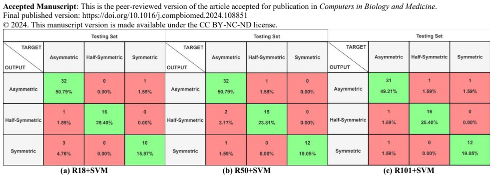
The paper's central claim is that lesion-shape asymmetry, the first criterion of the ABCDE melanoma rule, can be measured by a pixel-count ratio rather than by holistic visual judgment. GSAA divides a binary mask into four quadrants A, B, C, and D at the lesion centroid, counts white pixels in each, and forms four ratios ($A_p/B_p$, $A_p/D_p$, $B_p/C_p$, $C_p/D_p$); each ratio in the window 0.9 to 1.1 counts as a match. If all four ratios fail to match, the lesion is asymmetric; one or two matches means half-symmetric; three or more means symmetric. On PH2 this rule reproduces expert labels with 99.00% accuracy (kappa 98.2%), and on the newly expert-labeled ISIC2016 set with 99.06% accuracy (kappa 98.5%). The paper then argues that features extracted from the layer before ResNet101's classification layer, fed to a one-versus-one linear SVM, classify the three shape classes with 94% kappa, 95% macro F1, and 97% weighted F1 on APH2, and that this combination outperforms the decision-tree, histogram, neural-network, and prior CNN baselines included in the comparison.
Load-bearing premise
The reported accuracies assume the ISIC2016 ground truth was set by independent expert judgment, but the paper says the pixel-ratio imaging algorithm was used to augment those manual annotations, so the 99% agreement may partly compare the algorithm with labels it helped produce.
Editorial extensions
If this is right
- A non-expert can now point to a concrete, repeatable numerical rule for the asymmetry criterion instead of relying only on subjective visual inspection.
- The labeled ISIC2016 shape classes (667 asymmetric, 344 half-symmetric, 268 symmetric) become a training resource for future dermoscopic shape models.
- A pretrained ResNet with a linear SVM can classify three shape classes with high agreement using a small 5% test split, suggesting a data-efficient path for shape classification.
- GSAA can generate ground-truth shape labels for unannotated datasets, reducing the bottleneck of expert annotation when training data are scarce.
Reading between the lines
- If the ISIC2016 labels were co-produced by the same pixel-ratio logic, the 99%-class agreement figures reflect consistency between GSAA and its own label-augmentation step as much as agreement with independent experts; a blind re-annotation study would separate the two.
- The 0.9-to-1.1 threshold is a free parameter, so sweeping it against blind expert labels could produce a different optimal operating point and the reported rates are not invariant to that choice.
- The paper's own suggested eight-sector split is a natural stress test: if 45-degree sectors reproduce the three classes with similar agreement, the method generalizes, and if not, the four-quadrant rule may be tuned to the datasets used.
- Because GSAA operates on binary masks, its ceiling is set by segmentation quality; on imperfect segmentations the pixel-ratio method will inherit the segmenter's errors even if the ratio logic itself is sound.
Editorial analysis
A structured set of objections, weighed in public.
Referee Report
Summary. The manuscript proposes a two-stage method for classifying dermoscopic lesion shape into three classes (asymmetric, half-symmetric, symmetric). First, a pixel-count-based geometric algorithm (GSAA) divides binary lesion masks into four quadrants at the centroid and thresholds pairwise white-pixel ratios to assign a shape label; it is applied to PH2 and to a newly annotated ISIC2016 symmetry set. Second, features from three pretrained ResNets are fed to a multiclass linear SVM trained on augmented PH2 (APH2) and ISIC2016, with experiments in four train/test configurations. The headline results are 99.00% GSAA accuracy on PH2, 99.06% on ISIC2016, and 94% Kappa / 95% Macro F1 / 97% Weighted F1 for R101+SVM on the APH2 same-dataset test.
Significance. If the reported numbers are valid, the paper would provide a useful explainable support tool for non-experts and the first three-class symmetry annotation for ISIC2016, with a straightforward CNN-SVM pipeline. The PH2 evaluation is a genuine external check for GSAA, and the confusion matrices allow verification of the aggregate metrics. However, the ISIC2016 ground truth may have been influenced by GSAA itself, and the APH2 evaluation suffers from image-level leakage; until these are resolved, the empirical support for the central claims is limited. The paper's strength is its transparency: the GSAA rules are fully specified, the datasets are public, and the CNN-SVM protocol is reproducible.
major comments (4)
- [Section 3.1, Table 5] The ISIC2016 ground truth is not demonstrably independent of the GSAA algorithm. Section 3.1 states that "to augment our manual annotations, we developed an imaging algorithm designed to quantitatively assess lesion symmetry based on pixel counts within the segmented masks," and Section 3.3 later evaluates GSAA against this same ground truth, reporting 99.06% accuracy in Table 5. If the final labels were influenced by GSAA during labeling, the agreement is circular and the ISIC2016 rows of Tables 5, 6, 7, 10, and 11 are correspondingly inflated. The sentence in Section 3.3 that "the output of GSAA is not used" for CNN training does not address the possibility that the labels themselves incorporate GSAA. The authors must either disclose a labeling protocol in which GSAA played no role in the final expert decisions, or re-annotate a held-out subset for independent evaluation.
- [Section 3.1, Table 8] The APH2 evaluation uses a random split after mirror augmentation, which leaks identical lesions across the train/test boundary. Each of the 200 PH2 images is mirrored vertically and horizontally to produce 600 images, so each original lesion appears in three near-duplicate copies. If the 95/5 split is performed on the 600 images, the same original lesion can appear in both training and test sets; with 600 images and a 5% test fraction, most of the 200 originals will be split across the sets. This makes the same-dataset results in Fig. 8 and Table 8 (97% weighted F1, 94% Kappa) unreliable as estimates of generalization. The split should be stratified at the original-lesion level, e.g., by assigning all mirrored copies of each PH2 image to the same fold.
- [Section 4.1, Tables 6-9] The test sets are small and the results are given for a single random split. APH2 contributes 31 test images and ISIC2016 63 test images; with three classes, the per-class counts are in single digits for some classes (e.g., the half-symmetric class in the APH2 test set appears to have only 4-5 instances). The headline Kappa and F1 values therefore have very wide confidence intervals, and the repeated conclusion that "R101+SVM consistently emerges as the top-performing model" is not supported by any significance test. Please report results over multiple random splits (e.g., 5-fold or repeated 95/5 splits) with means and standard deviations, or at least provide the exact test-set CMs and class counts so readers can assess the uncertainty.
- [Section 4.2, Table 10] The comparative study does not support the claim that the proposed method "outperforms state-of-the-art methods." The proposed method's numbers are taken from different experimental configurations (the table itself cites "From tables 7&9"), while the comparison methods use their own datasets, class numbers (2 vs 3), and evaluation protocols. Accuracy values for different class counts are not directly comparable, and the APH2 accuracy is inflated by the leakage described above. A fair comparison requires a common benchmark and protocol, or at least matching class definitions and held-out splits.
minor comments (4)
- [Section 3.2, Eq. (7)] Equation (7) defines accuracy in binary terms (TP, TN, P, N) but is applied to three-class confusion matrices; please state that accuracy is computed as the fraction of correctly classified samples on the diagonal.
- [Section 3.4] The test set size for APH2 is given as 31 images, but 5% of 600 is 30; please verify the rounding or the augmentation count.
- [Table 10] Table 10 does not state which of Tables 6-9 each proposed-method accuracy value comes from; please add explicit table references so the reader can trace the provenance of each number.
- [Section 3.3] The GSAA ratio threshold of 0.90-1.10 is introduced without justification; please explain how this tolerance was chosen and whether the reported PH2 accuracy is sensitive to it.
Circularity Check
ISIC2016 ground-truth labels were augmented by the same pixel-count algorithm later scored at 99.06% on them, so the ISIC2016 GSAA accuracy and the CNN results measured against those labels are not fully external.
-
self definitional
[Section 3.1 (Data Acquisition); GSAA evaluation in Section 3.3, Table 5]
"Furthermore, to augment our manual annotations, we developed an imaging algorithm designed to quantitatively assess lesion symmetry based on pixel counts within the segmented masks. This algorithm splits each lesion into four distinct sections, calculating the proportion of pixels within each segment relative to the total lesion area."
This imaging algorithm is the same four-quadrant pixel-count method later presented as GSAA in Section 3.3. The ISIC2016 ground-truth labels were therefore not independent expert labels: they were augmented by GSAA during construction. When GSAA is then scored against that ground truth, as reported in Section 3.3 ('These outputs match the ground truth with an accuracy of 99.06%, as presented in the CM in Table 5'), the comparison is between the algorithm and a benchmark it helped create. The 99.06% ISIC2016 accuracy is therefore partly determined by the labeling procedure itself rather than by fully external clinical consensus.
-
other
[Section 3.1 vs. Section 3.3 and Section 4.1 (Tables 6 and 7)]
"By combining manual annotations from clinical dermatologists with computational techniques, we provide ground truths about lesion shape and introduce a methodology for objectively quantifying symmetry characteristics. ... Since PH2 and ISIC2016 have clinical-based ground truth, which are more reliable than GSAA generated ground truth. For this reason, the clinical-based ground truth of PH2 and ISIC2016 are used to train and evaluate the proposed CNN."
The second passage claims that the ISIC2016 CNN labels are clinical-based ground truth, but the first passage states that the ISIC2016 ground truths were produced by combining expert annotations with a computational imaging algorithm, i.e., the same pixel-count approach later called GSAA. Consequently, the CNN in-dataset ISIC2016 results in Table 6 and the ISIC2016-testing leg in Table 7 are evaluated against labels that GSAA helped define. This does not make the CNN scores tautological, but it removes their status as independent external validation; the ISIC2016 CNN numbers inherit the circularity of the ISIC2016 ground-truth construction.
full rationale
The circularity is real but partial. The PH2 path is independent: PH2 has pre-existing clinical lesion-shape ground truth, so the 99.00% GSAA agreement on PH2 (Table 3) and the best CNN headline figures from the APH2 in-dataset experiment (Table 8: 94% Kappa, 95% Macro F1, 97% Weighted F1) do not reduce to the algorithm's own construction. The problem is confined to ISIC2016: Section 3.1 explicitly says the authors augmented their manual annotations with a pixel-count imaging algorithm, and Section 3.3 then reports 99.06% GSAA agreement against those augmented labels and uses them as 'ground truth' for CNN training and evaluation. The later statement that 'the output of GSAA is not used' for CNN training does not resolve the ambiguity, because the labels themselves may have been shaped by GSAA. Cross-dataset experiments (APH2-to-ISIC2016 and ISIC2016-to-APH2) provide some independent evidence, since one leg uses independent PH2-derived labels, but the ISIC2016 leg still depends on possibly algorithm-augmented labels. There are no load-bearing self-citations or imported uniqueness theorems; the cited prior work is external. Overall, the ISIC2016 GSAA success and the ISIC2016 CNN scores are partially circular, giving a score of 6 rather than a higher score because the PH2-based claims remain externally grounded.
Assumptions & free parameters
free parameters (3)
- GSAA quadrant ratio tolerance =
0.90 to 1.10
- GSAA quadrant pairing scheme =
Ap/Bp, Ap/Dp, Bp/Cp, Cp/Dp
- Train/test split ratio =
95/5
assumptions (4)
- domain assumption Binary lesion masks accurately represent the lesion shape relevant to clinical asymmetry.
- domain assumption The centroid-based split into four quadrants is a valid operationalization of dermatological symmetry.
- domain assumption ImageNet-pretrained features transfer to dermoscopic lesion shape classification.
- domain assumption Expert labels for PH2 and ISIC2016 are correct and independent of the proposed algorithm.
Cite this review
Pith. "Pith review of Asymmetric Lesion Detection with Geometric Patterns and CNN-SVM Classification." pith.science (2026). https://pith.science/paper/DIFYZ363
@misc{pith2026250717185,
  author       = {Pith},
  title        = {Pith review of: Asymmetric Lesion Detection with Geometric Patterns and CNN-SVM Classification},
  year         = {2026},
  howpublished = {\url{https://pith.science/paper/DIFYZ363}},
  note         = {Machine review of arXiv:2507.17185}
}
read the original abstract
In dermoscopic images, which allow visualization of surface skin structures not visible to the naked eye, lesion shape offers vital insights into skin diseases. In clinically practiced methods, asymmetric lesion shape is one of the criteria for diagnosing melanoma. Initially, we labeled data for a non-annotated dataset with symmetrical information based on clinical assessments. Subsequently, we propose a supporting technique, a supervised learning image processing algorithm, to analyze the geometrical pattern of lesion shape, aiding non-experts in understanding the criteria of an asymmetric lesion. We then utilize a pre-trained convolutional neural network (CNN) to extract shape, color, and texture features from dermoscopic images for training a multiclass support vector machine (SVM) classifier, outperforming state-of-the-art methods from the literature. In the geometry-based experiment, we achieved a 99.00% detection rate for dermatological asymmetric lesions. In the CNN-based experiment, the best performance is found with 94% Kappa Score, 95% Macro F1-score, and 97% Weighted F1-score for classifying lesion shapes (Asymmetric, Half-Symmetric, and Symmetric).
Figures


Figures from the paper (5 more)



Reference graph
Works this paper leans on
-
[1]
ABCDE —An Evolving Concept in the Early Detection of Melanoma
Rigel, D.S., Friedman, R.J., Kopf, A.W., Polsky, D., 2005. ABCDE —An Evolving Concept in the Early Detection of Melanoma. Archives of Dermatology 141. https://doi.org/10.1001/archderm.141.8.1032
-
[2]
Three -point checklist of dermoscopy: an open internet study
Zalaudek, I., Argenziano, G., Soyer, H.P., Corona, R., Sera, F., Blum, A., Braun, R.P., Cabo, H., Ferrara, G., Kopf, A.W., La ngford, D., Menzies, S.W., Pellacani, G., Peris, K., Seidenari, S., 2005. Three -point checklist of dermoscopy: an open internet study. British Journal of Dermatology 154, 431–437. https://doi.org/10.1111/j.1365-2133.2005.06983.x
-
[3]
CASH Algo rithm for Dermoscopy Revisited
Henning, J.S., Stein, J.A., Yeung, J., Dusza, S.W., Marghoob, A.A., Rabinovitz, H.S., Polsky, D., Kopf, A.W., 2008. CASH Algo rithm for Dermoscopy Revisited. Archives of Dermatology 144. https://doi.org/10.1001/archderm.144.4.554
-
[4]
F.; Celebi, Emre; Helba, Brian; Marchetti, Michael; Mishra, Nabin; Halpern, Allan
Gutman, David; Codella, Noel C. F.; Celebi, Emre; Helba, Brian; Marchetti, Michael; Mishra, Nabin; Halpern, Allan. "Skin Lesi on Analysis toward Melanoma Detection: A Challenge at the International Symposium on Biomedical Imaging (ISBI) 2016, hosted by the Intern ational Skin Imaging Collaboration (ISIC)". eprint arXiv:1605.01397. 2016. Accepted Manuscrip...
arXiv 2016
-
[5]
Lorentzen, H.F., Weismann, K., Larsen, F.G., 2001. Structural asymmetry as a dermatoscopic indicator of malignant melanoma – a latent class analysis of sensitivity and classification errors. Melanoma Research 11, 495–501. https://doi.org/10.1097/00008390-200110000-00009
-
[6]
Determining the asymmetry of skin lesion with fuzzy borders
Ng, V.T.Y., Fung, B.Y.M., Lee, T.K., 2005. Determining the asymmetry of skin lesion with fuzzy borders. Computers in Biology and Medicine 35, 103–120. https://doi.org/10.1016/j.compbiomed.2003.11.004
-
[7]
Automatic boundary detection and symmetry calculation in dermoscopy images of skin lesions
7.Sirakov, N.M., Mete, M., Chakrader, N.S., 2011. Automatic boundary detection and symmetry calculation in dermoscopy images of skin lesions. 2011 18th IEEE International Conference on Image Processing. https://doi.org/10.1109/icip.2011.6115757
-
[8]
Chakravorty R, Liang S, Abedini M, Garnavi R. Dermatologist -like feature extraction from skin lesion for improved asymmetry classification in PH2 database. Proceedings of the Annual International Conference of the IEEE Engineering in Medicine and Biology Society, EMB S, vol. 2016- October, Institute of Electrical and Electronics Engineers Inc.; 2016, p. ...
Show all 24 references
-
[9]
Skin lesions dermatological shape asymmetry measures,
P. Milczarski, Z. Stawska and P. Maślanka , "Skin lesions dermatological shape asymmetry measures," 2017 9th IEEE International Conference on Intelligent Data Acquisition and Advanced Computing Systems: Technology and Applications (IDAACS), 2017, pp. 1056 -1061, doi: 10.1109/I...
2017
-
[10]
Quantitative evaluation of binary digital region asymmetry with application to skin lesion detection
Sancen-Plaza, A., Santiago-Montero, R., Sossa, H., Perez -Pinal, F.J., Martinez-Nolasco, J.J., Padilla-Medina, J.A., 2018. Quantitative evaluation of binary digital region asymmetry with application to skin lesion detection. BMC Medical Informatics and Decision Making 18. http...
2018 doi
-
[11]
Towards the automatic detection of skin lesion shape asymmetry, color variegation and diameter in dermoscopic images
Ali AR, Li J, O’Shea SJ. Towards the automatic detection of skin lesion shape asymmetry, color variegation and diameter in dermoscopic images. PLoS One 2020;15. https://doi.org/10.1371/journal.pone.0234352
2020 doi
-
[12]
Skin Lesions Asymmetry Estimation Using Artificial Neural Networks
Damian FA, Moldovanu S, Moraru L. Skin Lesions Asymmetry Estimation Using Artificial Neural Networks. 2021 25th International Conference on System Theory, Control and Computing, ICSTCC 2021 - Proceedings, Institute of Electrical and Electronics Engineers Inc.; 2021, p. 64 –7. ...
2021
-
[13]
Asymmetry analysis of melanoma based on ABCD rule
Zhang, G., Guo, S., 2021. Asymmetry analysis of melanoma based on ABCD rule. Journal of Physics: Conference Series 1883, 0120 70. https://doi.org/10.1088/1742-6596/1883/1/012070
2021 doi
-
[14]
A novel approach for skin lesion symmetry classification with a deep learning model
Talavera-Martínez, L., Bibiloni, P., Giacaman, A., Taberner, R., Hernando, L.J.D.P., González -Hidalgo, M., 2022. A novel approach for skin lesion symmetry classification with a deep learning model. Computers in Biology and Medicine 145, 105450. https://doi.org/10.1016/j.compb...
2022
-
[15]
Ferreira, Jorge Marques, Andre R
Teresa Mendonça, Pedro M. Ferreira, Jorge Marques, Andre R. S. Marcal, Jorge Rozeira. PH² - A dermoscopic image database for research and benchmarking, 35th International Conference of the IEEE Engineering in Medicine and Biology Society, July 3-7, 2013, Osaka, Japan
2013
-
[16]
Imagenet: A large -scale hierarchical image database
Deng, Jia, et al. "Imagenet: A large -scale hierarchical image database." Computer Vision and Pattern Recognition, 2009. CVPR 2009. IEEE Conference on. IEEE, 2009
2009
-
[17]
Deep Residual Learning for Image Recognition
He, K., Zhang, X., Ren, S., Sun, J., 2016. Deep Residual Learning for Image Recognition. 2016 IEEE Conference on Computer Vis ion and Pattern Recognition (CVPR). https://doi.org/10.1109/cvpr.2016.90
2016 doi
-
[18]
ImageNet classification with deep convolutional neural networks
Krizhevsky, A., Sutskever, I., Hinton, G.E., 2017. ImageNet classification with deep convolutional neural networks. Communica tions of the ACM 60, 84–90. https://doi.org/10.1145/3065386
2017 doi
-
[19]
Review of deep learning: concepts, CNN architectures, challenges, applications, future directions
Alzubaidi, L., Zhang, J., Humaidi, A.J., Al-Dujaili, A., Duan, Y., Al-Shamma, O., Santamaría, J., Fadhel, M.A., Al-Amidie, M., Farhan, L., 2021. Review of deep learning: concepts, CNN architectures, challenges, applications, future directions. Journal of Big Data 8. https://do...
2021 doi
-
[20]
and Darrell, T., 2014, January
Donahue, J., Jia, Y., Vinyals, O., Hoffman, J., Zhang, N., Tzeng, E. and Darrell, T., 2014, January. Decaf: A deep convolutio nal activation feature for generic visual recognition. In International conference on machine learning (pp. 647-655). PMLR
2014
-
[21]
Minimizing Average of Loss Functions Using Gradient Descent and Stochastic Gradient Desc ent
Arefin, M.R., Asadujjaman, M., 2016. Minimizing Average of Loss Functions Using Gradient Descent and Stochastic Gradient Desc ent. Dhaka University Journal of Science 64, 141–145. https://doi.org/10.3329/dujs.v64i2.54490
2016 doi
-
[22]
An improved multiclass LogitBoost using adaptive -one-vs-one
Sun, P., Reid, M.D., Zhou, J., 2014. An improved multiclass LogitBoost using adaptive -one-vs-one. Machine Learning 97, 295 –326. https://doi.org/10.1007/s10994-014-5434-3
2014 doi
-
[23]
Skin Lesion Analysis Toward Melanoma Detection 2018: A Challenge Hosted by the International Skin Imaging Collaboration (ISIC)
Noel Codella, Veronica Rotemberg, Philipp Tschandl, M. Emre Celebi, Stephen Dusza, David Gutman, Brian Helba, Aadi Kalloo, Ko nstantinos Liopyris, Michael Marchetti, Harald Kittler, Allan Halpern: "Skin Lesion Analysis Toward Melanoma Detection 2018: A Challenge Hosted by the ...
2018 arXiv
-
[24]
The HAM10000 dataset, a large collection of multi -sourcedermatoscopic images of common pigmented skin lesions
Tschandl, P., Rosendahl, C., Kittler, H., 2018. The HAM10000 dataset, a large collection of multi -sourcedermatoscopic images of common pigmented skin lesions. Scientific Data 5. https://doi.org/10.1038/sdata.2018.161
2018 doi
Reviewed August 6, 2026 · model on record in the stance chip above.
Discussion (0). Sign in to comment.