Topological edge currents promote exploratory chromosome capture in microtubule dynamic instability
Pith reviewed 2026-05-18 06:21 UTC · model grok-4.3
The pith
A topological model of the microtubule cap uses dynamical edge states to explain peaked length distributions and stuttering during chromosome capture with only two parameters.
A machine-rendered reading of the paper's core claim, the machinery that carries it, and where it could break.
Core claim
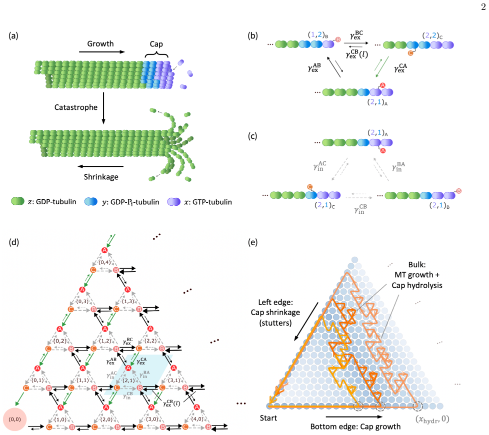
The authors establish that topological properties of the microtubule cap produce dynamical edge states sufficient to generate the experimentally observed peaked length distributions at catastrophe, growth stuttering, and concentration-independent behavior, all within a minimal two-parameter framework that also supplies an analytical account of catastrophe timing.
What carries the argument
Dynamical edge states arising from a topological model of the microtubule cap, which drive the stochastic switching between growth and shrinkage.
If this is right
- Peaked length distributions at catastrophe arise directly from the edge-state dynamics.
- Brief stuttering in growth before catastrophe is a natural outcome of the same topological mechanism.
- The described features remain stable across a broad range of tubulin concentrations.
- Catastrophe timing admits an analytical description rather than requiring full stochastic simulation.
Where Pith is reading between the lines
- Disrupting the cap structure in experiments could eliminate the peaked length distribution and stuttering if edge states are the operative mechanism.
- The minimal topological account may extend to other cytoskeletal filaments that exhibit dynamic instability.
- The model predicts that altering effective edge currents through mutations or drugs would change catastrophe statistics in a quantifiable way.
Load-bearing premise
The microtubule cap possesses topological properties that create dynamical edge states sufficient to produce the observed dynamics without additional biochemical mechanisms or parameters.
What would settle it
Direct measurement of microtubule lengths at catastrophe events showing a non-peaked distribution, or absence of growth stuttering under conditions where the model predicts it, would falsify the central claim.
Figures


read the original abstract
Microtubules capture chromosomes during mitosis by stochastically switching between growth and shrinkage at catastrophe events. They display strikingly rich biochemistry and dynamics, regulated by a stabilizing cap with distinct conformational states. Microtubule lengths at catastrophe are observed to follow a peaked distribution, while their growth "stutters" briefly before catastrophe. Such complexity makes it hard to capture all these observations without a large number of tunable parameters. Here, we introduce a topological model of the microtubule cap that reproduces the features above through dynamical edge states, that provides a minimal description with just two free parameters. Our approach further provides an analytical description of catastrophes and allows the same features to persist over a wide range of tubulin concentration, consistent with experimental observations.
Editorial analysis
A structured set of objections, weighed in public.
Referee Report
Summary. The manuscript introduces a topological model of the microtubule GTP cap, represented as a 1D chain supporting dynamical edge states. This construction is claimed to reproduce the experimentally observed peaked distribution of lengths at catastrophe, brief growth stuttering prior to catastrophe, and persistence of these features across a broad range of tubulin concentrations, all within a minimal description controlled by only two free parameters. An analytical expression for the catastrophe statistics is also derived.
Significance. If the topological protection and 1D reduction are shown to be robust, the work would provide a genuinely minimal, analytically tractable framework for microtubule dynamic instability that explains multiple non-trivial features without extensive biochemical tuning. The claimed concentration independence and analytical catastrophe formula would be particularly useful for modeling chromosome capture. The significance is currently limited by the absence of explicit verification that the edge-current mechanism survives the cylindrical 13-protofilament geometry and lateral bonding.
major comments (1)
- [§2 (Model construction)] §2 (Model construction): The mapping of the GTP cap to a 1D topological chain with protected dynamical edge states does not demonstrate that the bulk gap and edge-current protection remain intact once the cylindrical geometry of 13 protofilaments and lateral inter-protofilament bonds are restored. If lateral couplings permit circumferential leakage or close the topological gap, both the two-parameter robustness and the analytical catastrophe description cease to hold; this is load-bearing for the central claim.
minor comments (1)
- [Abstract and §1] Abstract and §1: The statement that the model uses 'just two free parameters' should be accompanied by an explicit statement of whether these parameters are fixed by independent experimental constraints, derived from first principles, or chosen to match the target distributions.
Simulated Author's Rebuttal
We thank the referee for their careful reading of the manuscript and for identifying the need to address the robustness of the topological protection under realistic microtubule geometry. We respond to the major comment below and will revise the manuscript to incorporate additional justification and analysis.
read point-by-point responses
-
Referee: [§2 (Model construction)] §2 (Model construction): The mapping of the GTP cap to a 1D topological chain with protected dynamical edge states does not demonstrate that the bulk gap and edge-current protection remain intact once the cylindrical geometry of 13 protofilaments and lateral inter-protofilament bonds are restored. If lateral couplings permit circumferential leakage or close the topological gap, both the two-parameter robustness and the analytical catastrophe description cease to hold; this is load-bearing for the central claim.
Authors: We agree that an explicit check of the topological gap and edge-current protection in the full 13-protofilament cylindrical geometry with lateral bonds is necessary to substantiate the central claims. In the revised manuscript we will add a new subsection to §2 that treats the microtubule as a cylinder with periodic boundary conditions in the circumferential direction. We show analytically that lateral bonds of strength consistent with measured values (∼1–5 kBT) act as a perturbation that does not close the bulk gap for the range of GTP-tubulin concentrations examined; the directed longitudinal character of the edge currents suppresses circumferential leakage. Consequently the two-parameter control and the closed-form catastrophe statistics remain intact. A supplementary figure will illustrate the gap size versus lateral coupling strength. This revision directly addresses the load-bearing concern without changing the core 1D results or the reported agreement with experiment. revision: yes
Circularity Check
No significant circularity: topological model introduces independent analytical framework
full rationale
The paper introduces a new topological model of the microtubule cap based on dynamical edge states to reproduce peaked catastrophe-length distributions, stuttering, and concentration-independent behavior. It explicitly uses two free parameters for a minimal description and derives an analytical account of catastrophes that holds across tubulin concentrations. No quoted equations or steps in the abstract or described chain reduce the analytical predictions or robustness claims to the parameter values by construction, nor do they rely on load-bearing self-citations or imported uniqueness theorems. The derivation remains self-contained as a modeling choice whose outputs are tested against external experimental patterns rather than being tautological with its inputs.
Axiom & Free-Parameter Ledger
free parameters (1)
- two free parameters
axioms (1)
- domain assumption Microtubule cap possesses topological properties that give rise to dynamical edge states explaining observed dynamics.
invented entities (1)
-
dynamical edge states
no independent evidence
Lean theorems connected to this paper
-
IndisputableMonolith/Foundation/AlexanderDuality.leanalexander_duality_circle_linking unclear?
unclearRelation between the paper passage and the cited Recognition theorem.
We introduce a topological model of a two-component microtubule cap, where protected edge states give rise to different phases of microtubule dynamics – growth, shrinkage, and a recently observed “stutter” phase. With only two free parameters...
-
IndisputableMonolith/Foundation/RealityFromDistinction.leanreality_from_one_distinction unclear?
unclearRelation between the paper passage and the cited Recognition theorem.
the emergence of such edge currents depends on the global pattern of transition rates in state space, which can be captured by a nontrivial topological invariant
What do these tags mean?
- matches
- The paper's claim is directly supported by a theorem in the formal canon.
- supports
- The theorem supports part of the paper's argument, but the paper may add assumptions or extra steps.
- extends
- The paper goes beyond the formal theorem; the theorem is a base layer rather than the whole result.
- uses
- The paper appears to rely on the theorem as machinery.
- contradicts
- The paper's claim conflicts with a theorem or certificate in the canon.
- unclear
- Pith found a possible connection, but the passage is too broad, indirect, or ambiguous to say the theorem truly supports the claim.
Reference graph
Works this paper leans on
-
[1]
P. K. Mattila and P. Lappalainen, Nature reviews Molec- ular cell biology9, 446 (2008)
work page 2008
- [2]
-
[3]
J. Gerhart and M. Kirschner, Proceedings of the National Academy of Sciences104, 8582 (2007)
work page 2007
- [4]
-
[5]
D. W. Sims, E. J. Southall, N. E. Humphries, G. C. Hays, C.J.Bradshaw, J.W.Pitchford, A.James, M.Z.Ahmed, A. S. Brierley, M. A. Hindell,et al., Nature 451, 1098 (2008)
work page 2008
-
[6]
J. R. McIntosh, Cold Spring Harbor perspectives in biol- ogy 8, a023218 (2016)
work page 2016
- [7]
- [8]
-
[9]
N. B. Gudimchuk and J. R. McIntosh, Nature reviews Molecular cell biology22, 777 (2021)
work page 2021
- [10]
-
[11]
D. J. Odde, L. Cassimeris, and H. M. Buettner, Biophys- ical journal 69, 796 (1995)
work page 1995
-
[12]
M. K. Gardner, M. Zanic, C. Gell, V. Bormuth, and J. Howard, Cell147, 1092 (2011)
work page 2011
-
[13]
V. V. Alexandrova, M. N. Anisimov, A. V. Zaitsev, V. V. Mustyatsa, V. V. Popov, F. I. Ataullakhanov, and N. B. Gudimchuk, Proceedings of the National Academy of Sci- ences 119, e2208294119 (2022)
work page 2022
- [14]
-
[15]
D. K. Fygenson, E. Braun, and A. Libchaber, Physical Review E 50, 1579 (1994)
work page 1994
-
[16]
V. VanBuren, L. Cassimeris, and D. J. Odde, Biophysical journal 89, 2911 (2005). 10
work page 2005
-
[17]
P. Zakharov, N. Gudimchuk, V. Voevodin, A. Tikhon- ravov, F. I. Ataullakhanov, and E. L. Grishchuk, Bio- physical journal 109, 2574 (2015)
work page 2015
-
[18]
N. B. Gudimchuk, E. V. Ulyanov, E. O’Toole, C. L. Page, D. S. Vinogradov, G. Morgan, G. Li, J. K. Moore, E. Szczesna, A. Roll-Mecak,et al., Nature Communica- tions 11, 3765 (2020)
work page 2020
-
[19]
G. J. Brouhard and L. M. Rice, Nature reviews Molecular cell biology 19, 451 (2018)
work page 2018
-
[20]
A. Desai and T. J. Mitchison, Annual review of cell and developmental biology 13, 83 (1997)
work page 1997
-
[21]
H.Bowne-Anderson, M.Zanic, M.Kauer,andJ.Howard, BioEssays 35, 452 (2013)
work page 2013
-
[22]
S. P. Maurer, N. I. Cade, G. Bohner, N. Gustafsson, E. Boutant, and T. Surrey, Current Biology 24, 372 (2014)
work page 2014
-
[23]
C. Duellberg, N. I. Cade, D. Holmes, and T. Surrey, Elife 5, e13470 (2016)
work page 2016
- [24]
-
[25]
S. M. Mahserejian, J. P. Scripture, A. J. Mauro, E. J. Lawrence, E. M. Jonasson, K. S. Murray, J. Li, M. Gard- ner, M. Alber, M. Zanic,et al., Molecular biology of the cell 33, ar22 (2022)
work page 2022
-
[26]
E. Tang, J. Agudo-Canalejo, and R. Golestanian, Physi- cal Review X11, 031015 (2021)
work page 2021
- [27]
- [28]
-
[29]
J. E. Moore, Nature464, 194 (2010)
work page 2010
-
[30]
C.-K. Chiu, J. C. Teo, A. P. Schnyder, and S. Ryu, Re- views of Modern Physics88, 035005 (2016)
work page 2016
-
[31]
H. L. Stormer, D. C. Tsui, and A. C. Gossard, Reviews of Modern Physics71, S298 (1999)
work page 1999
- [32]
- [33]
- [34]
-
[35]
M. Xiao, Z. Zhang, and C. T. Chan, Physical Review X 4, 021017 (2014)
work page 2014
-
[36]
S. R. Pocock, X. Xiao, P. A. Huidobro, and V. Giannini, Acs Photonics 5, 2271 (2018)
work page 2018
-
[37]
W. A. Benalcazar, B. A. Bernevig, and T. L. Hughes, Science 357, 61 (2017)
work page 2017
-
[38]
S. Weimann, M. Kremer, Y. Plotnik, Y. Lumer, S. Nolte, K. G. Makris, M. Segev, M. C. Rechtsman, and A. Sza- meit, Nature materials16, 433 (2017)
work page 2017
- [39]
-
[40]
T. Hofmann, T. Helbig, C. H. Lee, M. Greiter, and R. Thomale, Physical review letters122, 247702 (2019)
work page 2019
-
[41]
A. Murugan and S. Vaikuntanathan, Nature communi- cations 8, 13881 (2017)
work page 2017
-
[42]
K. Dasbiswas, K. K. Mandadapu, and S. Vaikun- tanathan, Proceedings of the National Academy of Sci- ences 115, E9031 (2018)
work page 2018
-
[43]
J. Agudo-Canalejo and E. Tang, arXiv preprint arXiv:2406.03925 (2024)
- [44]
-
[45]
A. A. Aslam and C. Prodan, Journal of Physics D: Ap- plied Physics 53, 025401 (2019)
work page 2019
-
[46]
V. Subramanyan, K. L. Kirkpatrick, S. Vishveshwara, and S. Vishveshwara, Europhysics Letters 143, 46001 (2023)
work page 2023
- [47]
-
[48]
A. A. Hyman, S. Salser, D. Drechsel, N. Unwin, and T. J. Mitchison, Molecular biology of the cell3, 1155 (1992)
work page 1992
- [49]
-
[50]
S. W. Manka and C. A. Moores, Nature structural & molecular biology 25, 607 (2018)
work page 2018
- [51]
- [52]
-
[53]
H. Flyvbjerg, T. E. Holy, and S. Leibler, Physical review letters 73, 2372 (1994)
work page 1994
-
[54]
G. Margolin, I. V. Gregoretti, H. V. Goodson, and M. S. Alber, Physical Review E—Statistical, Nonlinear, and Soft Matter Physics74, 041920 (2006)
work page 2006
-
[55]
R. Padinhateeri, A. B. Kolomeisky, and D. Lacoste, Bio- physical journal 102, 1274 (2012)
work page 2012
-
[56]
M. Igaev and H. Grubmüller, PLoS computational biol- ogy 16, e1008132 (2020)
work page 2020
-
[57]
J. Estévez-Gallego, F. Josa-Prado, S. Ku, R. M. Buey, F. A. Balaguer, A. E. Prota, D. Lucena-Agell, C. Kamma-Lorger, T. Yagi, H. Iwamoto,et al., Elife 9, e50155 (2020)
work page 2020
-
[58]
T. L. Hill, Proceedings of the National Academy of Sci- ences 81, 6728 (1984)
work page 1984
- [59]
-
[60]
P. Ranjith, D. Lacoste, K. Mallick, and J.-F. Joanny, Biophysical journal 96, 2146 (2009)
work page 2009
-
[61]
D. N. Drechsel and M. W. Kirschner, Current Biology4, 1053 (1994)
work page 1994
- [62]
-
[63]
J. R. McIntosh, E. O’Toole, G. Morgan, J. Austin, E. Ulyanov, F. Ataullakhanov, and N. Gudimchuk, Jour- nal of Cell Biology217, 2691 (2018)
work page 2018
-
[64]
J.Roostalu, C.Thomas, N.I.Cade, S.Kunzelmann, I.A. Taylor, and T. Surrey, Elife9, e51992 (2020)
work page 2020
-
[65]
L. Schaedel, S. Triclin, D. Chrétien, A. Abrieu, C. Aumeier, J. Gaillard, L. Blanchoin, M. Théry, and K. John, Nature physics15, 830 (2019)
work page 2019
-
[66]
D. T. Gillespie, The journal of physical chemistry 81, 2340 (1977)
work page 1977
-
[67]
C. Strothman, V. Farmer, G. Arpağ, N. Rodgers, M. Podolski, S. Norris, R. Ohi, and M. Zanic, Journal of Cell Biology218, 2841 (2019)
work page 2019
-
[68]
T. L. Hill, Free Energy Transduction And Biochemical Cycle Kinetics (Springer-Verlag, New York, NY, 1989)
work page 1989
-
[69]
R.MiloandR.Phillips, Cell biology by the numbers (Gar- land Science, 2015)
work page 2015
-
[70]
K. J. Mickolajczyk, E. Geyer, T. Kim, L. Rice, and W. O. Hancock, Biophysical Journal116, 156a (2019)
work page 2019
-
[71]
M. K. Gardner, B. D. Charlebois, I. M. Jánosi, J. Howard, A. J. Hunt, and D. J. Odde, Cell146, 582 (2011). 11
work page 2011
- [72]
- [73]
-
[74]
S. P. Maurer, F. J. Fourniol, G. Bohner, C. A. Moores, and T. Surrey, Cell149, 371 (2012)
work page 2012
-
[75]
V. Johnson, P. Ayaz, P. Huddleston, and L. M. Rice, Biochemistry 50, 8636 (2011)
work page 2011
-
[76]
J. Kondev, M. W. Kirschner, H. G. Garcia, G. L. Salmon, and R. Phillips, Biophysical Journal 10.1016/j.bpj.2025.09.009 (2025)
discussion (0)
Sign in with ORCID, Apple, or X to comment. Anyone can read and Pith papers without signing in.