REVIEW 4 major objections 5 minor 1 cited by
Split the background to improve lesion segmentation.
Reviewed by Pith at T0; open to challenge. T0 means a machine referee read the full paper against a public rubric. the ladder, T0–T4 →
T0 review · deepseek-v4-flash
2026-08-03 20:28 UTC pith:UNVGD7FA
load-bearing objection Useful empirical trick with an over-reach: the theory is a known identity and doesn't directly cover the experiments, but the application is solid enough to warrant review. the 4 major comments →
BackSplit: The Importance of Sub-dividing the Background in Biomedical Lesion Segmentation
The pith
A machine-rendered reading of the paper's core claim, the machinery that carries it, and where it could break.
Core claim
The paper's central claim is that sub-dividing the background class during training is a provably information-preserving form of supervision. The theorem states that for every parameter value, the expected Fisher information of the full multi-class label is the expected Fisher information of the collapsed binary label plus the expected conditional variance of the multi-class score given the binary label and input; hence the binary information is always dominated in the Loewner order. Through the delta method, the paper shows that any smooth functional of the parameters—including the predicted probability of the target lesion—has no larger asymptotic variance under the multi-class estimator,
What carries the argument
The load-bearing object is a label-coarsening decomposition of the expected Fisher information: I_Y(θ) = I_Z(θ) + E_θ[Var(s_Y(θ)|Z,X)], with Lemma 1 identifying the coarsened score s_Z as the conditional expectation (L2 projection) of the full score s_Y given (Z,X). For softmax networks, Proposition 1 gives a closed-form expression for the information gap: (1−η_c) J_fᵀ (Diag(π)−ππᵀ) J_f, which is positive whenever at least two non-target classes have non-zero probability and their relative proportions depend on the parameters. This decomposition converts the intuition "anatomical context helps" into a statement about estimator variance, and it predicts exactly when the gain vanishes: when th
Load-bearing premise
The load-bearing premise is that gradient-descent-trained segmentation networks can be treated as approximate maximum-likelihood estimators that converge to a neighborhood of the best parameter values, so that the usual statistical limit theorems apply—even though actual training uses surrogate losses, data augmentation, and early stopping.
What would settle it
Train BackSplit and a binary baseline on data whose auxiliary labels are randomly permuted organ masks, holding architecture and compute identical: if Dice gains persist, the Fisher-information mechanism cannot be the explanation; if they vanish, the mechanism is confirmed. More directly, compute the empirical conditional variance E[Var(s_Y|Z,X)] at a fixed checkpoint on a real dataset—if it is zero while BackSplit still outperforms the binary baseline, the theorem would predict no gain, contradicting the claimed mechanism.
If this is right
- Models trained with subdivided backgrounds are asymptotically at least as statistically efficient as binary-trained models for any differentiable target functional, so prediction variances and confidence regions shrink no more slowly.
- Whenever two or more non-target classes are non-degenerate and influence parameters differently, the efficiency gain is strict—information is literally discarded by collapsing them into one background label.
- The softmax closed form shows the benefit is tied to learning the relative proportions of non-target structures, not merely separating target from rest.
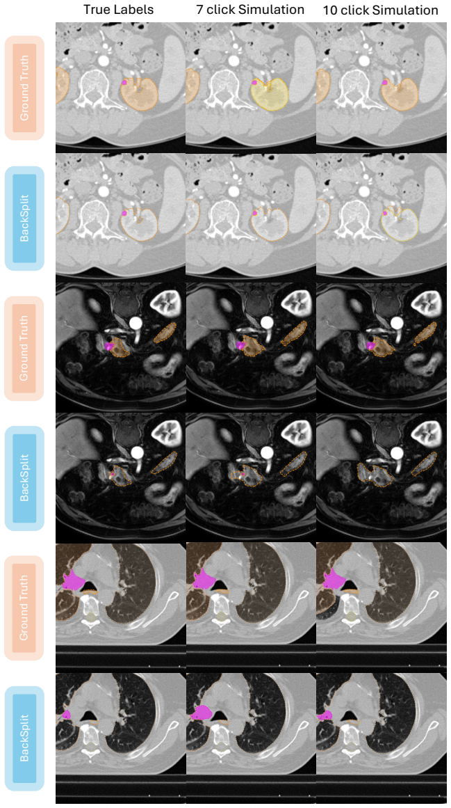
- The gain persists when auxiliary labels come from pretrained or interactive segmentation models and are noisy, as long as they are roughly spatially aligned with the target.
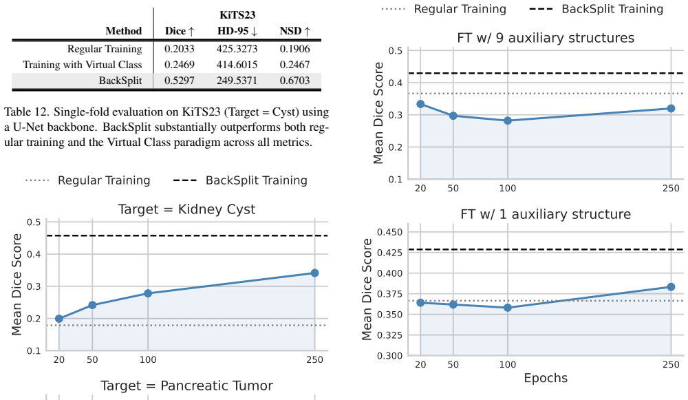
- Fine-tuning a pretrained binary model with the split background recovers most of the gain; with many auxiliary classes, training budgets may need to be lengthened so the model does not spend too many epochs on easier auxiliary structures first.
Where Pith is reading between the lines
- A testable extension, consistent with the paper's own supplementary remark, is that the gain should scale with background heterogeneity: datasets with many distinct non-target tissues should benefit far more than near-homogeneous backgrounds, and one could quantify the gain against an entropy measure of the auxiliary label distribution.
- Because the equality condition is a clean observable (all non-target classes sharing the same gradient direction given X), the framework suggests a cheap pre-training diagnostic: estimate the conditional variance term on a validation batch and predict which tasks will gain from background splitting before running full training.
- Since BackSplit is supervision-level only, it can be stacked on top of any architecture, loss, or augmentation pipeline; one could reasonably expect it to compound with other small-lesion techniques rather than replace them, though the paper itself does not test this interaction.
- The fine-tuning experiment with many auxiliary classes points to a practical scheduling question that the paper leaves open: whether a curriculum that introduces auxiliary classes gradually would avoid the observed early-training slowdown while preserving the eventual gains.
Editorial analysis
A structured set of objections, weighed in public.
Referee Report
Summary. BackSplit proposes to improve lesion segmentation by training with fine-grained auxiliary background classes (e.g., organs surrounding the lesion) in addition to the lesion target, while at inference time predicting only the target. The paper makes two contributions: (i) a theoretical result (Theorem 1 and Corollary 1) showing that, for a fixed parameterization, the expected Fisher information of the multiclass likelihood dominates that of the coarsened binary likelihood, with a softmax instantiation in Proposition 1; and (ii) an extensive empirical evaluation across five datasets, three architectures, and several auxiliary-label sources (manual, model-derived, interactive), reporting large Dice/HD-95/NSD gains. The theory is proved in the supplement via standard score-projection and law-of-total-variance arguments; the experiments are broad, but the interpretation of the results is complicated by a mismatch between the theoretical comparison (same parameter vector, coarsened labels) and the experimental comparison (separately parameterized binary vs. multiclass networks).
Significance. If the central claims hold, the paper offers a simple, architecture-agnostic, inference-free-lunch training modification with a plausible theoretical rationale, and it provides one of the first extensive empirical studies of background sub-division for small-lesion segmentation. The theoretical core is correct but is essentially the classical missing-information decomposition (Louis 1982; Oakes 1999); the paper's incremental contribution is the application to deep medical-image segmentation and the large empirical sweep. The empirical results are often striking (e.g., KiTS23 cyst Dice 0.18→0.46), and the robustness checks with automatically generated auxiliary labels are valuable. However, the headline 'consistent improvements' claim is contradicted by specific entries in Tables 1 and 2, and the theoretical justification does not directly apply to the experimental setup as currently formulated. The paper ships machine-checkable proofs in the supplement and detailed training configurations, which is a strength.
major comments (4)
- [Sec. 3.2 vs. Sec. 4.2 and Supp. D.1] Theory–experiment mismatch: Theorem 1 compares two likelihoods that share the same parameter vector θ—the coarsened likelihood is obtained by marginalizing the same K-class model. In experiments, the 'binary baseline' is a separately parameterized network with its own 2-channel output head, so its parameters live in a different space. Corollary 1's Loewner ordering and delta-method variance comparison therefore do not transfer to the reported binary-vs-BackSplit comparison. Gains could stem from the additional output head, optimization dynamics, or label structure rather than from the missing-information mechanism. A controlled experiment (e.g., deriving binary predictions from the K-class model's η_c, or matching output heads) is needed to support the claimed explanation.
- [Tab. 2(b) and Tab. 1(b)] The claim that BackSplit 'consistently improves' performance is overstated. Table 2(b) shows Kidney Stone Dice dropping from 0.195 to 0.1639, HD-95 increasing from 471.47 to 484.32, and NSD dropping from 0.6247 to 0.5401. Table 1(b) shows PANTHER ResEncU-Net HD-95 worsening from 66.26 to 70.55. These are not small fluctuations and directly contradict the 'consistent improvements' language in Sec. 4.2 and the abstract. Please report per-lesion variability and significance (e.g., bootstrap or paired tests), and soften the generality claim.
- [Supp. A.1, Assumption 1.3] The efficiency guarantee (Corollary 1) relies on treating SGD-trained deep segmentation networks as approximate MLEs whose iterations converge to stationary points near the optimum and obey standard MLE asymptotics. Actual training uses a Dice+CE loss, data augmentation, early stopping, and non-convex optimization; the approximate-MLE premise is asserted without evidence. Please either provide empirical diagnostics (e.g., descent behavior, closeness to optimum, influence of loss choice) or explicitly frame the theory as an idealized motivation rather than a quantitative explanation of the reported gains.
- [Sec. 5 and Supp. A.6/A.7/J] The supplementary limitations materially qualify the main claims. A.6 states that for heavily imbalanced classification the Fisher ordering may not be a useful description; A.7 states that for natural images or uniform backgrounds the gap is minimal; and Supp. J shows that fine-tuning AutoPET with 9 auxiliary structures fails to beat baseline unless the support set is reduced. These caveats should be moved into the main text and integrated with the 'broad applicability' claims, because they indicate the paradigm's benefits are context-dependent rather than universal.
minor comments (5)
- [General] Numerous small typos and formatting issues: 'for i=i' in Sec. 3.1, duplicated sentence in Supp. A.5 ('Therefore the score function...' appears twice), inconsistent capitalization of 'BackSplit', and Table 3/4 single-fold results reported without confidence intervals or fold selection details.
- [Notation] The notation I_Y(θ) is used for both the expected Fisher information and the observed information matrix; this is potentially confusing. Please use different symbols or disambiguate explicitly.
- [References] The related-work discussion of auxiliary background supervision cites [37] but does not position the empirical contribution relative to methods that already use organ-aware multi-class training (e.g., multi-organ/tumor joint training). A short discussion would clarify novelty.
- [Sec. 4.6 and Fig. 3] The fine-tuning and partial-supervision experiments are reported for a single dataset and a single fold; please state this in the main text and add error bars or at least indicate robustness.
- [Supp. D.5] The '0.02% parameter overhead' claim is unclear: does it refer to the change in output-layer parameters? If so, say so explicitly; otherwise it is misleading.
Circularity Check
No circularity: the Fisher-information theorem is a standard missing-information identity proven from cited external results, and the empirical comparisons are independent; the main caveats are theory-experiment parameterization mismatch and overclaimed consistency, not circularity.
full rationale
The claimed derivation chain is not circular. Lemma 1 is explicitly imported from the external Missing Information Principle (citations [43,47]), and Theorem 1 follows directly from the law of total variance: E[s_Y s_Y^T] = E[s_Z s_Z^T] + E[Var(s_Y|Z,X)], with Lemma 1 used to identify E[s_Y|Z,X] = s_Z. No parameter is fitted to any outcome to obtain the inequality; the Loewner ordering is a mathematical identity given the definitions of the full and coarsened likelihoods. Proposition 1 is a direct softmax/Bernoulli calculation, and Corollary 1 is standard MLE asymptotics and the delta method under the paper's stated regularity assumptions. The load-bearing caveats are non-circular: (i) Assumption 1.2 in Supplementary A.1 requires the compared models to share the same parameter space, but the experiments compare a K-class output head against a separately parameterized 2-channel binary head, so the experimental binary model's Fisher information is not I_Z(theta) of Theorem 1; this is a theory-experiment mismatch rather than equivalence-by-construction. (ii) The paper's own results qualify the 'consistently boosts' claim — Table 2(b) shows Kidney Stone Dice degrading from 0.195 to 0.1639, and Supplementary J reports AutoPET fine-tuning failing to surpass the baseline — but these are overclaiming or limitation issues, not circularity. Supplementary A.6 also concedes that the ordering may not describe highly imbalanced classification settings. No self-citation chain is load-bearing: the theoretical identity rests on external, standard references, and the author self-citations in related work are not used to force the main result.
Axiom & Free-Parameter Ledger
free parameters (1)
- Support-structure selection
axioms (6)
- standard math Score projection / missing information principle: E[s_Y(θ)|Z,X] = s_Z(θ), attributed to Louis (1982) and Oakes (1999).
- standard math Law of total variance: E[UU^T] = E[E[U|V]E[U|V]^T] + E[Var(U|V)].
- domain assumption Assumption 1.1: The parametric model is correctly specified at θ*, twice continuously differentiable, and Fisher information matrices exist and are non-singular.
- domain assumption Assumption 1.2: Both multiclass and binary models share the same architecture and parameterization up to the final classification layer.
- ad hoc to paper Assumption 1.3: SGD training converges to stationary points near MLEs, so standard asymptotic efficiency applies to deep networks.
- domain assumption Non-target classes induce distinct gradient directions, making the inequality in Theorem 1 strict.
read the original abstract
Segmenting small lesions in medical images remains notoriously difficult. Most prior work tackles this challenge by either designing better architectures, loss functions, or data augmentation schemes; and collecting more labeled data. We take a different view, arguing that part of the problem lies in how the background is modeled. Common lesion segmentation collapses all non-lesion pixels into a single "background" class, ignoring the rich anatomical context in which lesions appear. In reality, the background is highly heterogeneous-composed of tissues, organs, and other structures that can now be labeled manually or inferred automatically using existing segmentation models. In this paper, we argue that training with fine-grained labels that sub-divide the background class, which we call BackSplit, is a simple yet powerful paradigm that can offer a significant performance boost without increasing inference costs. From an information theoretic standpoint, we prove that BackSplit increases the expected Fisher Information relative to conventional binary training, leading to tighter asymptotic bounds and more stable optimization. With extensive experiments across multiple datasets and architectures, we empirically show that BackSplit consistently boosts small-lesion segmentation performance, even when auxiliary labels are generated automatically using pretrained segmentation models. Additionally, we demonstrate that auxiliary labels derived from interactive segmentation frameworks exhibit the same beneficial effect, demonstrating its robustness, simplicity, and broad applicability.
Figures




Forward citations
Cited by 1 Pith paper
-
Scaling up fine-grained intracranial vessel annotations in computed tomography angiography
SemanticVessel is a new CTA vessel segmentation dataset using intensity-guided region growing, expert labeling of 20 arterial classes, and multi-phase label reuse, with reported gains from including a generic minor-ar...
Reference graph
Works this paper leans on
-
[1]
A novel focal tversky loss function with improved attention u-net for lesion segmentation
Nabila Abraham and Naimul Mefraz Khan. A novel focal tversky loss function with improved attention u-net for lesion segmentation. In2019 IEEE 16th international symposium on biomedical imaging (ISBI 2019), pages 683–687. IEEE,
2019
-
[2]
Data from nsclc-radiomics-genomics
Hugo JWL Aerts, Emmanuel Rios Velazquez, Ralph TH Lei- jenaar, Chintan Parmar, Patrick Grossmann, Sara Carvalho, Johan Bussink, Ren ´e Monshouwer, Benjamin Haibe-Kains, Derek Rietveld, et al. Data from nsclc-radiomics-genomics. (No Title), 2015. 6, 17, 18
2015
-
[3]
Panther challenge: Pub- lic training dataset, 2025
Amparo Soeli Betancourt Tarifa, Faisal Mahmood, Uffe Bernchou, and Peter Jan Koopmans. Panther challenge: Pub- lic training dataset, 2025. 6, 17, 18, 20
2025
-
[4]
M Jorge Cardoso, Wenqi Li, Richard Brown, Nic Ma, Eric Kerfoot, Yiheng Wang, Benjamin Murrey, Andriy Myro- nenko, Can Zhao, Dong Yang, et al. Monai: An open-source framework for deep learning in healthcare.arXiv preprint arXiv:2211.02701, 2022. 16
Pith/arXiv arXiv 2022
-
[5]
Vir- tual class enhanced discriminative embedding learning.Ad- vances in Neural Information Processing Systems, 31, 2018
Binghui Chen, Weihong Deng, and Haifeng Shen. Vir- tual class enhanced discriminative embedding learning.Ad- vances in Neural Information Processing Systems, 31, 2018. 19
2018
-
[6]
Jieneng Chen, Yongyi Lu, Qihang Yu, Xiangde Luo, Ehsan Adeli, Yan Wang, Le Lu, Alan L Yuille, and Yuyin Zhou. Transunet: Transformers make strong encoders for medi- cal image segmentation.arXiv preprint arXiv:2102.04306,
-
[7]
Encoder-decoder with atrous separable convolution for semantic image segmentation
Liang-Chieh Chen, Yukun Zhu, George Papandreou, Florian Schroff, and Hartwig Adam. Encoder-decoder with atrous separable convolution for semantic image segmentation. In Proceedings of the European conference on computer vision (ECCV), pages 801–818, 2018. 1
2018
-
[8]
Med3d: Trans- fer learning for 3d medical image analysis.arXiv preprint arXiv:1904.00625, 2019
Sihong Chen, Kai Ma, and Yefeng Zheng. Med3d: Trans- fer learning for 3d medical image analysis.arXiv preprint arXiv:1904.00625, 2019. 2
Pith/arXiv arXiv 1904
-
[9]
3d u-net: learning dense volumetric segmentation from sparse annotation
¨Ozg¨un C ¸ ic ¸ek, Ahmed Abdulkadir, Soeren S Lienkamp, Thomas Brox, and Olaf Ronneberger. 3d u-net: learning dense volumetric segmentation from sparse annotation. In International conference on medical image computing and computer-assisted intervention, pages 424–432. Springer,
-
[10]
On label granularity and object localization
Elijah Cole, Kimberly Wilber, Grant Van Horn, Xuan Yang, Marco Fornoni, Pietro Perona, Serge Belongie, Andrew Howard, and Oisin Mac Aodha. On label granularity and object localization. InEuropean Conference on Computer Vision, pages 604–620. Springer, 2022. 3
2022
-
[11]
Tugba Akinci D’Antonoli, Lucas K Berger, Ashraya K In- drakanti, Nathan Vishwanathan, Jakob Weiß, Matthias Jung, Zeynep Berkarda, Alexander Rau, Marco Reisert, Thomas K¨ustner, et al. Totalsegmentator mri: Robust sequence- independent segmentation of multiple anatomic structures in mri.arXiv preprint arXiv:2405.19492, 2024. 7, 17
Pith/arXiv arXiv 2024
-
[12]
Omni-seg: A scale-aware dynamic network for renal pathological image segmentation.IEEE Transactions on Biomedical Engineering, 70(9):2636–2644,
Ruining Deng, Quan Liu, Can Cui, Tianyuan Yao, Jun Long, Zuhayr Asad, R Michael Womick, Zheyu Zhu, Agnes B Fogo, Shilin Zhao, et al. Omni-seg: A scale-aware dynamic network for renal pathological image segmentation.IEEE Transactions on Biomedical Engineering, 70(9):2636–2644,
-
[13]
Hats: Hierarchical adaptive taxonomy segmentation for panoramic pathology image analysis
Ruining Deng, Quan Liu, Can Cui, Tianyuan Yao, Juming Xiong, Shunxing Bao, Hao Li, Mengmeng Yin, Yu Wang, Shilin Zhao, et al. Hats: Hierarchical adaptive taxonomy segmentation for panoramic pathology image analysis. In International Conference on Medical Image Computing and Computer-Assisted Intervention, pages 155–166. Springer,
-
[14]
Prpseg: Universal proposition learning for panoramic renal pathology segmentation
Ruining Deng, Quan Liu, Can Cui, Tianyuan Yao, Jialin Yue, Juming Xiong, Lining Yu, Yifei Wu, Mengmeng Yin, Yu Wang, et al. Prpseg: Universal proposition learning for panoramic renal pathology segmentation. InProceedings of the IEEE/CVF conference on computer vision and pattern recognition, pages 11736–11746, 2024. 3
2024
-
[15]
Learning multi- class segmentations from single-class datasets
Konstantin Dmitriev and Arie E Kaufman. Learning multi- class segmentations from single-class datasets. InProceed- ings of the IEEE/CVF conference on computer vision and pattern recognition, pages 9501–9511, 2019. 2
2019
-
[16]
Multi-organ segmentation over partially labeled datasets with multi-scale feature abstrac- tion.IEEE Transactions on Medical Imaging, 39(11):3619– 3629, 2020
Xi Fang and Pingkun Yan. Multi-organ segmentation over partially labeled datasets with multi-scale feature abstrac- tion.IEEE Transactions on Medical Imaging, 39(11):3619– 3629, 2020. 2
2020
-
[17]
Efficient algorithms for learning from coarse labels
Dimitris Fotakis, Alkis Kalavasis, Vasilis Kontonis, and Christos Tzamos. Efficient algorithms for learning from coarse labels. InConference on Learning Theory, pages 2060–2079. PMLR, 2021. 3
2060
-
[18]
Data augmentation for medical imaging: A system- atic literature review.Computers in biology and medicine, 152:106391, 2023
Fabio Garcea, Alessio Serra, Fabrizio Lamberti, and Lia Morra. Data augmentation for medical imaging: A system- atic literature review.Computers in biology and medicine, 152:106391, 2023. 1
2023
-
[19]
A whole-body fdg-pet/ct dataset with manu- ally annotated tumor lesions.Scientific Data, 9(1):601, 2022
Sergios Gatidis, Tobias Hepp, Marcel Fr ¨uh, Christian La Foug `ere, Konstantin Nikolaou, Christina Pfannenberg, Bernhard Sch ¨olkopf, Thomas K ¨ustner, Clemens Cyran, and Daniel Rubin. A whole-body fdg-pet/ct dataset with manu- ally annotated tumor lesions.Scientific Data, 9(1):601, 2022. 6, 7, 19, 20
2022
-
[20]
Vibesegmentator: full body mri segmentation for the nako and uk biobank.European Radiology, pages 1–15, 2025
Robert Graf, Paul Platzek, Evamaria Olga Riedel, Constanze Ramsch¨utz, Sophie Starck, Hendrik K M ¨oller, Matan Atad, Henry V ¨olzke, Robin B ¨ulow, Carsten Oliver Schmidt, et al. Vibesegmentator: full body mri segmentation for the nako and uk biobank.European Radiology, pages 1–15, 2025. 7, 17
2025
-
[21]
Softseg: Advantages of soft versus binary training for image segmentation.Medical image analysis, 71:102038, 2021
Charley Gros, Andreanne Lemay, and Julien Cohen-Adad. Softseg: Advantages of soft versus binary training for image segmentation.Medical image analysis, 71:102038, 2021. 2
2021
-
[22]
Swin unetr: Swin transformers for semantic segmentation of brain tumors in mri images
Ali Hatamizadeh, Vishwesh Nath, Yucheng Tang, Dong Yang, Holger R Roth, and Daguang Xu. Swin unetr: Swin transformers for semantic segmentation of brain tumors in mri images. InInternational MICCAI brainlesion workshop, pages 272–284. Springer, 2021. 1, 16, 19 9
2021
-
[23]
Unetr: Transformers for 3d med- ical image segmentation
Ali Hatamizadeh, Yucheng Tang, Vishwesh Nath, Dong Yang, Andriy Myronenko, Bennett Landman, Holger R Roth, and Daguang Xu. Unetr: Transformers for 3d med- ical image segmentation. InProceedings of the IEEE/CVF winter conference on applications of computer vision, pages 574–584, 2022. 1
2022
-
[24]
Multi-task learning for the segmentation of organs at risk with label dependence.Medical Image Analysis, 61:101666,
Tao He, Junjie Hu, Ying Song, Jixiang Guo, and Zhang Yi. Multi-task learning for the segmentation of organs at risk with label dependence.Medical Image Analysis, 61:101666,
-
[25]
The kits21 chal- lenge: Automatic segmentation of kidneys, renal tumors, and renal cysts in corticomedullary-phase ct, 2023
Nicholas Heller, Fabian Isensee, Dasha Trofimova, Re- sha Tejpaul, Zhongchen Zhao, Huai Chen, Lisheng Wang, Alex Golts, Daniel Khapun, Daniel Shats, Yoel Shoshan, Flora Gilboa-Solomon, Yasmeen George, Xi Yang, Jian- peng Zhang, Jing Zhang, Yong Xia, Mengran Wu, Zhiyang Liu, Ed Walczak, Sean McSweeney, Ranveer Vasdev, Chris Hornung, Rafat Solaiman, Jamee S...
2023
-
[26]
Atrous convolution for binary semantic segmentation of lung nodule
Mohammad Hesam Hesamian, Wenjing Jia, Xiangjian He, and Paul J Kennedy. Atrous convolution for binary semantic segmentation of lung nodule. InICASSP 2019-2019 IEEE International Conference on Acoustics, Speech and Signal Processing (ICASSP), pages 1015–1019. IEEE, 2019. 2
2019
-
[27]
A deep multi-task learning framework for brain tumor segmentation.Frontiers in oncology, 11:690244, 2021
He Huang, Guang Yang, Wenbo Zhang, Xiaomei Xu, Weiji Yang, Weiwei Jiang, and Xiaobo Lai. A deep multi-task learning framework for brain tumor segmentation.Frontiers in oncology, 11:690244, 2021. 2
2021
-
[28]
nnu-net: a self-configuring method for deep learning-based biomedical image segmen- tation.Nature methods, 18(2):203–211, 2021
Fabian Isensee, Paul F Jaeger, Simon AA Kohl, Jens Pe- tersen, and Klaus H Maier-Hein. nnu-net: a self-configuring method for deep learning-based biomedical image segmen- tation.Nature methods, 18(2):203–211, 2021. 1, 6, 7, 16, 17, 18, 19, 20, 21
2021
-
[29]
nnu-net revisited: A call for rigorous validation in 3d medical image segmentation
Fabian Isensee, Tassilo Wald, Constantin Ulrich, Michael Baumgartner, Saikat Roy, Klaus Maier-Hein, and Paul F Jaeger. nnu-net revisited: A call for rigorous validation in 3d medical image segmentation. InInternational Confer- ence on Medical Image Computing and Computer-Assisted Intervention, pages 488–498. Springer, 2024. 6, 16, 19
2024
-
[30]
nninteractive: Redefining 3d promptable segmen- tation.arXiv preprint arXiv:2503.08373, 2025
Fabian Isensee, Maximilian Rokuss, Lars Kr ¨amer, Stefan Dinkelacker, Ashis Ravindran, Florian Stritzke, Benjamin Hamm, Tassilo Wald, Moritz Langenberg, Constantin Ul- rich, et al. nninteractive: Redefining 3d promptable segmen- tation.arXiv preprint arXiv:2503.08373, 2025. 7, 17
Pith/arXiv arXiv 2025
-
[31]
Anatomy-guided pathology segmentation
Alexander Jaus, Constantin Seibold, Simon Reiß, Lukas Heine, Anton Schily, Moon Kim, Fin Hendrik Bahnsen, Ken Herrmann, Rainer Stiefelhagen, and Jens Kleesiek. Anatomy-guided pathology segmentation. InInternational Conference on Medical Image Computing and Computer- Assisted Intervention, pages 3–13. Springer, 2024. 3
2024
-
[32]
Boundary loss for highly unbalanced segmentation
Hoel Kervadec, Jihene Bouchtiba, Christian Desrosiers, Eric Granger, Jose Dolz, and Ismail Ben Ayed. Boundary loss for highly unbalanced segmentation. InInternational confer- ence on medical imaging with deep learning, pages 285–296. PMLR, 2019. 1
2019
-
[33]
Weakly supervised learning for multi-class medical image segmentation via fea- ture decomposition.Computers in Biology and Medicine, 171:108228, 2024
Zhuo Kuang, Zengqiang Yan, and Li Yu. Weakly supervised learning for multi-class medical image segmentation via fea- ture decomposition.Computers in Biology and Medicine, 171:108228, 2024. 2
2024
-
[34]
Erwan Lecesne, Antoine Simon, Mireille Garreau, Gilles Barone-Rochette, and C ´eline Fouard. Segmentation of car- diac infarction in delayed-enhancement mri using probabil- ity map and transformers-based neural networks.Computer Methods and Programs in Biomedicine, 242:107841, 2023. 2
2023
-
[35]
Deeply-supervised nets
Chen-Yu Lee, Saining Xie, Patrick Gallagher, Zhengyou Zhang, and Zhuowen Tu. Deeply-supervised nets. InArti- ficial intelligence and statistics, pages 562–570. Pmlr, 2015. 18
2015
-
[36]
A tri-path boundary preserving network via context-aware aggregator for multi- organ medical image segmentation.Biomedical Signal Pro- cessing and Control, 104:107593, 2025
Bicao Li, Panpan Li, Zhoufeng Liu, Bei Wang, Chunlei Li, Xuwei Guo, Jing Wang, and Wei Li. A tri-path boundary preserving network via context-aware aggregator for multi- organ medical image segmentation.Biomedical Signal Pro- cessing and Control, 104:107593, 2025. 3
2025
-
[37]
Context label learning: Improving background class representations in semantic segmentation
Zeju Li, Konstantinos Kamnitsas, Cheng Ouyang, Chen Chen, and Ben Glocker. Context label learning: Improving background class representations in semantic segmentation. IEEE Transactions on Medical Imaging, 42(6):1885–1896,
-
[38]
Focal loss for dense object detection
Tsung-Yi Lin, Priya Goyal, Ross Girshick, Kaiming He, and Piotr Doll´ar. Focal loss for dense object detection. InPro- ceedings of the IEEE international conference on computer vision, pages 2980–2988, 2017. 1
2017
-
[39]
Semi-supervised medical image segmentation using cross-style consistency with shape-aware and local context constraints.IEEE Transactions on Medical Imaging, 43(4): 1449–1461, 2023
Jinhua Liu, Christian Desrosiers, Dexin Yu, and Yuanfeng Zhou. Semi-supervised medical image segmentation using cross-style consistency with shape-aware and local context constraints.IEEE Transactions on Medical Imaging, 43(4): 1449–1461, 2023. 3
2023
-
[40]
Shape- aware multi-task learning for semi-supervised 3d medical image segmentation
Shasha Liu, Yan Li, Xiaohu Li, and Guitao Cao. Shape- aware multi-task learning for semi-supervised 3d medical image segmentation. In2021 IEEE international conference on bioinformatics and biomedicine (BIBM), pages 1418–
-
[41]
A panoptic segmentation dataset and deep-learning approach for explainable scoring of tumor-infiltrating lymphocytes
Shangke Liu, Mohamed Amgad, Deeptej More, Muham- mad A Rathore, Roberto Salgado, and Lee AD Cooper. A panoptic segmentation dataset and deep-learning approach for explainable scoring of tumor-infiltrating lymphocytes. NPJ Breast Cancer, 10(1):52, 2024. 2
2024
-
[42]
Canet: Context aware network for brain glioma segmentation.IEEE Transactions on Medical Imag- ing, 40(7):1763–1777, 2021
Zhihua Liu, Lei Tong, Long Chen, Feixiang Zhou, Zheheng Jiang, Qianni Zhang, Yinhai Wang, Caifeng Shan, Ling Li, and Huiyu Zhou. Canet: Context aware network for brain glioma segmentation.IEEE Transactions on Medical Imag- ing, 40(7):1763–1777, 2021. 2
2021
-
[43]
Finding the observed information matrix when using the em algorithm.Journal of the Royal Statis- tical Society Series B: Statistical Methodology, 44(2):226– 233, 1982
Thomas A Louis. Finding the observed information matrix when using the em algorithm.Journal of the Royal Statis- tical Society Series B: Statistical Methodology, 44(2):226– 233, 1982. 4, 12
1982
-
[44]
Berger, Alexander Weers, Nico Stucki, Daniel Rueckert, Ulrich Bauer, and Johannes C
Laurin Lux, Alexander H. Berger, Alexander Weers, Nico Stucki, Daniel Rueckert, Ulrich Bauer, and Johannes C. Paet- zold. Topograph: An efficient graph-based framework for 10 strictly topology preserving image segmentation. InICLR,
-
[45]
V-net: Fully convolutional neural networks for volumetric medical image segmentation
Fausto Milletari, Nassir Navab, and Seyed-Ahmad Ahmadi. V-net: Fully convolutional neural networks for volumetric medical image segmentation. In2016 fourth international conference on 3D vision (3DV), pages 565–571. Ieee, 2016. 1
2016
-
[46]
3d mri brain tumor segmentation using autoencoder regularization
Andriy Myronenko. 3d mri brain tumor segmentation using autoencoder regularization. InInternational MICCAI brain- lesion workshop, pages 311–320. Springer, 2018. 6, 16
2018
-
[47]
Direct calculation of the information matrix via the em.Journal of the Royal Statistical Society: Series B (Statistical Methodology), 61(2):479–482, 1999
David Oakes. Direct calculation of the information matrix via the em.Journal of the Royal Statistical Society: Series B (Statistical Methodology), 61(2):479–482, 1999. 4, 12
1999
-
[48]
Pytorch: An im- perative style, high-performance deep learning library.Ad- vances in neural information processing systems, 32, 2019
Adam Paszke, Sam Gross, Francisco Massa, Adam Lerer, James Bradbury, Gregory Chanan, Trevor Killeen, Zeming Lin, Natalia Gimelshein, Luca Antiga, et al. Pytorch: An im- perative style, high-performance deep learning library.Ad- vances in neural information processing systems, 32, 2019. 16
2019
-
[49]
Abdomenatlas-8k: Anno- tating 8,000 ct volumes for multi-organ segmentation in three weeks.Advances in Neural Information Processing Systems, 36, 2023
Chongyu Qu, Tiezheng Zhang, Hualin Qiao, Yucheng Tang, Alan L Yuille, and Zongwei Zhou. Abdomenatlas-8k: Anno- tating 8,000 ct volumes for multi-organ segmentation in three weeks.Advances in Neural Information Processing Systems, 36, 2023. 7, 17, 19
2023
-
[50]
U- net: Convolutional networks for biomedical image segmen- tation
Olaf Ronneberger, Philipp Fischer, and Thomas Brox. U- net: Convolutional networks for biomedical image segmen- tation. InInternational Conference on Medical image com- puting and computer-assisted intervention, pages 234–241. Springer, 2015. 1, 6, 7, 16, 17, 18, 19, 20, 21
2015
-
[51]
cldice-a novel topology-preserving loss function for tubular structure seg- mentation
Suprosanna Shit, Johannes C Paetzold, Anjany Sekuboyina, Ivan Ezhov, Alexander Unger, Andrey Zhylka, Josien PW Pluim, Ulrich Bauer, and Bjoern H Menze. cldice-a novel topology-preserving loss function for tubular structure seg- mentation. InProceedings of the IEEE/CVF conference on computer vision and pattern recognition, pages 16560– 16569, 2021. 1
2021
-
[52]
Topologically faithful im- age segmentation via induced matching of persistence bar- codes
Nico Stucki, Johannes C Paetzold, Suprosanna Shit, Bjo- ern Menze, and Ulrich Bauer. Topologically faithful im- age segmentation via induced matching of persistence bar- codes. InInternational Conference on Machine Learning, pages 32698–32727. PMLR, 2023. 3
2023
-
[53]
Mesh-prompted anatomy segmentation
Dingjie Su, Yihao Liu, Lianrui Zuo, and Benoit Dawant. Mesh-prompted anatomy segmentation. InMedical Imaging with Deep Learning, 2025. 3
2025
-
[54]
Deep high-resolution repre- sentation learning for visual recognition.IEEE transactions on pattern analysis and machine intelligence, 43(10):3349– 3364, 2020
Jingdong Wang, Ke Sun, Tianheng Cheng, Borui Jiang, Chaorui Deng, Yang Zhao, Dong Liu, Yadong Mu, Mingkui Tan, Xinggang Wang, et al. Deep high-resolution repre- sentation learning for visual recognition.IEEE transactions on pattern analysis and machine intelligence, 43(10):3349– 3364, 2020. 1
2020
-
[55]
Abdominal multi-organ seg- mentation with organ-attention networks and statistical fu- sion.Medical image analysis, 55:88–102, 2019
Yan Wang, Yuyin Zhou, Wei Shen, Seyoun Park, Elliot K Fishman, and Alan L Yuille. Abdominal multi-organ seg- mentation with organ-attention networks and statistical fu- sion.Medical image analysis, 55:88–102, 2019. 2
2019
-
[56]
To- talsegmentator: robust segmentation of 104 anatomic struc- tures in ct images.Radiology: Artificial Intelligence, 5(5): e230024, 2023
Jakob Wasserthal, Hanns-Christian Breit, Manfred T Meyer, Maurice Pradella, Daniel Hinck, Alexander W Sauter, Tobias Heye, Daniel T Boll, Joshy Cyriac, Shan Yang, et al. To- talsegmentator: robust segmentation of 104 anatomic struc- tures in ct images.Radiology: Artificial Intelligence, 5(5): e230024, 2023. 7
2023
-
[57]
Mswal: 3d multi-class segmentation of whole abdominal lesions dataset
Zhaodong Wu, Qiaochu Zhao, Ming Hu, Yulong Li, Haochen Xue, Zhengyong Jiang, Angelos Stefanidis, Qi- ufeng Wang, Imran Razzak, Zongyuan Ge, et al. Mswal: 3d multi-class segmentation of whole abdominal lesions dataset. InInternational Conference on Medical Image Com- puting and Computer-Assisted Intervention, pages 378–388. Springer, 2025. 6, 7
2025
-
[58]
Co-seg: Mutual prompt-guided collaborative learning for tissue and nuclei segmentation
Qing Xu, Wenting Duan, and Zhen Chen. Co-seg: Mutual prompt-guided collaborative learning for tissue and nuclei segmentation. InInternational Conference on Medical Im- age Computing and Computer-Assisted Intervention, pages 130–140. Springer, 2025. 3
2025
-
[59]
Weakly supervised representation learning with coarse la- bels
Yuanhong Xu, Qi Qian, Hao Li, Rong Jin, and Juhua Hu. Weakly supervised representation learning with coarse la- bels. InProceedings of the IEEE/CVF International Con- ference on Computer Vision, pages 10593–10601, 2021. 3
2021
-
[60]
Multi-class dis- ease detection using deep learning and human brain medi- cal imaging.Biomedical Signal Processing and Control, 85: 104875, 2023
Fatima Yousaf, Sajid Iqbal, Nosheen Fatima, Tanzeela Kousar, and Mohd Shafry Mohd Rahim. Multi-class dis- ease detection using deep learning and human brain medi- cal imaging.Biomedical Signal Processing and Control, 85: 104875, 2023. 2
2023
-
[61]
Cutmix: Regu- larization strategy to train strong classifiers with localizable features
Sangdoo Yun, Dongyoon Han, Seong Joon Oh, Sanghyuk Chun, Junsuk Choe, and Youngjoon Yoo. Cutmix: Regu- larization strategy to train strong classifiers with localizable features. InProceedings of the IEEE/CVF international con- ference on computer vision, pages 6023–6032, 2019. 1
2019
-
[62]
Dodnet: Learning to segment multi-organ and tumors from multiple partially labeled datasets
Jianpeng Zhang, Yutong Xie, Yong Xia, and Chunhua Shen. Dodnet: Learning to segment multi-organ and tumors from multiple partially labeled datasets. InProceedings of the IEEE/CVF conference on computer vision and pattern recognition, pages 1195–1204, 2021. 2
2021
-
[63]
Yongtao Zhang, Haimei Li, Jie Du, Jing Qin, Tianfu Wang, Yue Chen, Bing Liu, Wenwen Gao, Guolin Ma, and Baiying Lei. 3d multi-attention guided multi-task learning network for automatic gastric tumor segmentation and lymph node classification.IEEE transactions on medical imaging, 40(6): 1618–1631, 2021. 2
2021
-
[64]
target vs. rest
Wentao Zhu, Yufang Huang, Liang Zeng, Xuming Chen, Yong Liu, Zhen Qian, Nan Du, Wei Fan, and Xiaohui Xie. Anatomynet: deep learning for fast and fully automated whole-volume segmentation of head and neck anatomy. Medical physics, 46(2):576–589, 2019. 1 11 BackSplit: The Importance of Sub-dividing the Background in Biomedical Lesion Segmentation Supplement...
2019
-
[65]
Including this structure allows the model to learn fine-grained vari- ations in local tissue appearance, which directly improves boundary precision and reduces ambiguity
Select the organ containing the lesion.The most effec- tive auxiliary structure is typically the organ in which the le- sion resides, as it fully encloses the target region. Including this structure allows the model to learn fine-grained vari- ations in local tissue appearance, which directly improves boundary precision and reduces ambiguity
-
[66]
These surrounding organs provide contextual cues that help the model disambiguate lesion boundaries and reduce drift into neighboring regions
Include adjacent or surrounding organs.Structures that spatially border the lesion serve as anatomical anchors. These surrounding organs provide contextual cues that help the model disambiguate lesion boundaries and reduce drift into neighboring regions. This is particularly useful for le- sions located near organ interfaces
-
[67]
3d fullres
Add organs prone to false positives.A third use- ful strategy is to incorporate organs whose appearance may cause confusion with the target lesion. These structures of- ten contain components or textures that resemble the lesion class, leading to false positives in standard training. By ex- 15 plicitly modeling these tissues, the network learns discrimi- ...
2033
discussion (0)
Sign in with ORCID, Apple, or X to comment. Anyone can read and Pith papers without signing in.