REVIEW 4 major objections 5 minor 48 references
PathoGen claims that augmenting histopathology training sets with diffusion-synthesized lesions improves segmentation accuracy more than geometric augmentations, and does so with automatic pixel-level annotations.
Reviewed by Pith at T0; open to challenge. T0 means a machine referee read the full paper against a public rubric. the ladder, T0–T4 →
T0 review · deepseek-v4-flash
2026-08-03 10:52 UTC pith:HRSPXBP3
load-bearing objection A sensible diffusion-based inpainting method for histopathology, but the abstract reports a pathologist study that isn't in the paper, and the augmentation experiments lack a copy-paste control, so the causal claim needs work. the 4 major comments →
Controllable Diffusion-Based Lesion Inpainting for Scalable Histopathology Data Augmentation
The pith
A machine-rendered reading of the paper's core claim, the machinery that carries it, and where it could break.
Core claim
The central discovery is that a diffusion model trained with self-supervised inpainting—reconstructing randomly masked regions of real tissue without requiring any lesion annotations—learns to synthesize histologically plausible lesions when conditioned on a masked benign image and a lesion-reference patch. The paper demonstrates this across four datasets, showing that the generated lesions carry morphology that transfers to downstream segmentation: a standard segmentation model trained on 50 real plus 150 synthetic patches outperforms one trained on 50 real patches alone (kidney glomeruli Dice 0.48 to 0.66; melanoma 0.38 to 0.56; breast tumor 0.36 to 0.60; prostate gland 0.44 to 0.65), with
What carries the argument
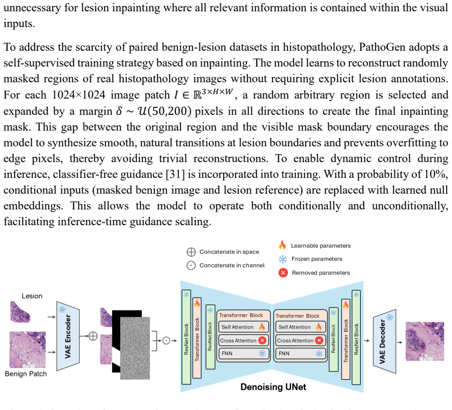
PathoGen is a latent diffusion model in which a frozen variational autoencoder maps both the masked benign image and the lesion-reference patch into a shared latent space; these latents and the downsampled mask are concatenated channel-wise and fed to a denoising network. Text cross-attention is removed because the task is purely visual, and only self-attention parameters are trained. Training is self-supervised on random irregular masks expanded by 50–200 pixels, which forces the model to learn smooth tissue transitions rather than trivial edge continuation, and classifier-free guidance is enabled by dropping the conditioning inputs 10% of the time. At inference, the user supplies a benign
Load-bearing premise
The load-bearing premise is that self-supervised inpainting of random masks over real tissue teaches the model genuine lesion morphology rather than texture continuation, so that at inference the lesion-reference patch carries the disease information; if a model trained on random masks cannot transfer to true lesions, the reported segmentation gains will not replicate.
What would settle it
Run the same data-augmentation experiment with the lesion-reference patch replaced by an unrelated tissue patch or with that conditioning channel ablated; if segmentation gains remain equal to the full PathoGen, the model is not using lesion information and the claimed transfer from inpainting to lesion synthesis fails. A second check is a fully specified blinded pathologist reader study comparing synthetic lesions to real ones, which the full text defers to future work.
If this is right
- Using PathoGen to augment small training sets improves segmentation Dice scores by 0.18–0.24 across kidney, skin, breast, and prostate datasets compared with training on original data alone.
- PathoGen outperforms geometric augmentations (rotation, flipping, translation, and combinations) and both a conditional GAN and a general-purpose diffusion baseline when each method adds the same number of synthetic samples.
- The augmentation benefit is largest when real annotated data is scarcest and narrows as more real data becomes available, so the method targets the regime where manual annotation is most costly.
- Because the inpainted region is exactly the mask, each synthetic image includes a ready-made pixel-level annotation without additional pathologist hours.
- The abstract reports near-chance expert distinguishability of synthetic versus real tissue, suggesting the generated lesions may carry diagnostic, not merely statistical, fidelity.
Where Pith is reading between the lines
- If the self-supervised inpainting transfer is as general as the paper suggests, the same recipe could generate lesion images for rare pathology subtypes by swapping the reference patch, without needing annotated examples of that subtype.
- Because synthetic images come with their own labels and are not patient-derived, large synthetic datasets could be shared across institutions with fewer privacy constraints than real whole-slide images.
- A natural testable extension is to vary the mask and reference patch to control lesion size, density, or grade, turning augmentation into targeted stress-testing for segmentation and classification models.
- The pathologist-realism result appears only in the abstract while the full text defers such a study to future work, so a fully specified blinded expert reader study is the direct way to confirm that the realism claim is backed by evidence.
Editorial analysis
A structured set of objections, weighed in public.
Referee Report
Summary. PathoGen is a latent diffusion model for controllable lesion inpainting in histopathology images. Given a benign tissue patch, a lesion mask, and a lesion reference patch, the model synthesizes a lesion within the mask. The model is trained with a self-supervised inpainting objective on random masked regions of unannotated images, with classifier-free guidance and a shared VAE. The authors evaluate on four public datasets (KPI, PUMA, TIGER, RING), reporting lower FID/KID than CGAN and StableDiffusion, and improved UNet segmentation Dice when augmenting a 50-patch training set with 150 synthetic samples. The abstract additionally claims a pathologist reader study with 57.75% accuracy and a 35.4% win rate, but this study is not described in the full text; the Discussion instead lists it as future work.
Significance. The practical goal — scalable generation of annotated lesion data for downstream segmentation — is of clear value to computational pathology. The authors provide code, weights, and use publicly available datasets, and the inclusion of four different tissue types is a strength. If the causal claim were rigorously established, PathoGen would be a useful augmentation tool. However, the current evidence does not yet support the central attribution: the augmentation protocol fails to specify the source of the lesion reference, an essential control (plain copy-paste or blending) is missing, and all segmentation results are point estimates with no repeated runs or statistical tests. The abstract/full-text discrepancy on the reader study also undermines confidence in the reported validation.
major comments (4)
- [Abstract vs. Discussion] The abstract reports a pathologist reader study: 'six expert pathologists... 57.75% accuracy' and '35.4% win rate.' The full text contains no methods, results, or analysis for this study; the Discussion explicitly says that 'Comprehensive reader studies... would strengthen validation' and lists blinded pathologist evaluation as 'future work.' This is a load-bearing inconsistency for the paper's credibility. Either include the full study with protocol and statistics, or remove these claims from the abstract.
- [Inference Process (Eq. 4–5) and 'Morphological augmentation outperforms geometric transformations'] The manuscript does not specify the source of the lesion reference I_l in the augmentation experiments. Eq. (4) defines I_l only as 'e.g., a patch from another image with the target lesion type,' and the benchmark in Figure 5 says only that 'each method augmented a common base dataset of 50 real histopathology patches with 150 synthetic samples.' If I_l is a real annotated lesion crop, PathoGen may be effectively transplanting real lesion texture into a benign context, making the method similar to copy-paste with diffusion-based blending. Without a control that replaces PathoGen with simple copy-paste (or alpha-blending) of the reference into the mask, the observed Dice gains cannot be causally attributed to the learned inpainting objective. Please report the exact reference-source protocol (e.g., drawn from the same 50 patch set, from training WSIs, or from held-out WSIs) and include a
- [Results, Figures 3–5] All segmentation Dice scores are reported as single point estimates (e.g., 0.48→0.66 for KPI). No repeated training runs, standard deviations, confidence intervals, or statistical significance tests are provided. UNet training and patch sampling are stochastic; without multi-seed evaluation, the observed improvements cannot be distinguished from noise. Even the learning curves in Figure 4 lack error bars. Please report mean±std over at least three seeds, with appropriate paired tests (e.g., paired t-test or Wilcoxon) across the augmentation conditions.
- [Methods, Architecture and Training Strategy] The training strategy is described as self-supervised inpainting 'without requiring explicit lesion annotations,' yet the inference and training inputs include a lesion reference I_l. The manuscript does not explain how random-region inpainting on unannotated images teaches the model to synthesize lesions rather than merely continue benign tissue texture. Specifically, it is unclear whether the target content inside the random mask is a benign region or a known lesion, and whether I_l is taken from the same image, a different image, or a lesion-labeled image. This ambiguity is load-bearing because it determines whether the model learns transferable lesion morphology or memorizes/copies arbitrary reference textures. Please clarify the training data construction and the role of I_l, and provide an ablation where I_l is omitted or replaced with a benign patch.
minor comments (5)
- [Abstract] The abstract states 'improves segmentation Dice scores by up to 0.18,' but the full text reports gains of 0.24 (TIGER, 0.36→0.60) and 0.21 (RING, 0.44→0.65). Correct the number to match the results.
- [Table 1] FID/KID values are reported without confidence intervals, making it difficult to judge the significance of the improvements over StableDiffusion. Consider adding bootstrap confidence intervals.
- [Results/Figure 5] The baseline 'vanilla' condition is not defined; please state whether it refers to the 50 real patches without augmentation. Also clarify whether the geometric augmentations include test-time augmentation or only training-time.
- [Implementation Details] The UNet segmentation model architecture is not described (depth, base channels, input resolution, optimizer, epochs). For reproducibility, provide these details or cite the exact implementation.
- [Methods, Datasets] For the segmentation experiments, it is not specified how the 50 real patches were selected from the 80% training split, or whether they include the same lesions used as references for PathoGen synthesis. Please specify the sampling procedure to avoid leakage between augmentation and evaluation.
Circularity Check
No significant circularity: PathoGen's derivation is self-contained and validated on held-out data.
full rationale
PathoGen's central derivation chain is self-contained. The model is trained with a self-supervised inpainting objective ('The model learns to reconstruct randomly masked regions of real histopathology images without requiring explicit lesion annotations'), so no lesion label is fitted into the generator. At inference, the lesion reference and mask are inputs, and the synthetic mask provides pixel labels by construction; this is standard weak supervision, not a predicted quantity that is also an input. The two quantitative validations are external to the fitted model: FID/KID use an Inception feature space against real images, and downstream Dice is measured on a held-out test set ('we reserved 20% of whole-slide images as a held-out test set'). The one self-citation [46] appears only in a limitation sentence and is not load-bearing. The undefined provenance of the lesion reference in augmentation experiments is a real methodology gap and could undermine causal attribution if it amounts to copying real lesions, but that is an independence/external-validity concern, not a circularity of the type where Eq. X reduces to Eq. Y by construction.
Axiom & Free-Parameter Ledger
free parameters (2)
- Inpainting mask margin δ =
U(50, 200) pixels
- Classifier-free guidance null-drop probability =
0.10
axioms (4)
- domain assumption Latent diffusion with a pretrained VAE operates faithfully on 1024×1024 histopathology patches.
- ad hoc to paper Random-region inpainting on unannotated images teaches transferable lesion morphology.
- domain assumption A single lesion reference patch provides sufficient morphological specification for the target lesion class across tissue contexts.
- domain assumption Inception-based FID/KID are valid proxies for histopathological realism.
read the original abstract
Expert-annotated training data remains the critical bottleneck for AI in histopathology, particularly for rare pathologies where even dozens of cases may be unavailable. While data augmentation offers a solution, existing methods fail to generate sufficiently realistic lesion morphologies that preserve tissue-specific architectures. Here we present PathoGen, a diffusion-based generative model enabling controllable, high-fidelity lesion inpainting into benign histopathology images. We validate PathoGen across four datasets representing kidney, skin, breast, and prostate pathology. Quantitative assessment confirms PathoGen outperforms state-of-the-art baselines in image fidelity and distributional similarity. Evaluation by six expert pathologists revealed that synthetic images by PathoGen were only marginally distinguished from real tissue image slightly above chance (57.75% accuracy), demonstrating strong perceptual realism of PathoGen-generated lesions. PathoGen achieved the highest win rate (35.4%) when pathologists ranked generation quality against all baselines. Crucially, augmenting training sets with PathoGen-synthesized lesions improves segmentation Dice scores by up to 0.18 compared to traditional augmentations, with maximum benefit in data-scarce regimes. By simultaneously generating realistic morphology and pixel-level annotations, PathoGen effectively addresses both data scarcity and annotation cost, two critical bottlenecks in computational pathology development.
Figures
Reference graph
Works this paper leans on
-
[1]
Artificial intelligence applications in histopathology,
C. D. Bahadir et al. , "Artificial intelligence applications in histopathology," Nature Reviews Electrical Engineering, vol. 1, no. 2, pp. 93-108, 2024
2024
-
[2]
Artificial intelligence in histopathology: enhancing cancer research and clinical oncology,
A. Shmatko, N. Ghaffari Laleh, M. Gerstung, and J. N. Kather, "Artificial intelligence in histopathology: enhancing cancer research and clinical oncology," Nature cancer, vol. 3, no. 9, pp. 1026-1038, 2022
2022
-
[3]
Deep learning for colon cancer histopathological images analysis,
A. B. Hamida et al. , "Deep learning for colon cancer histopathological images analysis," Computers in Biology and Medicine, vol. 136, p. 104730, 2021
2021
-
[4]
A deep-learning framework to predict cancer treatment response from histopathology images through imputed transcriptomics,
D.-T. Hoang et al., "A deep-learning framework to predict cancer treatment response from histopathology images through imputed transcriptomics," Nature cancer, vol. 5, no. 9, pp. 1305-1317, 2024
2024
-
[5]
Biological insights and novel biomarker discovery through deep learning approaches in breast cancer histopathology,
D. Mandair, J. S. Reis -Filho, and A. Ashworth, "Biological insights and novel biomarker discovery through deep learning approaches in breast cancer histopathology," NPJ breast cancer, vol. 9, no. 1, p. 21, 2023
2023
-
[6]
Learn like a pathologist: curriculum learning by annotator agreement for histopathology image classification,
J. Wei et al., "Learn like a pathologist: curriculum learning by annotator agreement for histopathology image classification," in Proceedings of the IEEE/CVF winter conference on applications of computer vision, 2021, pp. 2473-2483
2021
-
[7]
Machine learning in computational histopathology: Challenges and opportunities,
M. Cooper, Z. Ji, and R. G. Krishnan, "Machine learning in computational histopathology: Challenges and opportunities," Genes, Chromosomes and Cancer, vol. 62, no. 9, pp. 540-556, 2023
2023
-
[8]
Annotation practices in computational pathology: a European Society of Digital and Integrative Pathology (ESDIP) survey study,
D. Montezuma et al., "Annotation practices in computational pathology: a European Society of Digital and Integrative Pathology (ESDIP) survey study," Laboratory Investigation, vol. 105, no. 3, p. 102203, 2025
2025
-
[9]
Rare histological prostate cancer subtypes: Cancer-specific and other- cause mortality,
C. Siech et al., "Rare histological prostate cancer subtypes: Cancer-specific and other- cause mortality," Prostate cancer and prostatic diseases, vol. 28, no. 3, pp. 748 -754, 2025
2025
-
[10]
Why do errors arise in artificial intelligence diagnostic tools in histopathology and how can we minimize them?,
H. Evans and D. Snead, "Why do errors arise in artificial intelligence diagnostic tools in histopathology and how can we minimize them?," Histopathology, vol. 84, no. 2, pp. 279-287, 2024
2024
-
[11]
A systematic review of deep learning data augmentation in medical imaging: Recent advances and future research directions,
T. Islam, M. S. Hafiz, J. R. Jim, M. M. Kabir, and M. Mridha, "A systematic review of deep learning data augmentation in medical imaging: Recent advances and future research directions," Healthcare Analytics, vol. 5, p. 100340, 2024
2024
-
[12]
Image data augmentation approaches: A comprehensive survey and future directions,
T. Kumar, R. Brennan, A. Mileo, and M. Bendechache, "Image data augmentation approaches: A comprehensive survey and future directions," IEEE Access, 2024
2024
-
[13]
Histology image analysis of 13 healthy tissues reveals molecular - histological correlations,
Y . Gao et al. , "Histology image analysis of 13 healthy tissues reveals molecular - histological correlations," Scientific Reports, vol. 15, no. 1, p. 26812, 2025
2025
-
[14]
Robust histopathology image analysis: To label or to synthesize?,
L. Hou, A. Agarwal, D. Samaras, T. M. Kurc, R. R. Gupta, and J. H. Saltz, "Robust histopathology image analysis: To label or to synthesize?," in Proceedings of the IEEE/CVF conference on computer vision and pattern recognition , 2019, pp. 8533 - 8542
2019
-
[15]
Generative adversarial networks in digital histopathology: current applications, limitations, ethical considerations, and future directions,
S. A. Alajaji et al., "Generative adversarial networks in digital histopathology: current applications, limitations, ethical considerations, and future directions," Modern Pathology, vol. 37, no. 1, p. 100369, 2024
2024
-
[16]
NAS -SGAN: A semi -supervised generative adversarial network model for atypia scoring of breast cancer histopathological images,
A. Das, V . K. Devarampati, and M. S. Nair, "NAS -SGAN: A semi -supervised generative adversarial network model for atypia scoring of breast cancer histopathological images," IEEE Journal of Biomedical and Health Informatics, vol. 26, no. 5, pp. 2276-2287, 2021
2021
-
[17]
Generative adversarial networks in digital pathology: a survey on trends and future potential,
M. E. Tschuchnig, G. J. Oostingh, and M. Gadermayr, "Generative adversarial networks in digital pathology: a survey on trends and future potential," Patterns, vol. 1, no. 6, 2020
2020
-
[18]
Generative adversarial networks in medical image reconstruction: A systematic literature review,
J. Hussain, M. Båth, and J. Ivarsson, "Generative adversarial networks in medical image reconstruction: A systematic literature review," Computers in biology and medicine, vol. 191, p. 110094, 2025
2025
-
[19]
Diffusion models beat gans on image synthesis,
P. Dhariwal and A. Nichol, "Diffusion models beat gans on image synthesis," Advances in neural information processing systems, vol. 34, pp. 8780-8794, 2021
2021
-
[20]
A systematic review of generative AI approaches for medical image enhancement: Comparing GANs, transformers, and diffusion models,
C. Oulmalme, H. Nakouri, and F. Jaafar, "A systematic review of generative AI approaches for medical image enhancement: Comparing GANs, transformers, and diffusion models," International journal of medical informatics, p. 105903, 2025
2025
-
[21]
Unsupervised medical image translation with adversarial diffusion models,
M. Özbey et al., "Unsupervised medical image translation with adversarial diffusion models," IEEE Transactions on Medical Imaging, vol. 42, no. 12, pp. 3524-3539, 2023
2023
-
[22]
Diffusion models in medical imaging: A comprehensive survey,
A. Kazerouni et al., "Diffusion models in medical imaging: A comprehensive survey," Medical image analysis, vol. 88, p. 102846, 2023
2023
-
[23]
Adaptive latent diffusion model for 3d medical image to image translation: Multi -modal magnetic resonance imaging study,
J. Kim and H. Park, "Adaptive latent diffusion model for 3d medical image to image translation: Multi -modal magnetic resonance imaging study," in Proceedings of the IEEE/CVF Winter conference on applications of computer Vision, 2024, pp. 7604-7613
2024
-
[24]
Towards efficient diffusion -based image editing with instant attention masks,
S. Zou et al., "Towards efficient diffusion -based image editing with instant attention masks," in Proceedings of the AAAI Conference on Artificial Intelligence, 2024, vol. 38, no. 7, pp. 7864-7872
2024
-
[25]
Imagic: Text -based real image editing with diffusion models,
B. Kawar et al. , "Imagic: Text -based real image editing with diffusion models," in Proceedings of the IEEE/CVF conference on computer vision and pattern recognition, 2023, pp. 6007-6017
2023
-
[26]
Latentpaint: Image inpainting in latent space with diffusion models,
C. Corneanu, R. Gadde, and A. M. Martinez, "Latentpaint: Image inpainting in latent space with diffusion models," in Proceedings of the IEEE/CVF winter conference on applications of computer vision, 2024, pp. 4334-4343
2024
-
[27]
Histology image artifact restoration with lightweight transformer based diffusion model,
C. Wang, Z. He, J. He, J. Ye, and Y . Shen, "Histology image artifact restoration with lightweight transformer based diffusion model," in International Conference on Artificial Intelligence in Medicine, 2024: Springer, pp. 81-89
2024
-
[28]
Diffusion models for generative histopathology,
N. Sridhar, M. Elad, C. McNeil, E. Rivlin, and D. Freedman, "Diffusion models for generative histopathology," in International Conference on Medical Image Computing and Computer-Assisted Intervention, 2023: Springer, pp. 154-163
2023
-
[29]
High-resolution image synthesis with latent diffusion models,
R. Rombach, A. Blattmann, D. Lorenz, P. Esser, and B. Ommer, "High-resolution image synthesis with latent diffusion models," in Proceedings of the IEEE/CVF conference on computer vision and pattern recognition, 2022, pp. 10684-10695
2022
-
[30]
Catvton: Concatenation is all you need for virtual try-on with diffusion models,
Z. Chong et al., "Catvton: Concatenation is all you need for virtual try-on with diffusion models," arXiv preprint arXiv:2407.15886, 2024
Pith/arXiv arXiv 2024
-
[31]
Classifier -free diffusion guidance,
J. Ho and T. Salimans, "Classifier -free diffusion guidance," arXiv preprint arXiv:2207.12598, 2022
Pith/arXiv arXiv 2022
-
[32]
The KPI dataset [33] addresses chronic kidney disease (CKD) through Periodic acid-Schiff (PAS) stained whole -slide images containing annotated glomeruli
comprises digital pathology images of HER2 -positive and triple -negative breast cancer (TNBC) whole-slide images with expert annotations of tissue compartments, lymphocytes, and plasma cells. The KPI dataset [33] addresses chronic kidney disease (CKD) through Periodic acid-Schiff (PAS) stained whole -slide images containing annotated glomeruli. The PUMA ...
-
[33]
Kpis 2024 challenge: Advancing glomerular segmentation from patch- to slide-level,
R. Deng et al., "Kpis 2024 challenge: Advancing glomerular segmentation from patch- to slide-level," arXiv preprint arXiv:2502.07288, 2025
Pith/arXiv arXiv 2024
-
[34]
TIGER - Grand Challenge
grand-challenge.org. "TIGER - Grand Challenge." https://tiger.grand- challenge.org/Home/ (accessed 2025)
2025
-
[35]
A hybrid deep learning approach for gland segmentation in prostate histopathological images,
M. Salvi et al., "A hybrid deep learning approach for gland segmentation in prostate histopathological images," Artificial Intelligence in Medicine, vol. 115, p. 102076, 2021
2021
-
[36]
We assessed performance through both quantitative metrics and qualitative visual inspection across all four histopathology datasets
and StableDiffusion 1.5 [29]. We assessed performance through both quantitative metrics and qualitative visual inspection across all four histopathology datasets. Table 1. Quantitative evaluation of lesion synthesis quality. KPI TIGER RING PUMA Method FID ↓ KID ↓ FID ↓ KID ↓ FID ↓ KID ↓ FID ↓ KID ↓ CGAN 16.11 5.25 17.41 5.87 11.64 4.15 12.54 4.55 StableDi...
-
[37]
A novel dataset for nuclei and tissue segmentation in Melanoma with baseline nuclei segmentation and tissue segmentation benchmarks.[DOME-ML Annotations]. 2025,
M. Schuiveling, H. Liu, and D. Eek, "A novel dataset for nuclei and tissue segmentation in Melanoma with baseline nuclei segmentation and tissue segmentation benchmarks.[DOME-ML Annotations]. 2025," ed
2025
-
[38]
Conditional generative adversarial nets,
M. Mirza and S. Osindero, "Conditional generative adversarial nets," arXiv preprint arXiv:1411.1784, 2014
Pith/arXiv arXiv 2014
-
[39]
Gans trained by a two time -scale update rule converge to a local nash equilibrium,
M. Heusel, H. Ramsauer, T. Unterthiner, B. Nessler, and S. Hochreiter, "Gans trained by a two time -scale update rule converge to a local nash equilibrium," Advances in neural information processing systems, vol. 30, 2017
2017
-
[40]
On aliased resizing and surprising subtleties in gan evaluation,
G. Parmar, R. Zhang, and J. -Y . Zhu, "On aliased resizing and surprising subtleties in gan evaluation," in Proceedings of the IEEE/CVF conference on computer vision and pattern recognition, 2022, pp. 11410-11420
2022
-
[41]
Generative AI enables medical image segmentation in ultra low -data regimes,
L. Zhang et al., "Generative AI enables medical image segmentation in ultra low -data regimes," Nature Communications, vol. 16, no. 1, p. 6486, 2025
2025
-
[42]
Improving the Annotation Process in Computational Pathology: A Pilot Study with Manual and Semi -automated Approaches on Consumer and Medical Grade Devices,
G. Cazzaniga et al., "Improving the Annotation Process in Computational Pathology: A Pilot Study with Manual and Semi -automated Approaches on Consumer and Medical Grade Devices," Journal of Imaging Informatics in Medicine, vol. 38, no. 2, pp. 1112- 1119, 2025
2025
-
[43]
Dealing with multi-dimensional data and the burden of annotation: easing the burden of annotation,
B. R. Mitchell, M. C. Cohen, and S. Cohen, "Dealing with multi-dimensional data and the burden of annotation: easing the burden of annotation," The American Journal of Pathology, vol. 191, no. 10, pp. 1709-1716, 2021
2021
-
[44]
Generative Adversarial Network (GAN): A general review on different variants of GAN and applications,
M. Durgadevi, "Generative Adversarial Network (GAN): A general review on different variants of GAN and applications," in 2021 6th International Conference on Communication and Electronics Systems (ICCES), 2021: IEEE, pp. 1-8
2021
-
[45]
Domain generalization for medical image analysis: A review,
J. S. Yoon, K. Oh, Y . Shin, M. A. Mazurowski, and H.-I. Suk, "Domain generalization for medical image analysis: A review," Proceedings of the IEEE, 2024
2024
-
[46]
Histopathologic analysis of human kidney spatial transcriptomics data: toward precision pathology,
P. Isnard, D. Li, Q. Xuanyuan, H. Wu, and B. D. Humphreys, "Histopathologic analysis of human kidney spatial transcriptomics data: toward precision pathology," The American Journal of Pathology, vol. 195, no. 1, pp. 69-88, 2025
2025
-
[47]
Swiftbrush: One -step text-to-image diffusion model with variational score distillation,
T. H. Nguyen and A. Tran, "Swiftbrush: One -step text-to-image diffusion model with variational score distillation," in Proceedings of the IEEE/CVF Conference on Computer Vision and Pattern Recognition, 2024, pp. 7807-7816
2024
-
[48]
Generative AI in medical imaging: applications, challenges, and ethics,
M. Koohi-Moghadam and K. T. Bae, "Generative AI in medical imaging: applications, challenges, and ethics," Journal of Medical Systems, vol. 47, no. 1, p. 94, 2023
2023
discussion (0)
Sign in with ORCID, Apple, or X to comment. Anyone can read and Pith papers without signing in.