Rabies diagnosis in low-data settings: A comparative study on the impact of data augmentation and transfer learning
Pith reviewed 2026-05-10 05:35 UTC · model grok-4.3
The pith
EfficientNetB0 with geometric and color augmentations achieves optimal performance classifying rabies fluorescent images.
A machine-rendered reading of the paper's core claim, the machinery that carries it, and where it could break.
Core claim
The EfficientNetB0 model utilizing Geometric and Color augmentation and selected through stratified 3-fold cross-validation achieved optimal classification performance on cropped images, while TrivialAugmentWide was the most effective augmentation technique as it preserved critical fluorescent patterns while improving model robustness on the 155-image dataset.
What carries the argument
Transfer learning applied to pre-trained models on cropped fluorescent images, with data augmentation strategies compared to enhance generalization despite limited samples.
If this is right
- Fast and reliable rabies detection becomes possible without requiring constant expert interpretation on site.
- Deep learning proves viable for automating this diagnostic task even under constraints of small and imbalanced data.
- The deployed online tool provides immediate practical access for users in affected areas.
- The pipeline creates a reusable framework for similar medical imaging applications with limited training data.
Where Pith is reading between the lines
- The same combination of transfer learning and carefully chosen augmentations may support automated diagnosis for other fluorescence-based tests in comparable low-data environments.
- Augmentation methods that avoid distorting key visual markers such as fluorescent signals appear especially important when sample sizes are small.
- Testing the system on images from a wider range of laboratories would provide a direct check on how well the reported performance holds outside the original collection.
Load-bearing premise
A dataset of 155 images with noted class imbalance is representative enough for the trained models to generalize reliably to varied real-world conditions in low-resource laboratories.
What would settle it
If accuracy falls substantially when the best model is evaluated on a new collection of fluorescent images gathered from multiple independent low-resource labs in different regions, the generalization claim would not hold.
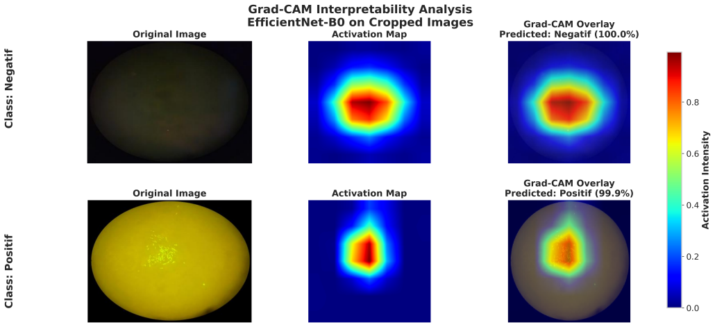
Figures


read the original abstract
Rabies remains a major public health concern across many African and Asian countries, where accurate diagnosis is critical for effective epidemiological surveillance. The gold standard diagnostic methods rely heavily on fluorescence microscopy, necessitating skilled laboratory personnel for the accurate interpretation of results. Such expertise is often scarce, particularly in regions with low annual sample volumes. This paper presents an automated, AI-driven diagnostic system designed to address these challenges. We developed a robust pipeline utilizing fluorescent image analysis through transfer learning with four deep learning architectures: EfficientNetB0, EfficientNetB2, VGG16, and Vision Transformer (ViTB16). Three distinct data augmentation strategies were evaluated to enhance model generalization on a dataset of 155 microscopic images (123 positive and 32 negative). Our results demonstrate that TrivialAugmentWide was the most effective augmentation technique, as it preserved critical fluorescent patterns while improving model robustness. The EfficientNetB0 model, utilizing Geometric & Color augmentation and selected through stratified 3fold cross-validation, achieved optimal classification performance on cropped images. Despite constraints posed by class imbalance and a limited dataset size, this work confirms the viability of deep learning for automating rabies diagnosis. The proposed method enables fast and reliable detection with significant potential for further optimization. An online tool was deployed to facilitate practical access, establishing a framework for future medical imaging applications. This research underscores the potential of optimized deep learning models to transform rabies diagnostics and improve public health outcomes.
Editorial analysis
A structured set of objections, weighed in public.
Referee Report
Summary. The manuscript presents an empirical comparison of four transfer learning architectures (EfficientNetB0, EfficientNetB2, VGG16, ViT-B16) paired with three data augmentation strategies for binary classification of rabies in a dataset of 155 fluorescent microscopy images (123 positive, 32 negative). Using stratified 3-fold cross-validation, it identifies EfficientNetB0 with Geometric & Color augmentation as optimal on cropped images and TrivialAugmentWide as the most effective augmentation technique overall. The work concludes that the approach demonstrates the viability of deep learning for automated rabies diagnosis in low-data settings and deploys an online tool for practical use.
Significance. If the internal performance estimates prove representative, the study could support AI tools for rabies diagnosis in resource-limited laboratories where expert microscopists are scarce. The comparative evaluation across architectures and augmentations, together with the deployed online tool, provides a concrete starting point for further medical imaging applications in low-resource contexts. The small dataset size and evaluation design, however, limit the strength of claims about real-world reliability and generalization.
major comments (3)
- [Methods] Methods section: The stratified 3-fold cross-validation is described without details on class-imbalance mitigation (e.g., loss weighting, oversampling, or threshold adjustment) or per-fold sensitivity/specificity values with confidence intervals. With only ~10–11 negative samples per fold, this omission makes it impossible to evaluate whether the reported optimality of EfficientNetB0 is robust or an artifact of the particular split.
- [Results] Results section: The central claim that EfficientNetB0 with Geometric & Color augmentation yields 'optimal classification performance' and that TrivialAugmentWide is 'most effective' rests entirely on internal 3-fold CV. No external held-out test set from different microscopes, laboratories, or geographies is reported, so the generalization argument for diverse low-resource settings lacks direct empirical support.
- [Discussion] Discussion/Conclusion: The statements that the method enables 'fast and reliable detection' and confirms 'viability' for real-world rabies diagnosis are not proportionate to the evidence; the 155-image dataset with acknowledged imbalance and no multi-center validation leaves open the possibility that performance reflects dataset-specific fluorescent artifacts rather than transferable pattern detection.
minor comments (2)
- [Abstract] Abstract: The claim of 'optimal classification performance' is stated without any numerical results (accuracy, sensitivity, specificity, or AUC), which weakens the abstract's utility as a standalone summary.
- [Methods] The manuscript would benefit from explicit reporting of the exact cropping coordinates or procedure and from qualitative examples showing how cropping affects fluorescent inclusion bodies.
Simulated Author's Rebuttal
We thank the referee for their constructive feedback on our manuscript. We have addressed each of the major comments in detail below and plan to revise the paper accordingly to improve clarity and temper our claims where appropriate.
read point-by-point responses
-
Referee: [Methods] Methods section: The stratified 3-fold cross-validation is described without details on class-imbalance mitigation (e.g., loss weighting, oversampling, or threshold adjustment) or per-fold sensitivity/specificity values with confidence intervals. With only ~10–11 negative samples per fold, this omission makes it impossible to evaluate whether the reported optimality of EfficientNetB0 is robust or an artifact of the particular split.
Authors: We acknowledge the need for greater transparency in our evaluation protocol. Our stratified 3-fold cross-validation ensured that each fold maintained the overall class distribution, but we did not implement additional imbalance mitigation strategies such as weighted loss or oversampling. We will revise the Methods section to document this explicitly. Additionally, we will report the per-fold performance metrics, including sensitivity and specificity, along with 95% confidence intervals estimated using bootstrap methods. This will enable assessment of the robustness of the EfficientNetB0 results across different splits, despite the small number of negative samples per fold. revision: yes
-
Referee: [Results] Results section: The central claim that EfficientNetB0 with Geometric & Color augmentation yields 'optimal classification performance' and that TrivialAugmentWide is 'most effective' rests entirely on internal 3-fold CV. No external held-out test set from different microscopes, laboratories, or geographies is reported, so the generalization argument for diverse low-resource settings lacks direct empirical support.
Authors: We agree that the absence of an external test set limits the strength of generalization claims. The study focuses on a comparative analysis within a constrained low-data regime using internal validation. We will modify the Results section to present the findings as internal performance estimates and will add explicit caveats regarding generalization to new settings. While we cannot add an external test set without new data, the online tool we deployed allows for prospective testing on external images, which we will highlight as a means for future validation. revision: partial
-
Referee: [Discussion] Discussion/Conclusion: The statements that the method enables 'fast and reliable detection' and confirms 'viability' for real-world rabies diagnosis are not proportionate to the evidence; the 155-image dataset with acknowledged imbalance and no multi-center validation leaves open the possibility that performance reflects dataset-specific fluorescent artifacts rather than transferable pattern detection.
Authors: We accept that some language in the Discussion and Conclusion may overstate the current evidence. We will revise these sections to use more measured terms, such as 'suggests the potential for' and 'provides initial support for the viability', and will include a new paragraph on limitations. This will address the small sample size, class imbalance, single-source data, and the possibility of capturing dataset-specific artifacts, while outlining plans for multi-center validation in future work. revision: yes
- Lack of external validation data from diverse sources, which cannot be provided without acquiring additional images from different laboratories or geographies.
Circularity Check
No circularity: purely empirical ML comparison with direct evaluation on fixed dataset
full rationale
The paper reports results from training and evaluating four transfer learning architectures (EfficientNetB0, EfficientNetB2, VGG16, ViTB16) plus three augmentation strategies on a fixed set of 155 images using stratified 3-fold cross-validation. No mathematical derivations, first-principles predictions, fitted parameters renamed as outputs, or self-citations that bear the central claim are present. Performance numbers and model optimality statements arise directly from standard training/evaluation loops on the provided data rather than any reduction by construction. The study is self-contained as an experimental benchmark.
Axiom & Free-Parameter Ledger
free parameters (2)
- Augmentation strategy selection
- Model architecture and cropping decisions
axioms (1)
- domain assumption Transfer learning from natural-image pretraining generalizes to fluorescent rabies microscopy images
Reference graph
Works this paper leans on
-
[1]
Magalhães, and Sunanda J
Abràmoff, Michael D., Paulo J. Magalhães, and Sunanda J. Ram. ”Image processing with ImageJ. ” Biophotonics interna- tional 11.7 (2004): 36-42
2004
-
[2]
”Image classification using deep learn- ing: A comparative study of vgg-16, inceptionv3 and efficient- net b7 models
Aggarwal, Shivam, et al. ”Image classification using deep learn- ing: A comparative study of vgg-16, inceptionv3 and efficient- net b7 models. ” 2023 3rd International Conference on Ad- vance Computing and Innovative Technologies in Engineering (ICACITE). IEEE, 2023
2023
-
[3]
”RoboFlow: A flow-based visual programming language for mobile manipulation tasks
Alexandrova, Sonya, Zachary Tatlock, and Maya Cakmak. ”RoboFlow: A flow-based visual programming language for mobile manipulation tasks. ” 2015 IEEE international conference on robotics and automation (ICRA). IEEE, 2015
2015
-
[4]
”A Child Surviving Rabies in Tunisia: A Case Report
Asma, Ben Halima, et al. ”A Child Surviving Rabies in Tunisia: A Case Report. ” Indian Journal of Pediatrics 91.3 (2024): 308- 308
2024
-
[5]
”QuPath: Open source software for digital pathology image analysis
Bankhead, Peter, et al. ”QuPath: Open source software for digital pathology image analysis. ” Scientific reports 7.1 (2017): 1-7
2017
-
[6]
”Molecular epidemiology of rabies in wild canidae in Tunisia
Bouslama, Zied, et al. ”Molecular epidemiology of rabies in wild canidae in Tunisia. ” Viruses 13.12 (2021): 2473
2021
-
[7]
Cliquet F, Aubert M, Sagné L. Development of a fuores- cent antibody virus neutralisation test (F A VN test) for the quantitation of rabies-neutralising antibody. J Immunol Meth- ods. 1998;212:79–87. https://doi.org/10.1016/S0022-1759(97) 00212-3
-
[8]
”Medical image data augmentation: techniques, comparisons and interpretations
Goceri, Evgin. ”Medical image data augmentation: techniques, comparisons and interpretations. ” Artificial intelligence review 56.11 (2023): 12561-12605
2023
-
[9]
”Alpha-divergence for classification, indexing and retrieval
Hero, Alfred O., et al. ”Alpha-divergence for classification, indexing and retrieval. ” Communication and Signal Processing Laboratory, Technical Report CSPL-328, U. Mich (2001)
2001
-
[10]
Huang, Liang-Kai, and Mao-Jiun J. Wang. ”Image thresholding by minimizing the measures of fuzziness. ” Pattern recognition 28.1 (1995): 41-51
1995
-
[11]
Sahoo, and Andrew KC Wong
Kapur, Jagat Narain, Prasanna K. Sahoo, and Andrew KC Wong. ”A new method for gray-level picture thresholding using the entropy of the histogram. ” Computer vision, graphics, and image processing 29.3 (1985): 273-285
1985
-
[12]
”Distinct Views Improve Generalization and Robustness: Combinations of Augmentations With Different Features
Kim, Keon, Hyun woo Kim, and Yong Suk Choi. ”Distinct Views Improve Generalization and Robustness: Combinations of Augmentations With Different Features. ” IEEE Access (2025)
2025
-
[13]
”EfficientNet
Koonce, Brett. ”EfficientNet. ” Convolutional neural networks with swift for Tensorflow: image recognition and dataset cate- gorization. Berkeley, CA: Apress, 2021. 109-123
2021
-
[14]
Li, Chun Hung, and C. K. Lee. ”Minimum cross entropy thresholding. ” Pattern recognition 26.4 (1993): 617-625
1993
-
[15]
”CellProfiler 3.0: Next-generation image processing for biology
McQuin, Claire, et al. ”CellProfiler 3.0: Next-generation image processing for biology. ” PLoS biology 16.7 (2018): e2005970
2018
-
[16]
”Trivialaugment: Tuning- free yet state-of-the-art data augmentation
Müller, Samuel G., and Frank Hutter. ”Trivialaugment: Tuning- free yet state-of-the-art data augmentation. ” Proceedings of the IEEE/CVF international conference on computer vision. 2021
2021
-
[17]
Otsu, N. (1975). A threshold selection method from gray-level histograms. Automatica, 11(285-296), 23-27
1975
-
[18]
Otsu, N. (1979). ”A Threshold Selection Method from Gray- Level Histograms. ” IEEE Transactions on Systems, Man, and Cybernetics 9(1): 62–66
1979
-
[19]
Ridler, Thomas Wilhelm, and S. Calvard. ”Picture thresholding using an iterative selection method. ” IEEE Trans. Syst. Man Cybern 8.8 (1978): 630-632
1978
-
[20]
Laboratory tech- niques in rabies
Rupprecht CE, Fooks AR, Abela-Ridder B. Laboratory tech- niques in rabies. 5th ed. 2018. p. 302
2018
-
[21]
Khoshgoftaar
Shorten, Connor, and Taghi M. Khoshgoftaar. ”A survey on image data augmentation for deep learning. ” Journal of big data 6.1 (2019): 1-48
2019
-
[22]
”A review on yolov8 and its advancements
Sohan, Mupparaju, Thotakura Sai Ram, and Ch Venkata Rami Reddy. ”A review on yolov8 and its advancements. ” International Conference on Data Intelligence and Cognitive Informatics. Springer, Singapore, 2024
2024
-
[23]
”Transfer learning using vgg-16 with deep convolutional neural network for classifying images
Tammina, Srikanth. ”Transfer learning using vgg-16 with deep convolutional neural network for classifying images. ” Interna- tional Journal of Scientific and Research Publications (IJSRP) 9.10 (2019): 143-150
2019
-
[24]
Laboratory techniques in rabies, volume 1, 5th ed
World Health Organization, Rupprecht, Charles E, Fooks, Anthony R & Abela-Ridder, Bernadette (2018). Laboratory techniques in rabies, volume 1, 5th ed. World Health Organi- zation. https://iris.who.int/handle/10665/310836. Licence: CC BY-NC-SA 3.0 IGO
2018
-
[25]
World Organisation for Animal Health
WOAH, Terrestrial Manual: Chapter 3.1.18 — Rabies (In- fection with rabies virus and other lyssaviruses), version adopted May 2023. World Organisation for Animal Health. [On- line]. A vailable: https://www.woah.org/fileadmin/Home/eng/ Health_standards/tahm/3.01.18_RABIES.pdf
2023
-
[26]
Rabies (infection with Rabies virus and other lyssaviruses)
World Organization of Animal Health. Rabies (infection with Rabies virus and other lyssaviruses). OIE Terrestrial Man- ual; 2018. p. 34. https://www.woah.org/fleadmin/Home/eng/ Health_standards/tahm/3.01.18_RABIES.pdf
2018
-
[27]
25, 2025
World Health Organization, ”Rabies — Epidemiology and burden of disease,” Control of Neglected Tropical Diseases, accessed: Sep. 25, 2025. [Online]. A vailable: https: //www.who.int/teams/control-of-neglected-tropical-diseases/ rabies/epidemiology-and-burden
2025
-
[28]
Rogers, and Samuel A
Zack, Gregory W., William E. Rogers, and Samuel A. Latt. ”Au- tomatic measurement of sister chromatid exchange frequency. ” Journal of Histochemistry & Cytochemistry 25.7 (1977): 741- 753
1977
discussion (0)
Sign in with ORCID, Apple, or X to comment. Anyone can read and Pith papers without signing in.